. 2023 Jun 5;155(6):e202213318.
doi: 10.1085/jgp.202213318. Epub 2023 Apr 17.

The contribution of N-terminal truncated cMyBPC to in vivo cardiac function

Affiliations

The contribution of N-terminal truncated cMyBPC to in vivo cardiac function

Katherine L Dominic et al. J Gen Physiol. .

Abstract

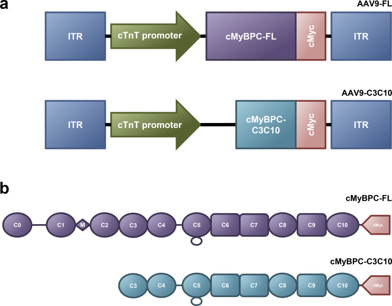
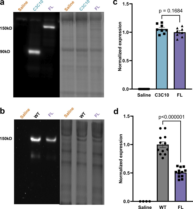
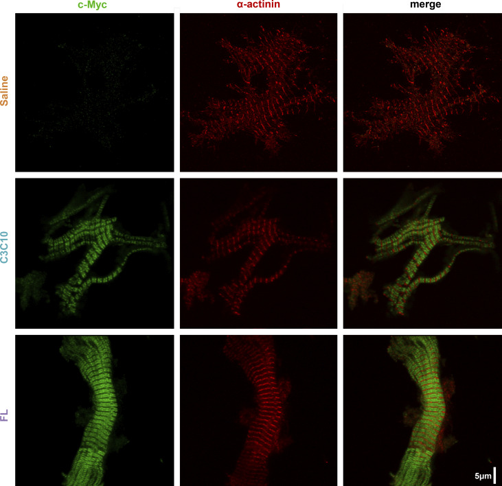
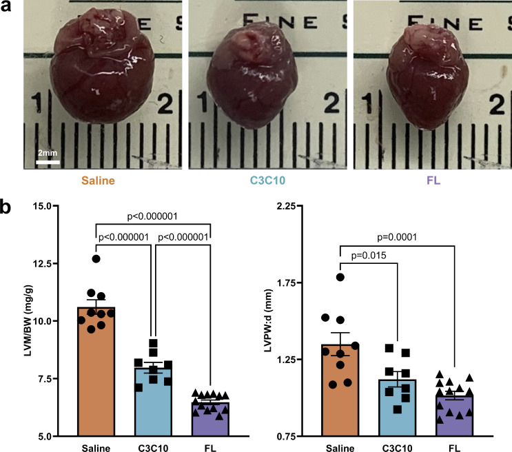
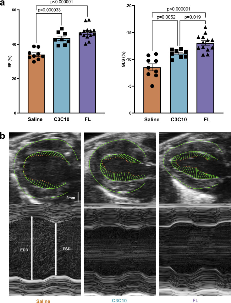
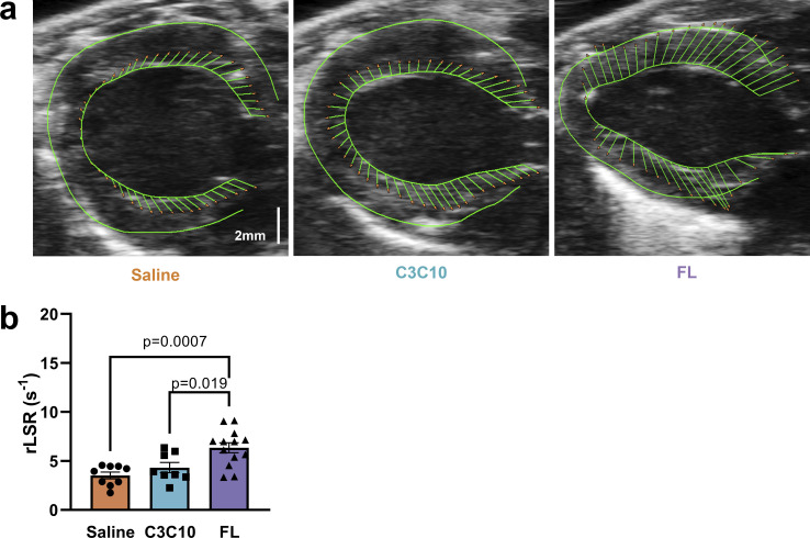
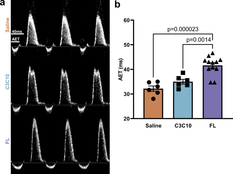
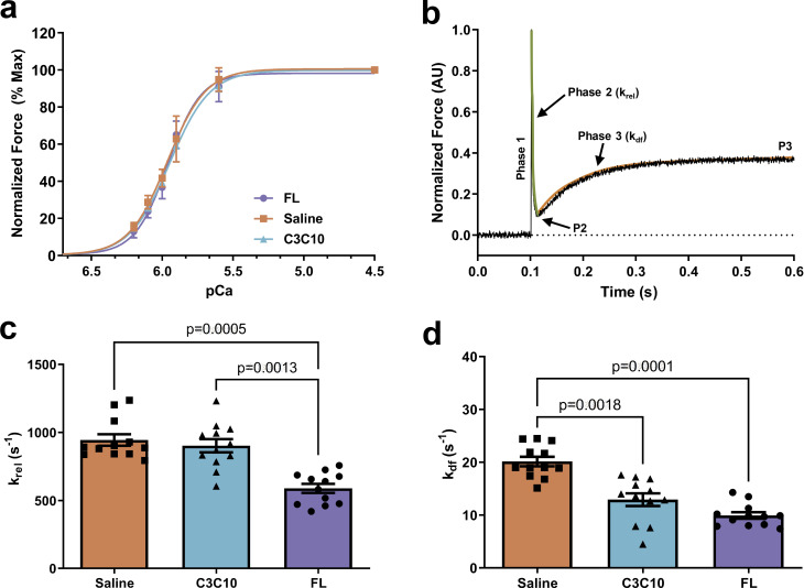
Cardiac myosin binding protein C (cMyBPC) is an 11-domain sarcomeric protein (C0-C10) integral to cardiac muscle regulation. In vitro studies have demonstrated potential functional roles for regions beyond the N-terminus. However, the in vivo contributions of these domains are mostly unknown. Therefore, we examined the in vivo consequences of expression of N-terminal truncated cMyBPC (C3C10). Neonatal cMyBPC-/- mice were injected with AAV9-full length (FL), C3C10 cMyBPC, or saline, and echocardiography was performed 6 wk after injection. We then isolated skinned myocardium from virus-treated hearts and performed mechanical experiments. Our results show that expression of C3C10 cMyBPC in cMyBPC-/- mice resulted in a 28% increase in systolic ejection fraction compared to saline-injected cMyBPC-/- mice and a 25% decrease in left ventricle mass-to-body weight ratio. However, unlike expression of FL cMyBPC, there was no prolongation of ejection time compared to saline-injected mice. In vitro mechanical experiments demonstrated that functional improvements in cMyBPC-/- mice expressing C3C10 were primarily due to a 35% reduction in the rate of cross-bridge recruitment at submaximal Ca2+ concentrations when compared to hearts from saline-injected cMyBPC-/- mice. However, unlike the expression of FL cMyBPC, there was no change in the rate of cross-bridge detachment when compared to saline-injected mice. Our data demonstrate that regions of cMyBPC beyond the N-terminus are important for in vivo cardiac function, and have divergent effects on cross-bridge behavior. Elucidating the molecular mechanisms of cMyBPC region-specific function could allow for development of targeted approaches to manipulate specific aspects of cardiac contractile function.


Figures

Figure 1.
Figure 1.
Vector design and construct maps. (a) Schematic of AAV9 vectors containing cMyBPC-FL and –C3C10 with C-terminal c-Myc tag under the control of a truncated chicken cardiac troponin T promoter. (b) Schematics of full-length (FL) cMyBPC and truncated cMyBPC consisting of domains C3 through C10 (C3C10), including the C-terminal c-Myc tag used to identify exogenously expressed cMyBPC.
Figure 2.
Figure 2.
Expression of AAV9-delivered constructs. (a) Expression of AAV9-FL and –C3C10 as determined by immunoblotting (left) against the c-Myc epitope, normalized to total protein loading (right). (b) Expression of endogenously produced cMyBPC (WT) versus AAV9-FL as determined by immunoblotting (left) against the N-terminus of cMyBPC, normalized to total protein loading (right). (c) Plot of mean ± SEM c-Myc expression normalized to the average c-Myc expression of the FL group, for n = 8 hearts per group. (d) Plot of mean ± SEM cMyBPC expression normalized to the average cMyBPC expression of the WT group. n = 12 hearts per group. Comparisons between C3C10 and FL, and WT and FL, made by one-tailed unpaired t test.
Figure 3.
Figure 3.
Incorporation of constructs into sarcomeres. Representative IHC of c-Myc- and α-actinin-stained myofibrils from respective hearts showing incorporation of AAV9-derived constructs in doublets between z-lines.
Figure 4.
Figure 4.
Morphology normalized by C3C10. (a) Representative images of hearts from mice injected with saline or AAV9-FL or –C3C10. (b) Plots of mean ± SEM LVM/BW and LVPW:d. Group comparison made by one-way ANOVA with post-hoc Tukey’s multiple comparisons test.
Figure 5.
Figure 5.
Contractile function improved by C3C10. (a) Plots of mean ± SEM EF (left) and GLS (right). Group comparison made by one-way ANOVA with post-hoc Tukey’s multiple comparisons test. (b) Representative PSLAX B-mode images (top) with vectors indicating longitudinal strain along the endocardial border. Representative SAX M-mode images (bottom) indicating end diastolic diameter (EDD) and end systolic diameter (ESD).
Figure 6.
Figure 6.
Diastolic impairment in the absence of NTDs. (a) Representative para-sternal long axis (PSLAX) B-mode images with vectors indicating longitudinal strain along the endocardial border. (b) Plot of mean ± SEM rLSR. Group comparison made by one-way ANOVA with post-hoc Tukey’s multiple comparisons test.
Figure 7.
Figure 7.
Truncated ejection in the absence of NTDs. (a) Representative pulsed wave Doppler traces depicting the velocity of blood flow through a region of interest positioned at the area of peak inflow during diastole. Velocities above the baseline represent ventricular inflow, and below baseline represent ventricular outflow. (b) AET is indicated, and plotted as mean ± SEM. Group comparison made by one-way ANOVA with post-hoc Tukey’s multiple comparisons test.
Figure 8.
Figure 8.
Effect of FL and C3C10 cMyBPC on myofilament pCa50 and cooperativity of force development (nH), and effect of FL and C3C10 cMyBPC on dynamic stretch activation parameters. (a) Force–pCa relationship of FL, C3C10, and saline-treated myocardium analyzed by plotting normalized forces generated at a range of pCa. No significant differences were observed between the groups (b). Representative force trace in response to a sudden 2% stretch in ML in an isometrically contracting FL myocardium preparation at an SL of 2.1 µm. The green line represents the rate of cross-bridge detachment (krel) and the orange line represents the rate of cross-bridge recruitment (kdf). (c) The rate of cross-bridge detachment, krel. (d) The rate of cross-bridge recruitment, kdf. Values are expressed as mean ± SEM. Data from n = 4 hearts per group with three fiber preparations per heart was analyzed in a hierarchical, nested manner using one-way repeated measures ANOVA followed by Tukey’s multiple comparisons test.

References

    1. Alfares, A.A., Kelly, M.A., McDermott, G., Funke, B.H., Lebo, M.S., Baxter, S.B., Shen, J., McLaughlin, H.M., Clark, E.H., Babb, L.J., et al. . 2015. Results of clinical genetic testing of 2,912 probands with hypertrophic cardiomyopathy: Expanded panels offer limited additional sensitivity. Genet. med. 17:880–888. 10.1038/gim.2014.205 - DOI - PubMed
    1. Cho, G.Y., Marwick T.H., Kim H.S., Kim M.K., Hong K.S., and Oh D.J.. 2009. Global 2-dimensional strain as a new prognosticator in patients with heart failure. J. Am. Coll. Cardiol. 54:618–624. 10.1016/j.jacc.2009.04.061 - DOI - PubMed
    1. de Lange, W.J., Grimes A.C., Hegge L.F., and Ralphe J.C.. 2013. Ablation of cardiac myosin-binding protein-C accelerates contractile kinetics in engineered cardiac tissue. J. Gen. Physiol. 141:73–84. 10.1085/jgp.201210837 - DOI - PMC - PubMed
    1. Doh, C.Y., Li J., Mamidi R., and Stelzer J.E.. 2019. The HCM-causing Y235S cMyBPC mutation accelerates contractile function by altering C1 domain structure. Biochim. Biophys. Acta Mol. Basis Dis. 1865:661–677. 10.1016/j.bbadis.2019.01.007 - DOI - PMC - PubMed
    1. Doh, C.Y., Bharambe N., Holmes J.B., Dominic K.L., Swanberg C.E., Mamidi R., Chen Y., Bandyopadhyay S., Ramachandran R., and Stelzer J.E.. 2022a. Molecular characterization of linker and loop-mediated structural modulation and hinge motion in the C4-C5 domains of cMyBPC. J. Struct. Biol. 214:107856. 10.1016/j.jsb.2022.107856 - DOI - PMC - PubMed

Publication types

Substances