

Bidirectional Communication Between the Brain and Other Organs: The Role of Extracellular Vesicles
- PMID: 37067749
- PMCID: PMC10106324
- DOI: 10.1007/s10571-023-01345-5
Bidirectional Communication Between the Brain and Other Organs: The Role of Extracellular Vesicles
Abstract
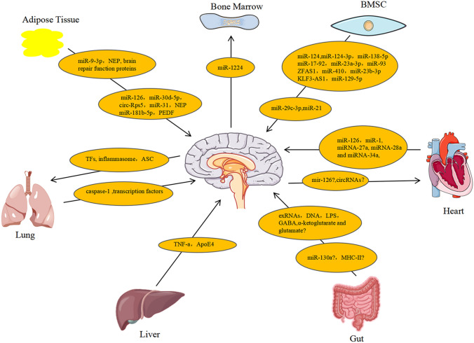
A number of substances released by the brain under physiological and pathological conditions exert effects on other organs. In turn, substances produced primarily by organs such as bone marrow, adipose tissue, or the heart may have an impact on the metabolism and function and metabolism of the healthy and diseased brain. Despite a mounting amount of evidence supports such bidirectional communication between the brain and other organs, research on the function of molecular mediators carried by extracellular vesicles (EVs) is in the early stages. In addition to being able to target or reach practically any organ, EVs have the ability to cross the blood-brain barrier to transport a range of substances (lipids, peptides, proteins, and nucleic acids) to recipient cells, exerting biological effects. Here, we review the function of EVs in bidirectional communication between the brain and other organs. In a small number of cases, the role has been explicitly proven; yet, in most cases, it relies on indirect evidence from EVs in cell culture or animal models. There is a dearth of research currently available on the function of EVs-carrying mediators in the bidirectional communication between the brain and bone marrow, adipose tissue, liver, heart, lungs, and gut. Therefore, more studies are needed to determine how EVs facilitate communication between the brain and other organs.
Keywords: Bidirectional communication; Brain; Exosomes; Extracellular vesicles; Microvesicles.
© 2023. The Author(s), under exclusive licence to Springer Science+Business Media, LLC, part of Springer Nature.
Conflict of interest statement
The authors declare that they have no conflict of interest to report.
Figures
Similar articles
-
Extracellular vesicle-mediated bidirectional communication between heart and other organs.Am J Physiol Heart Circ Physiol. 2022 May 1;322(5):H769-H784. doi: 10.1152/ajpheart.00659.2021. Epub 2022 Feb 18. Am J Physiol Heart Circ Physiol. 2022. PMID: 35179973 Free PMC article. Review.
-
Extracellular vesicle-mediated bidirectional communication between the liver and other organs: mechanistic exploration and prospects for clinical applications.J Nanobiotechnology. 2025 Mar 8;23(1):190. doi: 10.1186/s12951-025-03259-4. J Nanobiotechnology. 2025. PMID: 40055724 Free PMC article. Review.
-
Extracellular vesicles- crucial players in human pregnancy.Placenta. 2023 Sep 7;140:30-38. doi: 10.1016/j.placenta.2023.07.006. Epub 2023 Jul 26. Placenta. 2023. PMID: 37531747 Review.
-
Methods and Protocols for Using Extracellular Vesicles as Delivery Vehicles in Neuronal Research.Methods Mol Biol. 2023;2668:159-189. doi: 10.1007/978-1-0716-3203-1_13. Methods Mol Biol. 2023. PMID: 37140797
-
The Transport Mechanism of Extracellular Vesicles at the Blood-Brain Barrier.Curr Pharm Des. 2017;23(40):6206-6214. doi: 10.2174/1381612823666170913164738. Curr Pharm Des. 2017. PMID: 28914201 Review.
Cited by
-
Approximation of bone mineral density and subcutaneous adiposity using T1-weighted images of the human head.Imaging Neurosci (Camb). 2024 Dec 6;2:imag-2-00390. doi: 10.1162/imag_a_00390. eCollection 2024. Imaging Neurosci (Camb). 2024. PMID: 40800364 Free PMC article.
-
Comparison of Methods for Isolation and Characterization of Total and Astrocyte-Enriched Extracellular Vesicles From Human Serum and Plasma.J Extracell Biol. 2025 Feb 14;4(2):e70035. doi: 10.1002/jex2.70035. eCollection 2025 Feb. J Extracell Biol. 2025. PMID: 39958973 Free PMC article.
-
Brain-Periphery Axes: The Potential Role of Extracellular Vesicles-Delivered miRNAs.Biology (Basel). 2024 Dec 16;13(12):1056. doi: 10.3390/biology13121056. Biology (Basel). 2024. PMID: 39765723 Free PMC article. Review.
-
The Effect of Ionising Radiation on the Properties of Tumour-Derived Exosomes and Their Ability to Modify the Biology of Non-Irradiated Breast Cancer Cells-An In Vitro Study.Int J Mol Sci. 2025 Jan 4;26(1):376. doi: 10.3390/ijms26010376. Int J Mol Sci. 2025. PMID: 39796230 Free PMC article.
-
Role of Redox Homeostasis in the Communication Between Brain and Liver Through Extracellular Vesicles.Antioxidants (Basel). 2024 Dec 6;13(12):1493. doi: 10.3390/antiox13121493. Antioxidants (Basel). 2024. PMID: 39765821 Free PMC article. Review.
References
-
- Ay H, Koroshetz WJ, Benner T, Vangel MG, Melinosky C, Arsava EM, Ayata C, Zhu M, Schwamm LH, Sorensen AG (2006) Neuroanatomic correlates of stroke-related myocardial injury. Neurology 66(9):1325–1329. 10.1212/01.wnl.0000206077.13705.6d - PubMed
Publication types
MeSH terms
Grants and funding
LinkOut - more resources
Full Text Sources
