

Segregation of Neural Circuits Involved in Social Gaze and Non-Social Arrow Cues: Evidence from an Activation Likelihood Estimation Meta-Analysis
- PMID: 37067764
- PMCID: PMC11166804
- DOI: 10.1007/s11065-023-09593-4
Segregation of Neural Circuits Involved in Social Gaze and Non-Social Arrow Cues: Evidence from an Activation Likelihood Estimation Meta-Analysis
Abstract
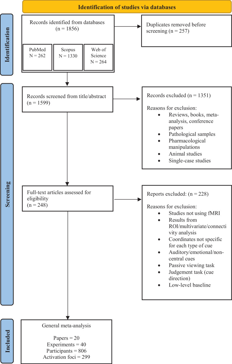
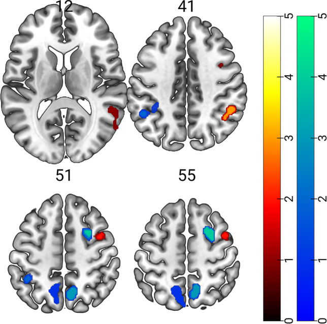
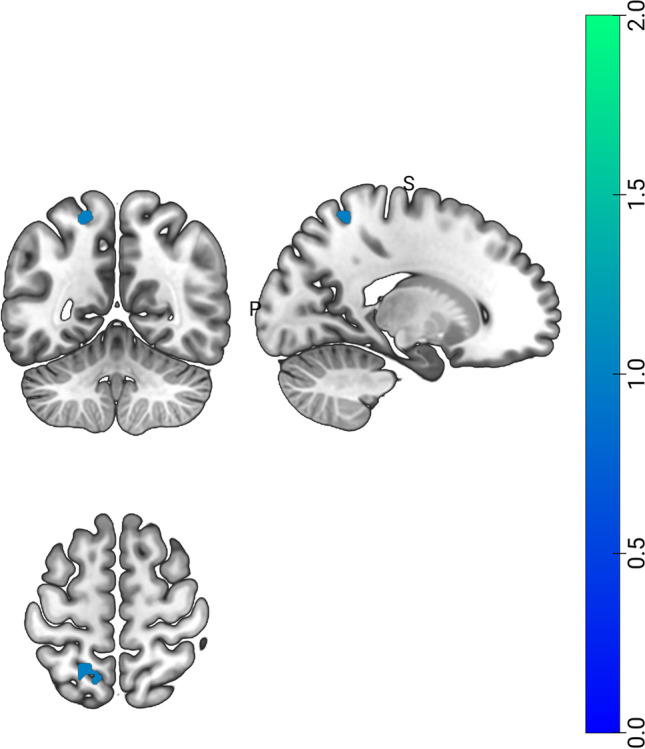
Orienting attention by social gaze cues shares some characteristics with orienting attention by non-social arrow cues, but it is unclear whether they rely on similar neural mechanisms. The present ALE-meta-analysis assessed the pattern of brain activation reported in 40 single experiments (18 with arrows, 22 with gaze), with a total number of 806 participants. Our findings show that the network for orienting attention by social gaze and by non-social arrow cues is in part functionally segregated. Orienting by both types of cues relies on the activity of brain regions involved in endogenous attention (the superior frontal gyrus). Importantly, only orienting by gaze cues was also associated with the activity of brain regions involved in exogenous attention (medial frontal gyrus), processing gaze, and mental state attribution (superior temporal sulcus, temporoparietal junction).
Keywords: ALE meta-analysis; Arrow; Frontal gyrus; Gaze; Superior temporal sulcus; Temporoparietal junction.
© 2023. The Author(s).
Conflict of interest statement
The authors have no competing interests to declare that are relevant to the content of this article.
Figures
Similar articles
-
Automatic attention orienting by social and symbolic cues activates different neural networks: an fMRI study.Neuroimage. 2006 Oct 15;33(1):406-13. doi: 10.1016/j.neuroimage.2006.06.048. Epub 2006 Sep 1. Neuroimage. 2006. PMID: 16949306
-
A common and specialized neural code for social attention triggered by eye gaze and biological motion.Neuroimage. 2024 Nov 1;301:120889. doi: 10.1016/j.neuroimage.2024.120889. Epub 2024 Oct 15. Neuroimage. 2024. PMID: 39419423
-
Attention orienting by eye gaze and arrows reveals flexibility to environmental changes.Acta Psychol (Amst). 2014 Jul;150:100-5. doi: 10.1016/j.actpsy.2014.05.003. Epub 2014 May 24. Acta Psychol (Amst). 2014. PMID: 24866453
-
The neural basis of eye gaze processing.Curr Opin Neurobiol. 2013 Jun;23(3):450-5. doi: 10.1016/j.conb.2012.11.014. Epub 2012 Dec 20. Curr Opin Neurobiol. 2013. PMID: 23266245 Review.
-
Neural mechanisms of social attention.Trends Cogn Sci. 2009 Mar;13(3):135-43. doi: 10.1016/j.tics.2008.12.006. Epub 2009 Feb 14. Trends Cogn Sci. 2009. PMID: 19223221 Review.
Cited by
-
Preserved learning of implicit regularities with predictive social cues in older adults.Front Psychiatry. 2024 Dec 10;15:1470649. doi: 10.3389/fpsyt.2024.1470649. eCollection 2024. Front Psychiatry. 2024. PMID: 39720433 Free PMC article.
References
-
- Baron-Cohen S. Mindblindness: An essay on autism and theory of mind. MIT Press; 1995.
Publication types
MeSH terms
Grants and funding
LinkOut - more resources
Full Text Sources
