. 2023 Aug 3;192(4):2871-2882.
doi: 10.1093/plphys/kiad227.

Maize β-amylase7 encodes 2 proteins using alternative transcriptional start sites: Nuclear BAM7 and plastidic BAM2

Affiliations

Maize β-amylase7 encodes 2 proteins using alternative transcriptional start sites: Nuclear BAM7 and plastidic BAM2

Kenan E Ozcan et al. Plant Physiol. .

Abstract

An unusual β-amylase7 (BAM7) gene in some angiosperms, including grasses such as maize (Zea mays), appears to encode 2 functionally distinct proteins: a nuclear-localized transcription factor (BAM7) and a plastid-localized starch hydrolase (BAM2). In Arabidopsis (Arabidopsis thaliana), these 2 proteins are encoded by separate genes on different chromosomes but their physiological functions are not well established. Using the maize BAM7 gene as a model, we detected 2 populations of transcripts by 5'-RACE which encode the predicted proteins. The 2 transcripts are apparently synthesized independently using separate core promoters about 1 kb apart, the second of which is located in the first intron of the full-length gene. The N-terminus of the shorter protein, ZmBAM7-S, begins near the 3' end of the first intron of ZmBAM7-L and starts with a predicted chloroplast transit peptide. We previously showed that ZmBAM7-S is catalytically active with properties like those of AtBAM2. Here, we report that ZmBAM7-S targets green fluorescent protein to plastids. The transcript encoding the longer protein, ZmBAM7-L, encodes an additional DNA-binding domain containing a functional nuclear localization signal. This putative dual-function gene originated at least 400 Mya, prior to the emergence of ferns, and has persisted in some angiosperms that lack a separate BAM2 gene. It appears to have been duplicated and subfunctionalized in at least 4 lineages of land plants, resulting in 2 genes resembling Arabidopsis BAM2 and BAM7. Targeting of 2 products from a single gene to different subcellular locations is not uncommon in plants, but it is unusual when they are predicted to serve completely different functions in the 2 locations.


Conflict of interest statement

Conflict of interest statement. The authors declare no conflict of interest.

Figures
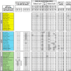
Figure 1.
Figure 1.
Selected amino acid comparisons of β-amylase2 (BAM2) (top set; yellow) and β-amylase7 BAM7 (middle set; cyan) proteins from 14 species that contain separate BAM2 and BAM7 genes and DF-BAM7 (bottom set; green) proteins from 15 species. Species and accession numbers are listed in Supplemental Table S2. Residue numbers are from soybean BAM5 and Arabidopsis BAM7. Nuclear localization signal residues were determined by Reinhold et al. (2011). Active site starch-binding residues in soybean BAM5 were determined by Laederach et al. (1999). Residues in the SBS were determined by Monroe et al. (2017, 2018), and subunit interface residues were determined by Monroe et al. (2018). White boxes indicate residues that are shared with either AtBAM7's DNA-binding domain or soybean BAM5's BAM domain. Gray boxes indicate residues that are not conserved.
Figure 2.
Figure 2.
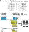
Alternative transcription of maize β-amylase7. A) Gene model of maize BAM7 (NP_001337631; GRMZM2G446515) illustrating exons (black bars), introns (black lines), and 2 putative transcriptional start sites (TSSs). N-terminal sequence of the predicted BAM7-S starting from in-frame Met residues nearest to the 3′ end of Intron 2 in 15 species containing putative DF-BAM7 genes and predictions (out of 1.0) for their chloroplast localization using 3 prediction programs (see Materials and methods for URLs). The amino acid that is highlighted gray in all species except Aegilops tauschii indicates the first residue of the gene's second exon. B) Predicted models of the 5′ ∼half of the long (ZmBAM7-L) and short (ZmBAM7-S) transcripts from maize BAM7. White bars indicate 5′ UTRs. The green region in the second transcript indicates 42 nucleotides of Intron 2 that encode the first part of the predicted chloroplast transit peptide (cTP). The binding site of the GSP used for 5′-RACE is indicated. Below the transcript models are sequences derived from 5′-RACE products aligned to the maize BAM7 gene. Cyan regions correspond to ZmBAM7-L and yellow regions correspond to ZmBAM7-S. Sequences of 5′-RACE clones are in Supplemental Table S3. Below the 5′ RACE-derived sequences are maps of 7 maize ESTs aligned to the maize BAM7 gene using the same color scheme. C) Agarose gel of 5′-RACE products. DNA in Lanes 1 and 2 underwent 25 and 35 cycles of polymerization, respectively. Brackets labeled A to D indicate regions of the gel that were excised for cloning.
Figure 3.
Figure 3.
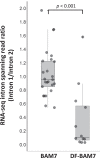
Ratios of Intron 1- to 2-spanning RNA-Seq reads for single-function (n = 25) and dual-function (n = 12) BAM7 genes from various angiosperms (listed in Supplemental Table S4). Within each box, the thick horizontal black lines indicate median values, and the boxes extend from the 25th to the 75th percentile of each set of values. Whiskers represent the values that are within 1.5× the interquartile range excluding the outliers. Transparent gray dots display the raw data. A Wilcoxon rank–sum test indicated that the means were significantly different (P < 0.001).
Figure 4.
Figure 4.
Subcellular localization of ZmBAM7-L DNA-binding domain (DBD) and ZmBAM7-S in Arabidopsis protoplasts. A) Protoplasts from wild-type Arabidopsis plants and from stable transgenic plants expressing the DNA-binding domain of ZmBAM7-L or full-length ZmBAM7-S fused to GFP. Chlorophyll autofluorescence is red, Hoechst 33342 fluorescence is cyan, and GFP fluorescence is green. Merged images combine all 3 single images. Images were collected at 20× magnification, and the scale bar represents 10 μm. B) Localization of GFP in ZmBAM7-S:GFP at 60× magnification.
Figure 5.
Figure 5.
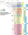
Evolution of β-amylase2 (BAM2) and β-amylase7 (BAM7) in and plants. Phylogenetic tree of Viridiplantae species ranging from charophyte algae to eudicots along with the presence of BAM2 (yellow), BAM7 (cyan), or DF-BAM7 (green) genes along with their relative genomic location: adjacent or separated. The green circle indicates the putative fusion event between the BZR1 transcription factor and BAM2. The 4 red circles indicate duplication and subfunctionalization events. Two cyan circles indicate a separation of the BAM2 and BAM7 gene onto separate chromosomes. The 3 orange circles indicate a loss of the BAM2 gene. The phylogenetic tree was generated using TimeTree: timetree.org (Kumar et al. 2017). The scale bar represents time in millions of years. Species and accession numbers are listed in Supplemental Table S4.

Comment in

Similar articles

Cited by

References

    1. Armenteros JJA, Salvatore M, Emanuelsson O, Winther O, Von Heijne G, Elofsson A, Nielsen H. Detecting sequence signals in targeting peptides using deep learning. Life Sci Alliance. 2019:2(5):e201900429. 10.26508/lsa.201900429 - DOI - PMC - PubMed
    1. Bergthorsson U, Andersson DI, Roth JR. Ohno's dilemma: evolution of new genes under continuous selection. Proc Natl Acad Sci USA. 2007:104(43):17004. 10.1073/pnas.0707158104 - DOI - PMC - PubMed
    1. Bruce BD. Chloroplast transit peptides: structure, function and evolution. Trends Cell Biol. 2000:10(10):440–447. 10.1016/S0962-8924(00)01833-X - DOI - PubMed
    1. Carrie C, Whelan J. Widespread dual targeting of proteins in land plants: when, where, how and why. Plant Signal Behav. 2013:8(8):e25034. 10.4161/psb.25034 - DOI - PMC - PubMed
    1. Chandrasekharan NP, Ravenburg CM, Roy IR, Monroe JD, Berndsen CE. Solution structure and assembly of β-amylase2 from Arabidopsis thaliana. Acta Crystallogr D Struct Biol. 2020:76(4):357–365. 10.1107/S2059798320002016 - DOI - PMC - PubMed

Publication types

Substances