Review
. 2023 Jul 1;35(4):226-234.
doi: 10.1097/BOR.0000000000000942. Epub 2023 Apr 14.

Imaging in pediatric spondyloarthritis

Affiliations
Review

Imaging in pediatric spondyloarthritis

Hallie A Carol et al. Curr Opin Rheumatol. .

Abstract

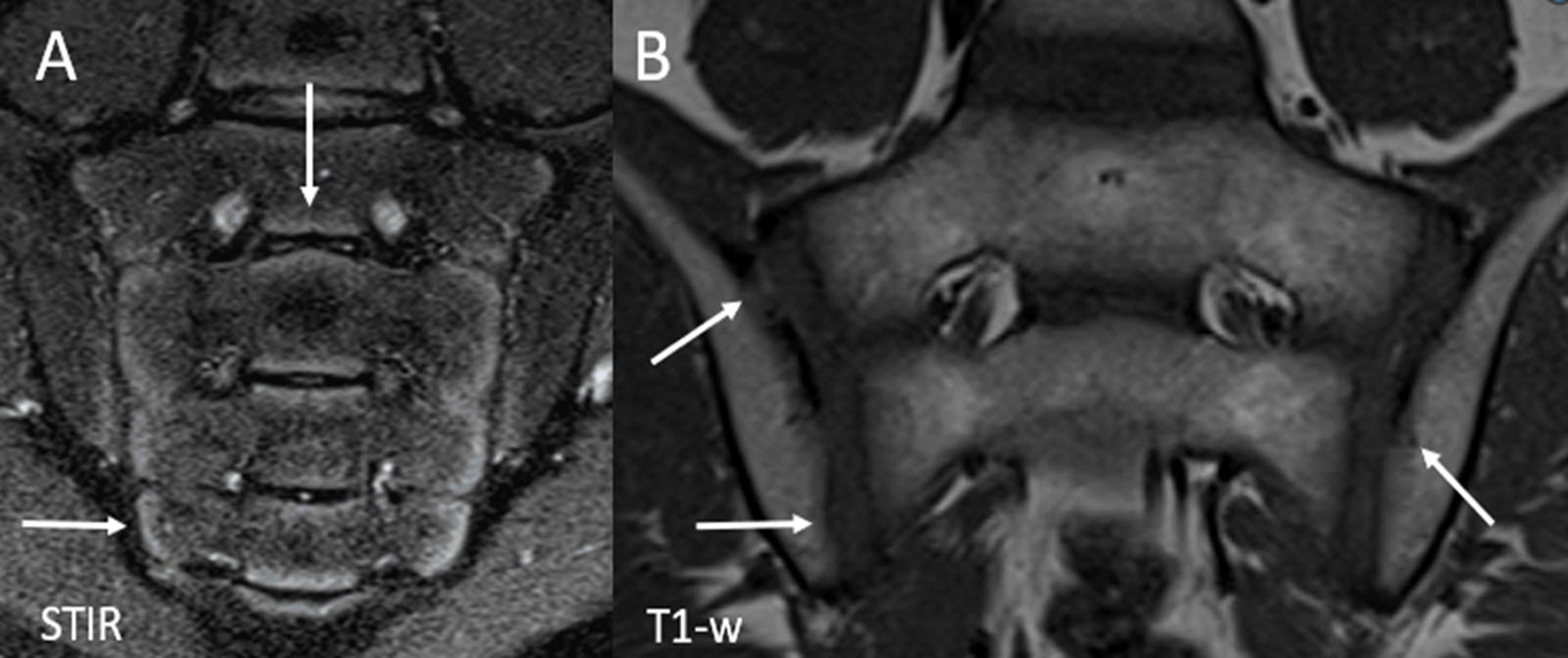
Purpose of review: Imaging is used in the diagnosis of peripheral and axial disease in juvenile spondyloarthritis (JSpA). Imaging of the joints and entheses in children and adolescents can be challenging for those unfamiliar with the appearance of the maturing skeleton. These differences are key for rheumatologists and radiologists to be aware of.

Recent findings: In youth, skeletal variation during maturation makes the identification of arthritis, enthesitis, and sacroiliitis difficult. A great effort has been put forward to define imaging characteristics seen in healthy children in order to more accurately identify disease. Additionally, there are novel imaging modalities on the horizon that are promising to further differentiate normal physiologic changes versus disease.

Summary: This review describes the current state of imaging, limitations, and future imaging modalities in youth, with key attention to differences in imaging interpretation of the peripheral joints, entheses, and sacroiliac joint in youth and adults.


Figures

Figure 1.
Figure 1.
Normal skeletal maturation. Long axis ultrasound images of the lateral aspect of the elbow in a healthy (A) 5-year-old girl, (B) 7-year-old girl and (C) 15-year-old girl. (A) Shows a completely cartilaginous capitellum, proximal radial epiphysis and lateral epicondylar apophysis. Normal vascularity is demonstrated by power Doppler with blood flow present in the unossified cartilage. (B) At age 7 years, there is partial ossification of the epiphyses and apophysis. The physes (arrows) remain open. (C) At age 15 years, the patient is skeletally mature with closure of the physes. D. Photograph of the elbow demonstrating transducer placement in the long axis along the lateral aspect of the elbow. C=capitellum, RE = proximal radial epiphysis, A= lateral epicondylar apophysis.
Figure 2.
Figure 2.
Dactylitis in a 29-month-old girl with pauciarticular JIA. (A) Short axis and (B) sagittal T2-weighted fat suppressed (FS) images show a large amount of fluid within the flexor tendon sheath of the third digit (solid arrows). There is adjacent soft tissue edema with resultant enlargement of the digit. A small amount of fluid is seen within the proximal interphalangeal joint (dashed arrow) and the distal interphalangeal joint. (C) Sagittal T1-weighted FS post contrast image shows diffuse enhancement of the tendon sheath (solid arrow) as well as the synovium of the proximal interphalangeal joint (dashed arrow) which is compatible with active tenosynovitis and joint synovitis, respectively.
Figure 3.
Figure 3.
Large versus small field of view imaging. (A) Coronal T2-weighted fat suppressed (FS) large field of view image of the pelvis shows a small amount of marrow edema within the right aspect of the sacrum (arrow). B. Small field of view coronal oblique STIR image of the sacroiliac joints shows the focus of bone marrow edema within the right aspect of the sacrum, confirming the periarticular location and better demonstrating the location within the mid to lower aspect of the right S2 level. STIR= Short tau inversion recovery.
Figure 4.
Figure 4.
16-year-old boy with juvenile spondyloarthritis. (A) Coronal oblique STIR image of the sacroiliac joints shows active inflammatory changes with bone marrow edema within right and left iliac bones (solid arrows) with edema within erosion cavities (dashed arrows). There is capsulitis along the anterior aspect of the right sacroiliac joint (arrowhead). (B) Coronal oblique T1-weighted image of the sacroiliac joints depicts chronic, structural changes with erosion cavities seen along the articular surfaces of the both iliac bones (solid arrows). There is a small amount of sclerosis along the superior aspect of the left iliac bone (dashed arrow). (C) Coned down coronal T2-weighted fat suppressed (FS) image of the right hip (obtained from large field of view pelvis image) shows active enthesitis at the ligamentous insertion of the greater trochanter (arrow). STIR = Short tau inversion recovery, FH= Femoral head.
Figure 5.
Figure 5.
Normal skeletal maturation in a healthy 10-year-old girl. (A) Coronal oblique STIR image of the sacroiliac joints demonstrates physiologic hyperintense metaphyseal equivalent signal along the periphery of the sacral apophyses (arrows) and to a lesser extent, the articular aspect of the iliac bones. The signal extends along the entire aspect of the sacrum (S4 and S5), inferior to the level of the joint space. (B) Coronal oblique T1-weighted image of the sacroiliac joints shows normal undulations and irregularities along the articular surface of the joints (arrows) which are more commonly found on the iliac side. STIR = Short tau inversion recovery.

Similar articles

Cited by

References

    1. Weiss PF, Xiao R, Biko DM, Chauvin NA. Assessment of Sacroiliitis at Diagnosis of Juvenile Spondyloarthritis by Radiography, Magnetic Resonance Imaging, and Clinical Examination. Arthritis Care Res (Hoboken) 2016. Feb;68(2):187–94. - PMC - PubMed
    1. Bollow M, Braun J, Biedermann T, Mutze S, Paris S, Schauer-Petrowskaja C, et al. Use of contrast-enhanced MR imaging to detect sacroiliitis in children. Skeletal Radiol 1998. Nov;27(11):606–16. - PubMed
    1. STOLL ML, BHORE R, DEMPSEY-ROBERTSON M, PUNARO M. Spondyloarthritis in a Pediatric Population: Risk Factors for Sacroiliitis. J Rheumatol 2010. Nov;37(11):2402–8. - PMC - PubMed
    1. Pagnini I, Savelli S, Matucci-Cerinic M, Fonda C, Cimaz R, Simonini G. Early predictors of juvenile sacroiliitis in enthesitis-related arthritis. J Rheumatol 2010. Nov;37(11):2395–401. - PubMed
    1. Taxter AJ, Chauvin NA, Weiss PF. Diagnosis and Treatment of Low Back Pain in the Pediatric Population. Phys Sportsmed 2014. Feb;42(1):94–104. - PMC - PubMed

Publication types