. 2023 Apr 25;120(17):e2215253120.
doi: 10.1073/pnas.2215253120. Epub 2023 Apr 17.

Reprogramming by drug-like molecules leads to regeneration of cochlear hair cell-like cells in adult mice

Affiliations

Reprogramming by drug-like molecules leads to regeneration of cochlear hair cell-like cells in adult mice

Yi-Zhou Quan et al. Proc Natl Acad Sci U S A. .

Abstract

Strategies to overcome irreversible cochlear hair cell (HC) damage and loss in mammals are of vital importance to hearing recovery in patients with permanent hearing loss. In mature mammalian cochlea, co-activation of Myc and Notch1 reprograms supporting cells (SC) and promotes HC regeneration. Understanding of the underlying mechanisms may aid the development of a clinically relevant approach to achieve HC regeneration in the nontransgenic mature cochlea. By single-cell RNAseq, we show that MYC/NICD "rejuvenates" the adult mouse cochlea by activating multiple pathways including Wnt and cyclase activator of cyclic AMP (cAMP), whose blockade suppresses HC-like cell regeneration despite Myc/Notch activation. We screened and identified a combination (the cocktail) of drug-like molecules composing of small molecules and small interfering RNAs to activate the pathways of Myc, Notch1, Wnt and cAMP. We show that the cocktail effectively replaces Myc and Notch1 transgenes and reprograms fully mature wild-type (WT) SCs for HC-like cells regeneration in vitro. Finally, we demonstrate the cocktail is capable of reprogramming adult cochlea for HC-like cells regeneration in WT mice with HC loss in vivo. Our study identifies a strategy by a clinically relevant approach to reprogram mature inner ear for HC-like cells regeneration, laying the foundation for hearing restoration by HC regeneration.

Keywords: RNAseq; adult mouse cochlea; hair cell regeneration; reprogramming; small moleules and siRNA.


Conflict of interest statement

The authors have organizational affiliations to disclose, Z.-Y.C. is a co-founder and a SAB member of Salubritas Therapeutics, which is developing treatments for hearing loss including genome editing, inner ear regeneration, novel delivery, and gene therapy. Z.-Y.C. has ownership of over 5% equity of Salubritas Therapeutics. Y.-Z.Q. and Z.-Y.C. are co-inventors on a patent application that has been filed based on the study by MEE. A patent application on the combination of small molecules/siRNAs in hair cell regeneration has been filed.

Figures

Fig. 1.
Fig. 1.
VPA/MYC synergistically reprogram adult SCs for HC-like cell regeneration. (A) A schematic diagram illustrating the experimental procedure of the cultured adult cochleae treated transiently by Dox to induce Myc and VPA to activate NICD (DV) and HC-like cell induction by Ad.Atoh1. (B and C) Vehicle (sterile water)/Ad.Atoh1 or Dox/VPA/Ad.Atoh1-treated adult (P30) rtTA/tet-Myc/Atoh1-GFP mice cochleae were labelled with MYO7A/GFP. Regenerated HC-like cells (MYO7A+/Atoh1-GFP+) were seen in the DV-treated group and occasional HCs were seen in the control sample. GFP+ cells were Ad.Atoh1 infected. (D and E) Quantification and comparison of regenerated HC-like cells and GFP+ cells in the apical turn of the cultured cochleae between Dox/VPA/Ad.Atoh1-treated and vehicle/Ad.Atoh1-treated groups. Significantly more HC-like cells were seen in the DV-treated than in control samples (D) and a similar number of GFP+ cells, i.e., the Ad.Atoh1-infected cells, were seen in the two groups (E). ***P < 0.001, two-tailed unpaired Student’s t test. Error bar, mean ± SEM, n = 5. (FI) Images of scanning electron microscopy (SEM) showing immature stereocilia from regenerated HC-like cells in rtTA/tet-Myc/Atoh1-GFP cochlea treated with Dox/VPA and Ad.Atoh1 in vitro. Arrows point to kinocilia. (JM) Regenerated HC-like cells (ESPN+) co-labeled with SOX2 (arrows) were seen in a cultured adult (P30) rtTA/tet-Myc mouse cochlea treated with Dox/VPA/Ad.Atoh1. Arrows point to the SOX2+/ESPN+ double-positive HC-like cells. SE: sensory epithelial region. (Scale bar in FI: 2 μm; JM: 10 μm.)
Fig. 2.
Fig. 2.
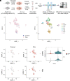
Single-cell RNA sequencing identified putative cell types across the sensory epithelium from adult mouse cochlear explant culture. (A) Schematic diagram depicting the experimental setup for collection and processing of cochlear explants for the 10× Genomics Chromium Single-Cell 3′ Gene Expression workflow. (B) UMAP plot showing the putative clusters from the integrated analysis of control and Dox groups. (C) UMAP plot of 2,395 cells displaying each cell colored according to putative cluster assignment. (D) UMAPs colored by the normalized log-transformed expression of Notch1 and Myc, showing Dox-induced transgene expression. The color key from gray to red indicates relative expression levels from low to high. (E) Violin plots illustrating normalized and Log-transformed expression profiles of Notch1 and Myc transgenes between control and Dox-induced cochleae.
Fig. 3.
Fig. 3.
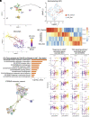
Computational lineage modeling analysis for the reprogramming trajectory for the interdental cell (IdC) cluster. (A) Subclustering of the interdental cells (IdC) showing distinct accumulation of control (red) and the surrounding Dox-induced sample-derived IdC cells (light blue). (B) Construction of a pseudotime trajectory of the IdC subcluster from control to Dox-treated samples, with the black line showing the path of pseudotime, and UMAP plots are colored according to pseudotime. (C) Expression changes of the co-regulated genes within each module generated by Monocle3 analysis. The modules are consisted of genes clustered according to the correlation of expression levels. (D) Gene ontology analysis of Module 4 by DAVID using the genes whose expression changed significantly over pseudotime. The GO terms with p value less than 0.05 were considered significantly enriched by the query genes. (E) Kinetic plots showing the relative expression profiles of ten representative genes associated with Response to cAMP or Wnt Signaling Pathway over the pseudotime. (F) A functional interaction network of the key genes from the Response to cAMP and Wnt signaling pathways. The nodes represent proteins, and the colored lines represent interactions between proteins. Presence of the more lines denotes a greater degree of connection.
Fig. 4.
Fig. 4.
A cocktail of siRNAs and small molecules to replace MYC/NICD in reprogramming for HC-like cell regeneration in adult cochlea in vitro. (A) A schematic diagram illustrating the experimental procedure of VLFsiFsiM/Ad.Atoh1 induced HC-like cell regeneration. (B and C) The cocktail (VLFsiFsiM)/Ad.Atoh1.mCherry or vehicle/Ad.Atoh1.mCherry treated adult (P30) WT mouse cochlear samples were labeled with MYO7A (blue)/POU4F3 (green) in the sensory epithelial region. Numerous HC-like cells (POU4F3+/MYO7A+) were seen in the apex of the VLFsiFsiM/Ad.Atoh1.mCherry-treated cochlea. (DG) Quantification and comparison of infected cells and regenerated HC-like cells in the apical turn of the cultured cochleae between VLFsiFsiM/Ad.Atoh1.mCherry-treated and vehicle/Ad.Atoh1.mCherry-treated groups. Significantly more HC-like cells were regenerated in the VLFsiFsiM/Ad.Atoh1-treated than vehicle/Ad.Atoh1-treated samples (E and G) in the sensory epithelial (SE) and the limbus (Lib) regions, respectively. The number of Ad.Atoh1.mCherry-infected cells between the two conditions was similar (D and F). (H) A schematic diagram illustrating the experimental procedure of lineage tracing for new HC-like cells after VLFsiFsiM/Ad.Atoh1 treatment. (I) In the cultured Sox2CreER/tdT adult cochlea, Tamoxifen (Tamo) induced tdT in the SCs which transdifferentiated to HC-like cells by VLFsiFsiM/Ad.Atoh1 treatment, shown by MYO7A+/Sox2-tdT+ labeling. The arrow indicates an existing HC, which was MYO7A+/Sox2-tdT. The arrowhead indicates a regenerated HC-like cell, shown by MYO7A+/Sox2-tdT+ labeling. (J) Enlarge inset from H to show the lineage traced HC-like cells (MYO7A+/Sox2-tdT+) (arrowheads). (K) VLFsiFsiM/Ad.Atoh1-treated adult (P30) Atoh1-GFP mouse cochlear samples showed infected cells (GFP+) that transdifferentiated to HC-like cell (MYO7A+) and were able to take up FM1-43. (L) A cluster of HC-like cells (MYO7A+) was seen in the VLFsiFsiM/Ad.Atoh1-treated adult cochlea. T, Tamo: 4-hydroxytamoxifen. V: VPA; L: LiCl; F: FSK; siF: siFIR; siM: siMxi1. SE: sensory epithelial region. Lib: Limbus Region. **P < 0.01, ***P < 0.001, Student’s t test. Error bar, mean ± SEM; N = 5 to 6 in each group. Source data are provided as a Source Data file. (Scale bars, 10 μm.)
Fig. 5.
Fig. 5.
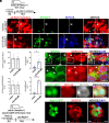
Characteristics of regenerated HC-like cells. (A) Kinocilia labeled with acetylated tubulin (Ac-TUBA4A) in WT cochlea reprogrammed by the cocktail and infected by ad.Atoh1.mCherry. (B) SLC26A5 (prestin) positive HC-like cells (arrows) in WT cochlea treated with the cocktail and ad.Atoh1.mCherry. (C) The ganglion neurites (labeled with NF-H) were in contact with the immature synaptic ribbons in regenerated HC-like cell (Atoh1-GFP+/PVALB+). (D and E) Low and high magnification pictures delineating the in vivo lineage tracing result. The Tamoxifen-treated adult Sox2-tdTomato mice (4-wk-old) were i.p. injected with Kanamycin/Furosemide (Kana/Furo) to kill hair cells for 7 d, with the subsequent delivery of the cocktail (VLFsiFsiM) into the middle ear space, followed by the injection of Ad.Atoh1-GFP into the inner ear by cochleostomy for 21 d. In the reprogrammed mice, Ad.Atoh1-GFP-treated adult cochleae showed that most regenerated HC-like cells were of SC origin (arrows, ESPN+/GFP+/Sox2-tdT+) in the outer hair cell region (OHCr) from the apex to the midturn. SE: sensory epithelial region.
Fig. 6.
Fig. 6.
Reprogramming and HC-like cell regeneration in a mouse model with HC loss in vivo. (A) A schematic diagram illustrating the experimental procedure that adult C57BL/6J mice were treated by Kanamycin/Furosemide (Kana/Furo) to kill hair cells, with the subsequent delivery of the cocktail (VLFsiFsiM) or vehicle (sterile water plus 0.1%DMSO) into the middle ear space, followed by the injection of Ad.Atoh1.mCherry into the inner ear by cochleostomy. (B) After Kana/Furo treatment, vehicles/Ad.Atoh1-mCherry-treated adult C57BL/6J cochleae showed scarce hair cells (arrows, ESPN+/mCherry+) in the outer hair cell region (OHCr) of the apex-middle turn. (C) After Kana/Furo treatment, cocktail/Ad.Atoh1.mCherry-treated adult C57BL/6j cochleae showed many hair cells (arrows, ESPN+/mCherry+) in the outer hair cell regions (OHCr) from the apex to the midturn. (D) Quantification and comparison of regenerated HC-like cells showed a significant increase in the HC-like cell number in the OHCr in the cocktail-treated samples compared to the vehicle-treated samples. (E) Quantification and comparison showed a comparable HC number in the IHC region in the cocktail-treated, vehicle-treated, and WT control cochlear samples. (F) Quantification and comparison showed significantly more ESPN+/Atoh1mCherry+ Cells in the cocktail-treated than vehicle-treated control cochlear samples. D: Dox; L: LiCl; F: FSK; siF: siFIR; siM: siMxi1. **P < 0.01, ***P < 0.001, ****P < 0.0001, two-tailed unpaired Student’s t test. Error bar, mean ± SEM; N = 5 to 6 in each group. Source data are provided as a Source Data file. (Scale bars, 10 μm.)
Fig. 7.
Fig. 7.
A schematic diagram summarizing the working model for SC reprogramming by the drug-like molecules and HC-like cell regeneration.

Similar articles

Cited by

References

    1. Corwin J. T., Cotanche D. A., Regeneration of sensory hair cells after acoustic trauma. Science 240, 1772–1774 (1988). - PubMed
    1. Ryals B. M., Rubel E. W., Hair cell regeneration after acoustic trauma in adult Coturnix quail. Science 240, 1774–1776 (1988). - PubMed
    1. Hudspeth A. J., How hearing happens. Neuron 19, 947–950 (1997). - PubMed
    1. Warchol M. E., Lambert P. R., Goldstein B. J., Forge A., Corwin J. T., Regenerative proliferation in inner ear sensory epithelia from adult guinea pigs and humans. Science 259, 1619–1622 (1993). - PubMed
    1. Forge A., Li L., Corwin J. T., Nevill G., Ultrastructural evidence for hair cell regeneration in the mammalian inner ear. Science 259, 1616–1619 (1993). - PubMed

Publication types