. 2023 Apr 25;120(17):e2216247120.
doi: 10.1073/pnas.2216247120. Epub 2023 Apr 17.

Ameliorating parkinsonian motor dysfunction by targeting histamine receptors in entopeduncular nucleus-thalamus circuitry

Affiliations

Ameliorating parkinsonian motor dysfunction by targeting histamine receptors in entopeduncular nucleus-thalamus circuitry

Jian-Ya Peng et al. Proc Natl Acad Sci U S A. .

Abstract

In Parkinson's disease (PD), reduced dopamine levels in the basal ganglia have been associated with altered neuronal firing and motor dysfunction. It remains unclear whether the altered firing rate or pattern of basal ganglia neurons leads to parkinsonism-associated motor dysfunction. In the present study, we show that increased histaminergic innervation of the entopeduncular nucleus (EPN) in the mouse model of PD leads to activation of EPN parvalbumin (PV) neurons projecting to the thalamic motor nucleus via hyperpolarization-activated cyclic nucleotide-gated (HCN) channels coupled to postsynaptic H2R. Simultaneously, this effect is negatively regulated by presynaptic H3R activation in subthalamic nucleus (STN) glutamatergic neurons projecting to the EPN. Notably, the activation of both types of receptors ameliorates parkinsonism-associated motor dysfunction. Pharmacological activation of H2R or genetic upregulation of HCN2 in EPNPV neurons, which reduce neuronal burst firing, ameliorates parkinsonism-associated motor dysfunction independent of changes in the neuronal firing rate. In addition, optogenetic inhibition of EPNPV neurons and pharmacological activation or genetic upregulation of H3R in EPN-projecting STNGlu neurons ameliorate parkinsonism-associated motor dysfunction by reducing the firing rate rather than altering the firing pattern of EPNPV neurons. Thus, although a reduced firing rate and more regular firing pattern of EPNPV neurons correlate with amelioration in parkinsonism-associated motor dysfunction, the firing pattern appears to be more critical in this context. These results also confirm that targeting H2R and its downstream HCN2 channel in EPNPV neurons and H3R in EPN-projecting STNGlu neurons may represent potential therapeutic strategies for the clinical treatment of parkinsonism-associated motor dysfunction.

Keywords: H2R; H3R; Parkinson’s disease; entopeduncular nucleus; histamine.


Conflict of interest statement

The authors declare no competing interest.

Figures

Fig. 1.
Fig. 1.
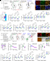
Histaminergic innervation from the TMN to the EPN excites EPN neurons by activating postsynaptic H2R, albeit its action is negatively regulated by activating presynaptic H3R. (A) Diagram depicting the AAV injection and histological reconstruction of the injection map in the TMN region of the mouse brain (n = 9). (B) Representative coronal images showing the AAV injection site and histaminergic neurons in the TMN. (C) Left panel: Representative coronal sections with NeuN staining revealed the reference map of the EPN region; Right four panels: Representative coronal images indicate that the histaminergic fibers from the TMN were localized close to the GABAergic EPN neurons (indicated by arrowheads). In these mouse brain sections, the EPN region was immediately below the internal capsule (ic) region. (D) Diagram depicting a sagittal mouse brain section displaying the location of the EPN between −1.06 and −1.58 mm from the bregma and the representative image of coronal brain slices exhibiting the area of the EPN and EPN neurons (indicated by arrowhead) investigated in this mouse. (E) In the voltage-clamp configuration, histamine (1 to 30 μM) induced an inward current in the recorded EPN neurons in a dose-dependent fashion in the presence of TTX (0.3 µM), and the concentration–response curves for histamine on the recorded EPN neurons (n = 10). (F) In the current clamp configuration, histamine and histamine together with IPP, potent and selective H3R antagonist, on the firing rate of a recorded EPN neuron and a representative discharge before, during, and after histamine and histamine together with IPP application. (G) The effect of histamine and IPP on the firing rate of the recorded EPN neurons over time and a comparison of the firing rate before histamine application and at the maximum after the application of histamine and histamine together with IPP (n = 10). Data are represented as mean ± SEM; ns, no statistical difference, ***P < 0.001.
Fig. 2
Fig. 2
CRISPR/Cas9-based downregulation of H2R in EPNPV neurons or H3R in EPN-projecting STNGlu neurons prevents the histaminergic agonist-induced promotion of motor performance. (A) Schematic of virus injection into the bilateral EPN and histological reconstruction of the injection map in LSL-Cas9-tdTomato::PV-Cre mice (n = 10). (B) Representative images depicting Cas9 and Hrh2 sgRNA expression on EPNPV neurons. (C) Relative quantification in numerical density of EPNPV neurons coexpressed with AAV-neg and AAV-Hrh2 sgRNA (n = 20). (D) Effect of dimaprit (30 μM) on the inward currents in EPNPV neurons from control, AAV-neg, and AAV-Hcn2 sgRNA-injected mice (n = 10). (E) Schematic diagram of the micro-osmotic pump implanted in the bilateral EPN, the micro-osmotic probe at the injection site (arrowheads) of the EPN, and the histological reconstruction of the injection map of the bilateral EPN across 10 animals after the behavioral test. (F) Bilateral microinfiltration of dimaprit (300 nM) into the EPN in the accelerating rotarod and balance beam tests in control, AAV-neg, and AAV-Hcn2 sgRNA-injected mice (n = 10). (G) Schematic of virus injection into bilateral STN or EPN and histological reconstruction of the injection map in LSL-Cas9-tdTomato::Vglut2-Cre mice (n = 10). (H) Representative images displaying Cas9 and Hrh3 sgRNA expression on STNGlu neurons. (I) Relative quantification in numerical density of STNGlu neurons coexpressed with AAV-neg and AAV-Hrh3 sgRNA (n = 20). (J) The effect of RAMH (10 μM) on mEPSC frequency and amplitude on EPNPV neurons in control-, AAV-neg-, and AAV-Hrh3 sgRNA-injected mice (n = 10). (K) Bilateral microinfiltration of RAMH (100 nM) into the EPN in the accelerating rotarod and balance beam tests in control-, AAV-neg-, and AAV-Hrh3 sgRNA-injected mice (n = 10). Data are represented as mean ± SEM; ns, no statistical difference, *P < 0.05, **P < 0.01, and ***P < 0.001.
Fig. 3.
Fig. 3.
Histamine levels are compensatorily elevated in the EPN, and histamine ameliorates motor dysfunction by activating H2R and H3R, and blocking HCN channels on EPNPV neurons prevents the effect of dimaprit by decreasing the neuronal firing rate and irregularizing the neuronal firing pattern in 6-OHDA-lesioned mice. (A) Histamine levels in the EPN from the same group of 10 mice at the various time points (0, 3, 7, 14, and 21 d) in normal and 6-OHDA-lesioned sides. (B) Area density of histaminergic afferent fibers in the EPN from different groups of 10 mice at each time point (0, 3, 7, 14, and 21 d) in normal and 6-OHDA-lesioned sides. (CE) Ipsilateral microinfiltration of saline, histamine (100 nM), dimaprit (300 nM), RAMH (100 nM), histamine (100 nM) together with ranitidine (3 nM), and histamine (100 nM) together with IPP (10 nM) into the EPN on motor performance in the adhesive removal test, gait test, and pole test in normal and 6-OHDA-lesioned mice (n = 10). (F) Diagram depicting the experimental timeline, virus injection into the EPN, and histological reconstruction of the injection map (n = 10 mice). (G) MEA integrated with optogenetic fiber implanted in the EPN of PV-Cre mice. (H) Representative images of the EPN neurons expressing ChR2. (I) ChR2-expressing EPNPV neurons excited by optogenetic stimulation were used for further analysis (n = 10). (J and K) Representative oscilloscope traces, firing rate, and ISI indicate the firing activity of EPNPV neurons when saline, dimaprit (300 nM), ZD7288 (100 nM), and ZD7288 (100 nM) together with dimaprit (300 nM) were microinfiltrated into the EPN using a micro-osmotic pump in normal and 6-OHDA-lesioned free-moving mice. (L) Group data indicating the effect of dimaprit, ZD7288, and ZD7288 together with dimaprit on the firing rate, CV of the ISI, and burst rate of EPNPV neurons in normal and 6-OHDA-lesioned mice (n = 10). Data are represented as mean ± SEM; ns, no statistical difference, *P < 0.05, **P < 0.01, and ***P < 0.001.
Fig. 4.
Fig. 4.
CRISPR/Cas9-based downregulation of HCN2 in EPNPV neurons aggravates motor dysfunction and prevents the ameliorating effect of dimaprit in 6-OHDA-lesioned mice. (A) Diagram depicting the experimental timeline, virus injection into the EPN, histological reconstruction of the injection map, and optogenetic stimulation-integrated MEA recordings of EPNPV neurons in 6-OHDA-lesioned LSL-Cas9-tdTomato::PV-Cre mice (n = 20). (B) Representative image of a coronal brain slice displaying expression of Cas9, sgRNA, and coexpression of Cas9 and sgRNA on the same EPNPV neurons. (C) ChR2-expressing EPNPV neurons excited by optogenetic activation were used for further analysis (n = 10). (DF) CRISPR/Cas9-based downregulation of HCN1-4 on the firing rate, CV of ISI, and burst rate of EPNPV neurons upon microinfiltration of saline or dimaprit into the EPN (n = 10). (GI) Downregulation of HCN1-4 in EPNPV neurons on motor dysfunction in the adhesive removal test, gait test, and pole test during microinfiltration of saline or dimaprit into the EPN (n = 10). Data are represented as mean ± SEM; ns, no statistical difference, **P < 0.01 and ***P < 0.001.
Fig. 5.
Fig. 5.
CRISPR/Cas9-based downregulation of H3R in EPN-projecting STNGlu neurons increases the firing rate of EPNPV neurons and aggravates the motor dysfunction, whereas pharmacological activation of H3R on EPN-projecting STNGlu neurons ameliorates motor dysfunction in 6-OHDA-lesioned mice. (A) Diagram depicting the experimental timeline, virus injection into the STN and EPN, histological reconstruction of the injection map, and optogenetic stimulation-integrated MEA recordings of EPNPV neurons in 6-OHDA-lesioned LSL-Cas9-tdTomato::PV-Cre mice (n = 20). (B) Upper panel: Representative image of the coronal brain slice displaying Cas9, sgRNA expression, and Cas9 and sgRNA coexpression on the same STN neurons; Lower panel: EPNPV neurons expressing ChR2, and EPN-projecting STNGlu fibers in the EPN. (C) ChR2-expressing EPNPV neurons excited by optogenetic activation were used for further analyses (n = 10). (D–F) CRISPR/Cas9-based downregulation of H3R in EPN-projecting STNGlu neurons on the firing rate, CV of ISI, and burst rate of EPNPV neurons during microinfiltration of saline or RAMH into the EPN (n = 10). (G–I) Downregulation of H3R in EPN-projecting STNGlu neurons on motor dysfunction in the adhesive removal test, gait test, and pole test during microinfiltration of saline or RAMH into the EPN (n = 10). (J) Diagram depicting the experimental timeline, virus injection into the STN and EPN, histological reconstruction of the injection map, and optogenetic activation of the axon terminal of EPN-projecting STNGlu neurons in vivo and in vitro in 6-OHDA-lesioned Vglut2-Cre mice (n = 20). (K) Representative image displaying EPNPV neurons, and the ChR2-expressing fibers of EPN-projecting STNGlu neurons in the EPN. (L) Representative images displaying EPNPV neurons and EPN-projecting STNGlu fibers under fluorescent field, and EPNPV neurons (arrowhead) under differential interference contrast field were selected for further voltage-clamp recordings. (M) Traces of recorded eEPSCs in EPNPV neurons evoked by the optogenetic activation of glutamatergic afferents were recorded at −70 and +40 mV in ACSF during application of RAMH (3 μM) and coapplication of NBQX (10 μM) and D-AP5 (20 μM). (N and O) Effect of RAMH on the amplitude of AMPA (N; n = 10) and NMDA eEPSCs (O; n = 10) in EPNPV neurons. (P–R) Microinfiltration of saline or RAMH into the EPN on the motor dysfunction in the adhesive removal test, gait test, and pole test upon optogenetic activation of the EPN-projecting STNGlu terminals in EPN (n = 10). Data are represented as mean ± SEM; ns, no statistical difference, *P < 0.05, **P < 0.01, and ***P < 0.001.

References

    1. Baker K. B., et al. , Somatotopic organization in the internal segment of the globus pallidus in Parkinson’s disease. Exp. Neurol. 222, 219–225 (2010). - PMC - PubMed
    1. Mullie Y., Arto I., Yahiaoui N., Drew T., Contribution of the entopeduncular nucleus and the globus pallidus to the control of locomotion and visually guided gait modifications in the cat. Cereb. Cortex 30, 5121–5146 (2020). - PMC - PubMed
    1. Fougere M., et al. , Optogenetic stimulation of glutamatergic neurons in the cuneiform nucleus controls locomotion in a mouse model of Parkinson’s disease. Proc. Natl. Acad. Sci. U.S.A. 118, e2110934118 (2021). - PMC - PubMed
    1. Adam E. M., Brown E. N., Kopell N., McCarthy M. M., Deep brain stimulation in the subthalamic nucleus for Parkinson’s disease can restore dynamics of striatal networks. Proc. Natl. Acad. Sci. U.S.A. 119, e2120808119 (2022). - PMC - PubMed
    1. Aristieta A., Ruiz-Ortega J. A., Morera-Herreras T., Miguelez C., Ugedo L., Acute L-DOPA administration reverses changes in firing pattern and low frequency oscillatory activity in the entopeduncular nucleus from long term L-DOPA treated 6-OHDA-lesioned rats. Exp. Neurol. 322, 113036 (2019). - PubMed

Publication types

Substances