. 2023 Apr 25;120(17):e2300376120.
doi: 10.1073/pnas.2300376120. Epub 2023 Apr 17.

Human airway and nasal organoids reveal escalating replicative fitness of SARS-CoV-2 emerging variants

Affiliations

Human airway and nasal organoids reveal escalating replicative fitness of SARS-CoV-2 emerging variants

Cun Li et al. Proc Natl Acad Sci U S A. .

Abstract

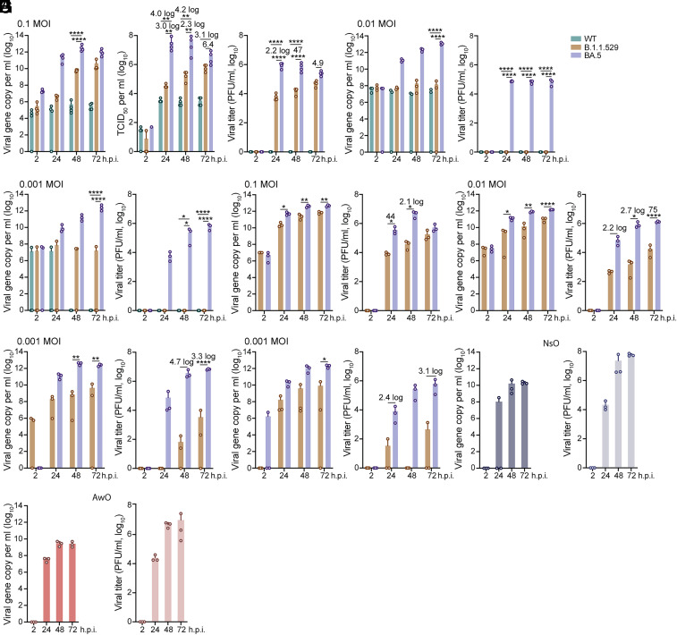
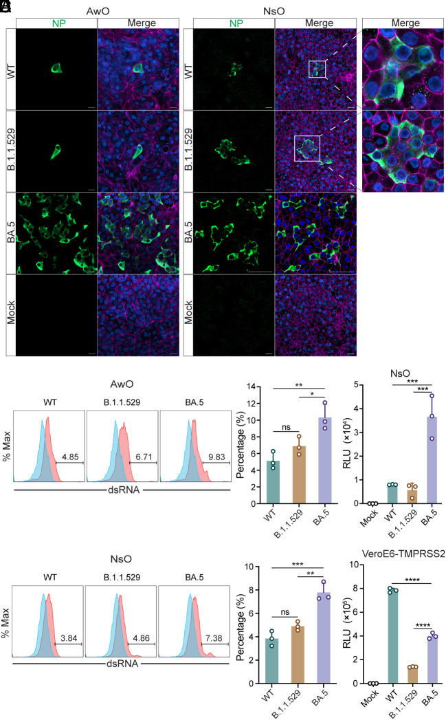
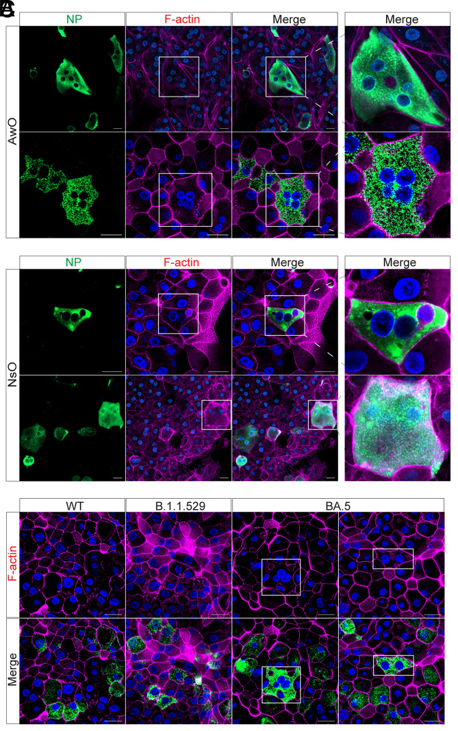
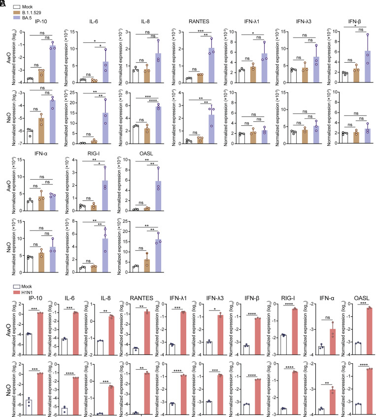
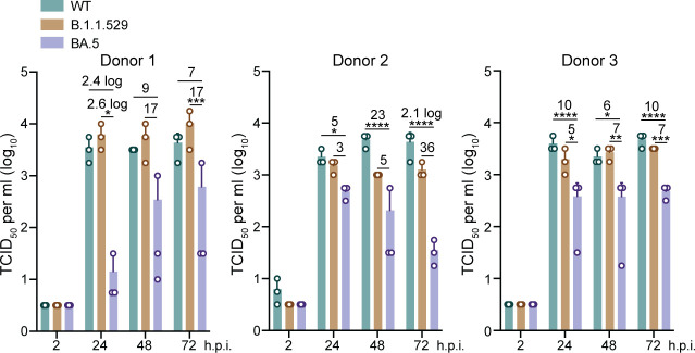
The high transmissibility of SARS-CoV-2 Omicron subvariants was generally ascribed to immune escape. It remained unclear whether the emerging variants have gradually acquired replicative fitness in human respiratory epithelial cells. We sought to evaluate the replicative fitness of BA.5 and earlier variants in physiologically active respiratory organoids. BA.5 exhibited a dramatically increased replicative capacity and infectivity than B.1.1.529 and an ancestral strain wildtype (WT) in human nasal and airway organoids. BA.5 spike pseudovirus showed a significantly higher entry efficiency than that carrying WT or B.1.1.529 spike. Notably, we observed prominent syncytium formation in BA.5-infected nasal and airway organoids, albeit elusive in WT- and B.1.1.529-infected organoids. BA.5 spike-triggered syncytium formation was verified by lentiviral overexpression of spike in nasal organoids. Moreover, BA.5 replicated modestly in alveolar organoids, with a significantly lower titer than B.1.1.529 and WT. Collectively, the higher entry efficiency and fusogenic activity of BA.5 spike potentiated viral spread through syncytium formation in the human airway epithelium, leading to enhanced replicative fitness and immune evasion, whereas the attenuated replicative capacity of BA.5 in the alveolar organoids may account for its benign clinical manifestation.

Keywords: SARS-CoV-2; organoids; syncytium formation; viral fitness.


Conflict of interest statement

The authors have the following patent filings to disclose: 1) C.L., M.C.C., H.C., K.-y.Y., and J.Z. Airway organoids, methods of making and uses thereof. Publication No: US-2021-0207081-A1. 2) C.L., M.C.C., K.-y.Y., and J.Z. Alveolar organoids, methods of making and uses thereof (patent registration number: 63/238,486). 3) C.L., M.C.C., K.-y.Y., and J.Z. Human nasal organoids and methods of making and methods of use thereof (patent registration number: 63/358,795).

Figures

Fig. 1.
Fig. 1.
SARS-CoV-2 infection and replicative fitness in human airway and nasal organoids. (AC) Airway organoids were inoculated with WT, B.1.1.529, and BA.5 at 0.1 multiplicity of infection (MOI) (A, n = 4), 0.01 MOI (B, n = 3), 0.001 MOI (C, n = 3). Culture media were harvested from infected airway organoids at the indicated hours postinoculation and applied to the detection of viral replication by the RT-PCR, TCID50, and plaque assay. Statistical significance was determined using two-way ANOVA with Tukey’s multiple comparisons test. (DG) Nasal organoids were inoculated with B.1.1.529 and BA.5 at an MOI of 0.1 (D), 0.01 (E), and 0.001 (F and G). At the indicated hours postinoculation, culture media were harvested and applied to viral load detection by RT-qPCR and viral titration by the plaque assay. n = 3. Statistical significance was determined using a two-tailed Student’s t test. Nasal organoids (H) and airway organoids (I) were inoculated with a pandemic H1N1 virus at 0.001 MOI. Culture media were harvested from infected organoids at the designated time points for viral load detection and viral titration by the plaque assay. Data represent mean and SD from a representative experiment. n = 3. *P < 0.05, **P < 0.01, ***P < 0.001, ****P < 0.0001.
Fig. 2.
Fig. 2.
Higher infection rate and entry efficiency of BA.5 in human airway and nasal organoids. (A and B) Airway (A) and nasal organoids (B) were inoculated with SARS-CoV-2 WT, B.1.1.529, and BA.5 at 0.01 MOI. At 24 hours postinoculation (h.p.i.), the infected organoids were fixed and applied to immunostaining of SARS-CoV-2 viral NP (green). Nuclei and actin filaments were counterstained with DAPI (blue) and Phalloidin-647 (purple), respectively. (Scale bar, 20 µm.) (C and D) WT-, B.1.1.529-, and BA.5-infected airway (C) or nasal (D) organoids were dissociated after fixation, then applied to immunostaining and flow cytometry to detect the percentage of virus-infected cells. Representative histograms are shown on the Left. Data presented on the right represent mean and SD from a representative experiment. n = 3. (E and F) Nasal organoids and VeroE6-TMPRSS2 cells were infected with lentiviruses pseudotyped with the spike of WT, B.1.1.529, and BA.4/5. At 72 h.p.i., cell lysates of the infected nasal organoids (E), and VeroE6-TMPRSS2 cells (F) were applied to the luciferase assay. Data represent mean and SD from a representative experiment. n = 3. Statistical significance was determined using one-way ANOVA with Tukey’s multiple comparisons test. *P < 0.05, **P < 0.01, ***P < 0.001, ****P < 0.0001. ns, not significant.
Fig. 3.
Fig. 3.
BA.5 infection and lentiviral overexpression of BA.5 spike-triggered syncytium formation in airway and nasal organoids. (A and B) Airway (A) and nasal organoids (B) were inoculated with SARS-CoV-2 BA.5 at 0.01 MOI. At 48 h.p.i., the infected organoids were fixed and applied to immunostaining of viral NP (green). Nuclei and actin filaments were counterstained with DAPI (blue) and Phalloidin-647 (purple), respectively. (Scale bar, 20 µm.) (C) Nasal organoids were transduced with lentiviruses overexpressing SARS-CoV-2 WT, B.1.1.529, and BA.4/5 spike. At 48 h posttransduction, the transduced organoids were fixed with 4% PFA and applied to immunostaining using an anti-FLAG antibody, followed by confocal imaging. Nuclei and actin filaments were counterstained with DAPI (blue) and Phalloidin-647 (purple), respectively. (Scale bar, 20 µm.)
Fig. 4.
Fig. 4.
Preferential utilization of TMPRSS2 for cellular entry and viral replication by three strains of viruses. (A) Nasal organoids were inoculated with lentiviruses pseudotyped with the spike of WT, B.1.1.529, and BA.4/5 in the presence or absence of Camostat and E64D. At 72 h.p.i., cell lysates of infected nasal organoids were applied to the luciferase assay. n = 4. (B) Nasal organoids were inoculated with SARS-CoV-2 WT, B.1.1.529, and BA.5 at 0.1 MOI in the presence or absence of Camostat and E64D. At 24 h.p.i., culture media were harvested for viral titration by the plaque assay. n = 3. Data represent mean and SD from a representative experiment. Statistical significance was determined using one-way ANOVA with Tukey’s multiple comparisons test. *P < 0.05, **P < 0.01, ***P < 0.001, ****P < 0.0001. ns, not significant.
Fig. 5.
Fig. 5.
Dampened innate immune response in SARS-CoV-2 infected human airway and nasal organoids. (A) At 48 h.p.i. of B.1.1.529, BA.5 (0.1 MOI) in the airway or nasal organoids, cell lysates were harvested to examine GAPDH normalized expression levels of IFNs, ISGs, and proinflammatory cytokines in the virus-infected and mock-infected organoids with the RT-qPCR assay. Data represent mean and SD from a representative experiment. n = 3. Statistical significance was determined using one-way ANOVA with Tukey’s multiple comparisons test. *P < 0.05, **P < 0.01, ***P < 0.001, ****P < 0.0001. ns, not significant. (B) At 48 h.p.i. of H1N1pdm (0.1 MOI) in the airway or nasal organoids, cell lysates were harvested to examine GAPDH-normalized expression levels of IFNs, ISGs, and proinflammatory cytokines in the virus-infected and mock-infected organoids with the RT-qPCR assay. Data represent the mean and SD of a representative experiment in one organoid line. Data represent mean and SD from a representative experiment. n = 3. Statistical significance was determined using the two-tailed Student’s t test. *P < 0.05, **P < 0.01, ***P < 0.001, ****P < 0.0001. ns, not significant.
Fig. 6.
Fig. 6.
Attenuated BA.5 replication in human alveolar organoids. Alveolar organoids from three donors were inoculated with SARS-CoV-2 WT, B.1.1.529, and BA.5 at 0.1 MOI. Culture media were harvested from infected organoids at the designated time points for viral titration by the TCID50 assay. Data represent mean and SD from a representative experiment in one line of organoids. n = 3. Statistical significance was determined using two-way ANOVA with Tukey’s multiple comparisons test. *P < 0.05, **P < 0.01, ***P < 0.001, ****P < 0.0001.

Similar articles

Cited by

References

    1. Brandal L. T., et al. , Outbreak caused by the SARS-CoV-2 Omicron variant in Norway, November to December 2021. Euro Surveill. 26, 2101147 (2021). - PMC - PubMed
    1. Viana R., et al. , Rapid epidemic expansion of the SARS-CoV-2 Omicron variant in southern Africa. Nature 603, 679–686 (2022). - PMC - PubMed
    1. Tegally H., et al. , Emergence of SARS-CoV-2 Omicron lineages BA.4 and BA.5 in South Africa. Nat. Med. 28, 1785–1790 (2022), 10.1038/s41591-022-01911-2. - DOI - PMC - PubMed
    1. Robinot R., et al. , SARS-CoV-2 infection induces the dedifferentiation of multiciliated cells and impairs mucociliary clearance. Nat. Commun. 12, 4354 (2021). - PMC - PubMed
    1. Han Y., et al. , Identification of SARS-CoV-2 inhibitors using lung and colonic organoids. Nature 589, 270–275 (2021). - PMC - PubMed

Publication types

Substances

Supplementary concepts