Review
. 2023 Jul;20(7):694-713.
doi: 10.1038/s41423-023-01019-8. Epub 2023 Apr 17.

Recent advancements in the B7/CD28 immune checkpoint families: new biology and clinical therapeutic strategies

Affiliations
Review

Recent advancements in the B7/CD28 immune checkpoint families: new biology and clinical therapeutic strategies

Marc C Pulanco et al. Cell Mol Immunol. 2023 Jul.

Abstract

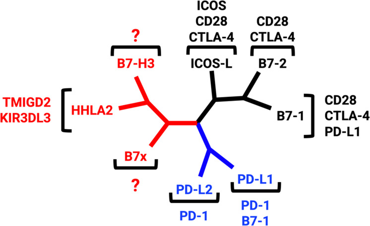
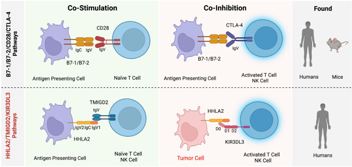
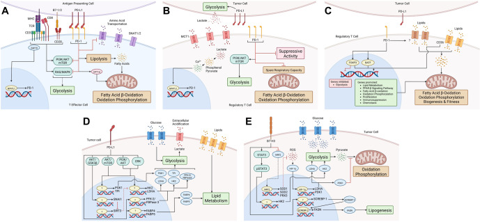
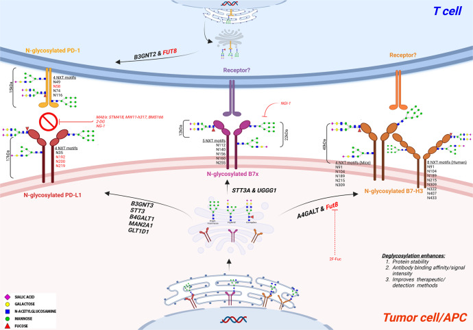
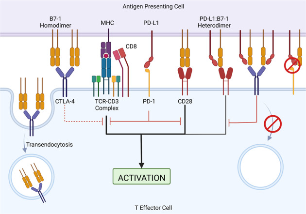
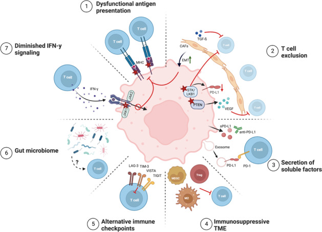
The B7/CD28 families of immune checkpoints play vital roles in negatively or positively regulating immune cells in homeostasis and various diseases. Recent basic and clinical studies have revealed novel biology of the B7/CD28 families and new therapeutics for cancer therapy. In this review, we discuss the newly discovered KIR3DL3/TMIGD2/HHLA2 pathways, PD-1/PD-L1 and B7-H3 as metabolic regulators, the glycobiology of PD-1/PD-L1, B7x (B7-H4) and B7-H3, and the recently characterized PD-L1/B7-1 cis-interaction. We also cover the tumor-intrinsic and -extrinsic resistance mechanisms to current anti-PD-1/PD-L1 and anti-CTLA-4 immunotherapies in clinical settings. Finally, we review new immunotherapies targeting B7-H3, B7x, PD-1/PD-L1, and CTLA-4 in current clinical trials.

Keywords: B7 family; glycobiology; immune checkpoints; metabolic regulators; therapy resistance.


Conflict of interest statement

XZ is the scientific co-founder of NextPoint Therapeutics. The other authors declare no competing interests.

Figures

Fig. 1
Fig. 1
The phylogenetic tree of the B7 family and their respective receptors was generated by Phylogenetic Analysis Using Parsimony (PAUP). Group I (black) consists of CD28/CTLA-4/B7-1/B7-2 and ICOS/ICOS-L. Group II (blue) contains PD-1/PD-L1/PD-L2. Group III (red) includes TMIGD2/KIR3DL3/HHLA2, B7-H3, and B7x
Fig. 2
Fig. 2
Comparison of the newest HHLA2/TMIGD2/KIR3DL3 immunoregulatory pathway and the prototype B7-1/B7-2/CD28/CTLA-4 pathway reveals some similarities and important differences. Both pathways contain ligands with dual roles (B7-1/B7-2 or HHLA2) that bind to costimulatory (CD28 or TMIGD2) or coinhibitory (CTLA-4 or KIR3DL3) receptors on T and NK cells. While CD28 or CTLA-4 binding to B7-1/2 is mutually exclusive, KIR3DL3 and TMIGD2 can simultaneously bind to different sites on HHLA2. HHLA2 but not B7-1/B7-2, which is highly expressed in various human cancers. While the B7-1/B7-2/CD28/CTLA-4 pathways are expressed in humans and mice, the HHLA2/TMIGD2/KIR3DL3 pathways are found in humans but not in mice
Fig. 3
Fig. 3
PD-1/PD-L1 and B7-H3 are metabolic regulators of effector and regulatory T cells (Tregs) and tumor cells. A In T effector cells, the TCR and CD28/B7-1/B7-2 costimulatory signals upregulate PD-1 via NFATc1 and activate RAS/MAPK and PI3K/AKT/mTOR, which stimulates glycolysis. PD-1/PD-L1 engagement inhibits the RAS/MAPK and PI3K/AKT/mTOR signaling pathways, thereby preventing the induction of glycolysis. It also inhibits the expression of the main glutamine transporters, SNAT1 and SNAT2, thus reducing amino acid uptake and metabolism. PD-1/PD-L1 promotes fatty acid β-oxidation (FAO) by increasing the expression of the mitochondrial FAO enzyme CPT1A and lipolysis, which generates fatty acids for FAO. B Tregs acquire lactate from the tumor microenvironment that is generated by tumor cell glycolysis, which is possibly regulated by PD-L1. The lactate transporter MCT1 transports lactate into Tregs, where lactate is converted into Ca2+, which induces NFATc1-mediated upregulation of PD-1. PD-1 promotes PI3K/AKT/mTOR signaling, thereby upregulating glycolysis. PD-1 inhibits the spare respiratory capacity and immunosuppressive activity of Tregs. C In intratumoral Tregs, PD-1 downstream signaling maintains the expression of FoxP3, inhibiting the expression of genes associated with glycolysis and promoting lipid metabolism, fatty acid β-oxidation (FAO), oxidation phosphorylation, peroxisome proliferator-activated receptor-β (PPAR-β) expression, proliferation, immunosuppression, and chemotaxis. These genes promote FAO, oxidation phosphorylation, and mitochondrial mass. PD-1 activates BATF to upregulate genes associated with the activation and function of Tregs. PD-1 maintains lipid uptake through unknown mechanisms. Tregs internalize lipids through CD36, activating the PPAR-β pathway to support mitochondrial biogenesis and fitness. PD-1 and CD36 may maintain the fitness, stability, and functions of intratumoral Tregs. D Tumor cells acquire glucose and lipids from the tumor microenvironment. PD-L1 promotes glucose metabolism by activating the AKT/GSK3β signaling pathway to upregulate the transcriptional repressor SNAI1 to inhibit SIRT3, which allows the expression of HK2 and LDHA. PD-L1 activates the AKT/mTOR pathway to upregulate the expression of PGK1, TPI, HK2, and LDHA. PD-L1 activates the PI3K/AKT and ERK pathways to induce the expression of HK2. Additionally, PD-L1 upregulates the expression of PFK-2/FBPase 3. Thus, PD-L1 promotes glucose metabolism by upregulating the expression of the glycolysis-related enzymes PGK1, TPI, HK2, LDHA, and PFK-2/FBPase 3. Lactate is produced from glycolysis and is exported out of the tumor cell, acidifying the tumor microenvironment. PD-L1 also promotes lipid uptake and metabolism by upregulating the expression of the fatty acid binding protein (FABP4) and FABP5, which are involved in lipid metabolism. E In tumor cells, B7-H3 inhibits the transcription factor NRF2, which governs the transcription of the antioxidant enzymes SOD1, SOD2, and PRX3; this leads to the accumulation of ROS, which stabilizes HIF-1α and increases the expression of LDHA and PDK1. LDHA participates in lactate production via glycolysis, and PDK1 inhibits pyruvate flux to the citric acid cycle. B7-H3 promotes the activation and phosphorylation of the STAT3 pathway, which upregulates the expression of HK2, which participates in glycolysis. B7-H3 promotes the transcription and translation of the transcription factor SCREBP-1, which regulates the mRNA and protein expression of fatty acid synthase (FASN), which is involved in lipogenesis
Fig. 4
Fig. 4
Glycosylation of the immune checkpoints PD-1/PD-L1, B7x, and B7-H3. N-glycosylation occurs at the consensus sequence/sequon Asn-X-Ser/Thr (NXT) in the endoplasmic reticulum (ER) lumen, followed by complete synthesis/maturation in the Golgi and ending in the plasma membrane, where it is either secreted or embedded in the membrane. Nearly 90% of the protein undergoes this co-translational modification. Major types of N-glycans are high-mannose, hybrid, and complex N-glycans. PD-1 contains 15 kDa N-glycan moieties with four glycosylation NXT motifs. B3GNT2 and Fut8 induce the glycosylation of PD-1, whereas its ligand PD-L1 contains approximately 17 kDa of N-glycan moieties and four conserved NXT motifs. The glycosylation of PD-L1 is induced by B3GNT3, STT3, B4GALT1, MAN2A1 and GLT1D1. Monoclonal antibodies include STM418, BMS166, and MW11 h317; small-molecule inhibitors such as NG-1 and the sugar analog 2DG can inhibit the interaction between PD-1/PD-L1. B7x has a differential N-glycosylation pattern ranging from highly glycosylated (~50 kDa) to less glycosylated (~40 kDa) with five NXT motifs. B7x glycosylation is induced by STT3A and UGGG1. The small-molecule inhibitor NG-1 was shown to inhibit the addition of N-glycans to B7x. B7-H3 consists of a 40 kDa N-glycan moiety with 8 NXT motif sites in humans and 4 NXT motif sites in mice. B7-H3 glycosylation is induced by A4GALT and Fut8. The sugar analog 2F-Fuc inhibits the B7-H3 core fucosylation required for its N-glycosylation. B3GNT: Beta-1,3-N-acetylglucosaminyltransferase; STT3: STT3 oligosaccharyltransferase complex catalytic subunit; UGGG1: UDP-glucose glycoprotein glucosyltransferase 1; A4GALT: Alpha 1,4-Galactosyltransferase; MAN2A1: Mannosidase α class II member; GLT1D1: Glycosyltransferase 1 containing domain 1; 2-DG: 2-Deoxy-D-glucose; NG-1: Aminobenzamide-sulfonamide inhibitor; BMS166: Cathepsin L monoclonal antibody; 2F-Fuc: 2-Fluoro-L-fucose
Fig. 5
Fig. 5
PD-L1/B7-1 cis-interaction in immune checkpoint pathways and T-cell activation. T-cell activation requires two signals: signal one is from the MHC-TCR complex and signal two is from the CD28/B7-1/B7-2 interaction. Inhibitory PD-1/PD-L1 signaling inhibits TCR and CD28 activation signals. CTLA-4 can bind to B7-1 to induce CTLA-4-mediated inhibition of the stimulatory signal. Additionally, CTLA-4 can sequester B7-1 from the cell surface of antigen-presenting cells by transendocytosis, thus removing available B7-1 from the system. The PD-L1/B7-1 heterodimer can bind to CD28 to induce a weak stimulatory signal and CTLA-4 to induce a weak inhibitory signal. CTLA-4 is unable to sequester the PD-L1/B7-1 heterodimer by transendocytosis, thereby allowing B7-1 to remain in the system. The PD-L1/B7-1 heterodimer is unable to bind to PD-1; thus, there is no PD-1-mediated inhibitory signal
Fig. 6
Fig. 6
Mechanisms of resistance to immune checkpoint blockade. The decreased efficacy of ICB in cancer patients can occur through tumor cell-intrinsic and -extrinsic factors as follows: (1) defects in antigen presentation due to the loss or reduced expression of MHC molecules; (2) the exclusion of T cells mediated by tumor-intrinsic genetic changes, CAF TGF-β signaling, and EMT; (3) the secretion of soluble and exosomal PD-L1 that potentially competes with drug binding; (4) the presence of immunosuppressive cell types in the TME that inhibit T-cell functions; (5) T-cell exhaustion and the expression of alternative inhibitory immune checkpoints; (6) a lack of bacterial diversity or the enrichment of specific ‘bad’ microbes in the gut microbiome; and (7) insensitivity to IFN-γ-mediated prevention of cancer cell apoptosis and the expression of MHC molecules. Stars denote loss of function or expression. ICB, immune checkpoint blockade; MHC, major histocompatibility complex; CAF, cancer-associated fibroblasts; sPD-L1, soluble PD-L1; M2, M2 macrophage; Treg, regulatory T-cell; MDSC, myeloid-derived suppressor cell

Similar articles

Cited by

References

    1. Ledford H, Else H, Warren M. Cancer immunologists scoop medicine Nobel prize. Nature. 2018;562:20–1. doi: 10.1038/d41586-018-06751-0. - DOI - PubMed
    1. Zang X. 2018 Nobel prize in medicine awarded to cancer immunotherapy: Immune checkpoint blockade - a personal account. Genes Dis. 2018;5:302–3. doi: 10.1016/j.gendis.2018.10.003. - DOI - PMC - PubMed
    1. Zhao R, Chinai JM, Buhl S, Scandiuzzi L, Ray A, Jeon H, et al. HHLA2 is a member of the B7 family and inhibits human CD4 and CD8 T-cell function. Proc Natl Acad Sci USA. 2013;110:9879–84. doi: 10.1073/pnas.1303524110. - DOI - PMC - PubMed
    1. Zang X, Loke P, Kim J, Murphy K, Waitz R, Allison JP. B7x: A widely expressed B7 family member that inhibits T cell activation. Proc Natl Acad Sci USA. 2003;100:10388–92. doi: 10.1073/pnas.1434299100. - DOI - PMC - PubMed
    1. Janakiram M, Shah UA, Liu W, Zhao A, Schoenberg MP, Zang X. The third group of the B7-CD28 immune checkpoint family: HHLA2, TMIGD2, B7x, and B7-H3. Immunol Rev. 2017;276:26–39. doi: 10.1111/imr.12521. - DOI - PMC - PubMed

Publication types