Review
. 2023 May;4(5):596-607.
doi: 10.1038/s43018-023-00546-7. Epub 2023 Apr 17.

Impact of context-dependent autophagy states on tumor progression

Affiliations
Review

Impact of context-dependent autophagy states on tumor progression

Mohamad Assi et al. Nat Cancer. 2023 May.

Abstract

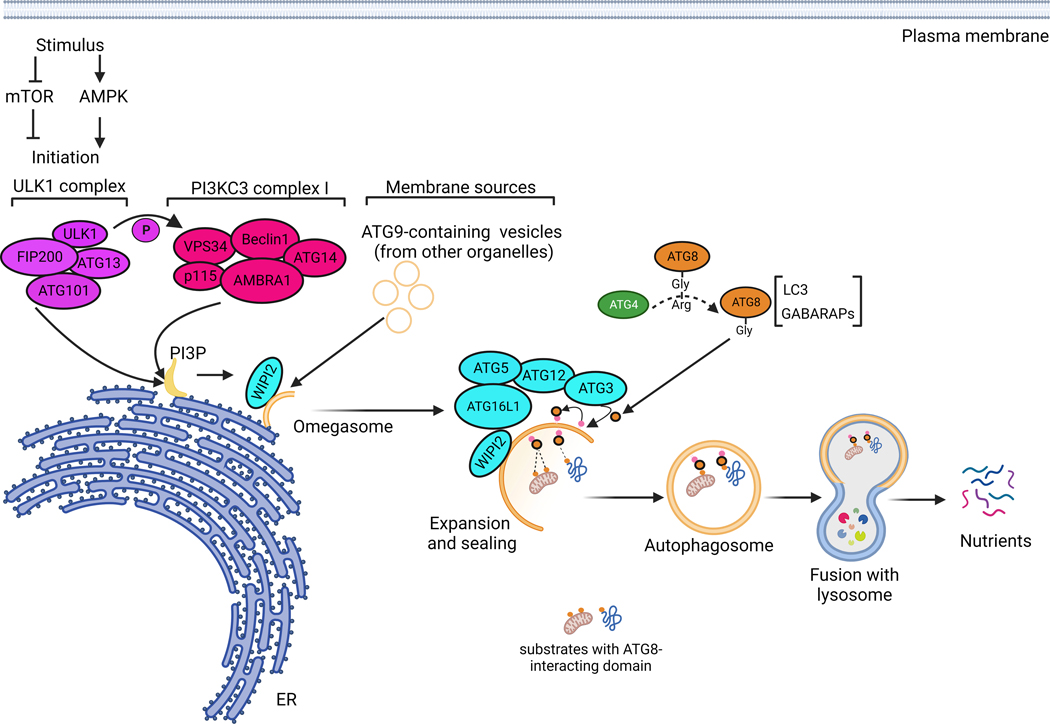
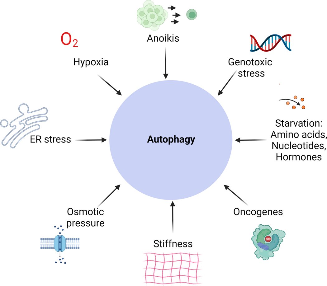
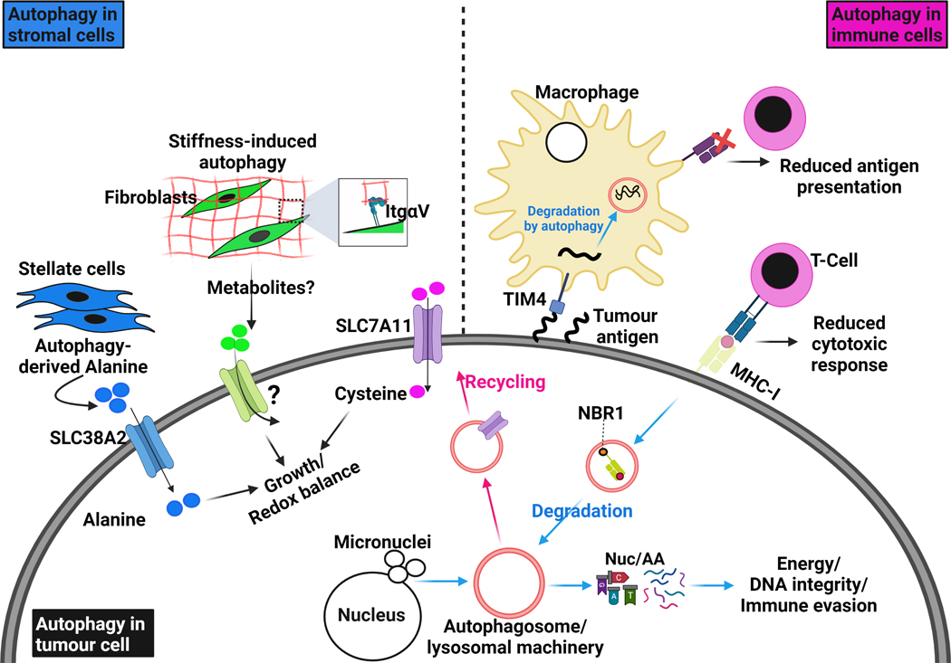
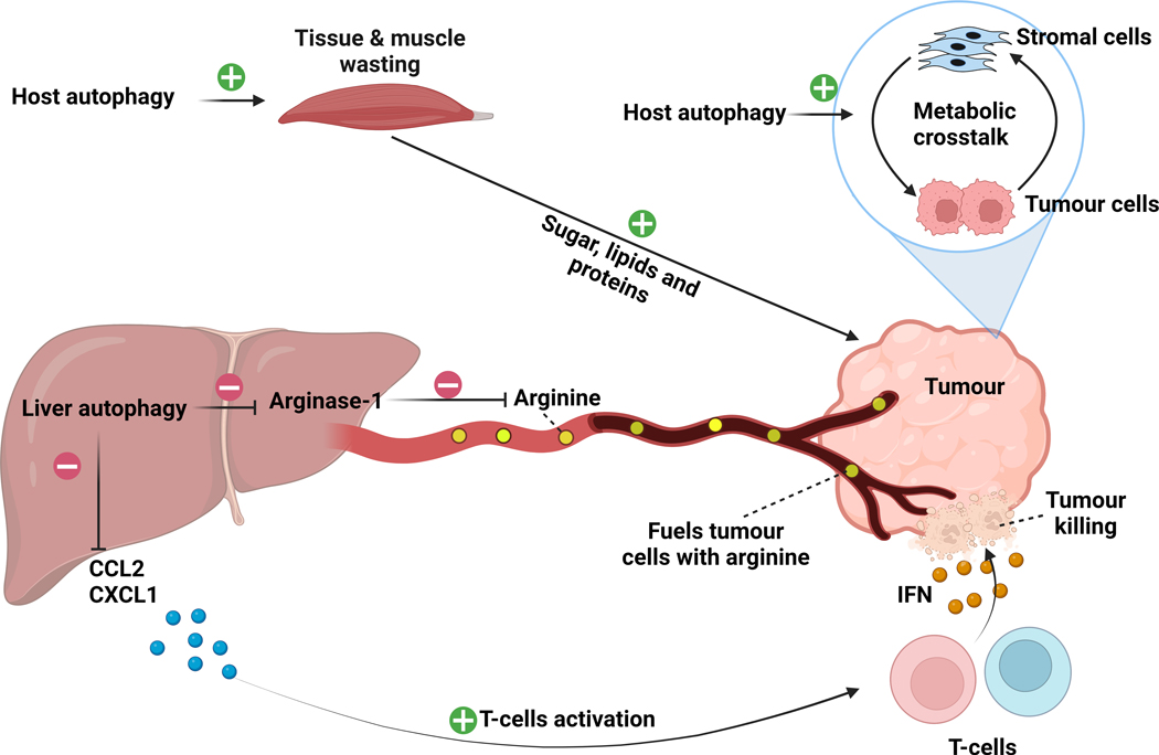
Macroautophagy is a cellular quality-control process that degrades proteins, protein aggregates and damaged organelles. Autophagy plays a fundamental role in cancer where, in the presence of stressors (for example, nutrient starvation, hypoxia, mechanical pressure), tumor cells activate it to degrade intracellular substrates and provide energy. Cell-autonomous autophagy in tumor cells and cell-nonautonomous autophagy in the tumor microenvironment and in the host converge on mechanisms that modulate metabolic fitness, DNA integrity and immune escape and, consequently, support tumor growth. In this Review, we will discuss insights into the tumor-modulating roles of autophagy in different contexts and reflect on how future studies using physiological culture systems may help to understand the complexity and open new therapeutic avenues.


Conflict of interest statement

Competing Interest

A.C.K. has financial interests in Vescor Therapeutics and is an inventor on patents pertaining to KRAS- regulated metabolic pathways and redox control pathways in pancreatic cancer, targeting GOT1 as a therapeutic approach, targeting alanine transport, and the autophagic control of iron metabolism. A.C.K. is on the scientific advisory board of Rafael/Cornerstone Pharmaceuticals, and is advisor for OncoRev, and has been a consultant for Deciphera and Abbvie. The other authors declare no competing interests. M.A. is postdoctoral fellow at New York University Langone Health.

Figures

Figure 1.
Figure 1.. Molecular mechanisms of autophagy.
In response to environmental stresses, autophagy initiation is thought to take place at the endoplasmic reticulum membranes, where the ULK1 complex activates the PI3KC3 complex by phosphorylation. The PI3KC3 complex initiates the production of phosphatidylinositol-3-phosphate (PI3P) on ER subdomains forming a structure called the omegasome. In addition to PI3P provided by PI3KC3 complex, ATG9-containing small vesicles, originating from the membranes of other organelles, are involved in autophagy initiation. From the omegasome structure, a series of reactions involving a large panel of ATGs will take place to allow autophagosome elongation, sealing, maturation and fusion with lysosomes. ATG4, including ATG4B, cleaves Pro-ATG8 forms, such as LC3 and GABARAPL, to allow their conjugation into major phospholipids on the forming autophagosome. This lipidation reaction is catalysed by a complex involving WIPI2, ATG16L1, ATG5, ATG12, ATG7 and ATG3. The lipidation of ATG8s, such as LC3, is important for autophagosome elongation and maturation, but also for the interaction with cargos destined for autophagic degradation; it is therefore a major and critical step in the autophagic process.
Figure 2.
Figure 2.. Cellular responses modulating autophagy.
A distinct set of stressors can activate autophagy in cancer cells. They do not act isolated from each other and may co-exist in the cell to activate autophagy, resulting in a highly complex signalling network.
Figure 3.
Figure 3.. Autophagy in the tumour and surrounding microenvironment.
Autophagy is involved in complex crosstalk between tumour cells and their surrounding microenvironment. Cell-autonomous autophagy in tumour cells is important to relieve replication stress and maintain DNA stability by removing damaged chromosomes and micronuclei, which are the result of aberrant proliferation. It also degrades and recycles essential components for redox homeostasis like the cystine transporter SLC7A11. As energy demands in tumour cells are progressively increasing, they produce factors that activate the stromal component in their microenvironment. Tumour-stimulated stromal cells activate autophagy to release amino acids, such as alanine, which in turn are up-taken by tumour cells through specific transporters, like SLC38A2, to fuel their metabolic networks. Integrin-mediated interaction with stiff extracellular matrix (ECM) directly activates autophagy in stromal cells, such as fibroblasts and stellate cells, and provides tumour cells with growth advantages. More studies are still needed to understand whether ECM stiffness modulates nutrients release from stromal cells. Therefore, autophagy in tumour cells and stromal cells provides the former with sufficient amino acids and nucleotide pools to survive intense episodes of stress. The degradation of surface proteins such as MHC-I by autophagy allows tumour cells to escape anti-tumor immunity; MHC-I degradation and delivery to autophagosomes is dependent on the cargo NBR1. Interestingly, autophagy in immune cells can also sense and degrade tumour antigens through TIM4 and reduce their presentation to cytotoxic T-cells. All these examples illustrate the autophagy-dependent crosstalk that exists between tumour cells and their surrounding microenvironment.
Figure 4.
Figure 4.. Autophagy in the tumour-bearing host.
Inhibition of host autophagy by expressing a dominant negative version of Atg4B impairs tumour growth by disrupting the metabolic crosstalk between tumour cells and stromal cells and may have other systemic effects in various tissues. In drosophila melanogaster, host autophagy has been shown to induce tissue and muscle wasting to fuel tumour cells with sugars, lipids and proteins. In another model, liver-specific inhibition of autophagy, through genetic Atg7 ablation, induced a stress response in the liver, leading to the release of Arginase-1 into the circulation. High circulating Arginase-1 promoted the degradation of the amino acid arginine that is necessary for the growth of a subset of arginine auxotroph tumours. In parallel, liver-specific autophagy inhibition induced the production and release of pro-inflammatory cytokines from the liver into the circulation. This pro-inflammatory state activated the CD4+ and CD8+ T-cell immune response, resulting in increased tumour killing through the IFN-γ pathway. Green positive sign indicates activation. Red negative sign indicates inhibition.

References

    1. Mizushima N. & Komatsu M. Autophagy: renovation of cells and tissues. Cell 147, 728–741, doi:10.1016/j.cell.2011.10.026 (2011). - DOI - PubMed
    1. Mizushima N, Yoshimori T. & Ohsumi Y. The role of Atg proteins in autophagosome formation. Annu Rev Cell Dev Biol 27, 107–132, doi:10.1146/annurev-cellbio-092910-154005 (2011). - DOI - PubMed
    1. Levine B. & Kroemer G. Autophagy in the pathogenesis of disease. Cell 132, 27–42, doi:10.1016/j.cell.2007.12.018 (2008). - DOI - PMC - PubMed
    1. Levine B. & Kroemer G. Biological Functions of Autophagy Genes: A Disease Perspective. Cell 176, 11–42, doi:10.1016/j.cell.2018.09.048 (2019). - DOI - PMC - PubMed
    1. Karsli-Uzunbas G. et al. Autophagy is required for glucose homeostasis and lung tumor maintenance. Cancer Discov 4, 914–927, doi:10.1158/2159-8290.CD-14-0363 (2014). - DOI - PMC - PubMed

Publication types