

Comparison of prostate volume measured by transabdominal ultrasound and MRI with the radical prostatectomy specimen volume: a retrospective observational study
- PMID: 37069539
- PMCID: PMC10111778
- DOI: 10.1186/s12894-023-01234-5
Comparison of prostate volume measured by transabdominal ultrasound and MRI with the radical prostatectomy specimen volume: a retrospective observational study
Abstract
Background: Few studies have compared the use of transabdominal ultrasound (TAUS) and magnetic resonance imaging (MRI) to measure prostate volume (PV). In this study, we evaluate the accuracy and reliability of PV measured by TAUS and MRI.
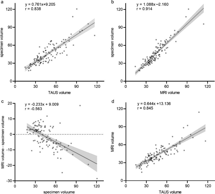
Methods: A total of 106 patients who underwent TAUS and MRI prior to radical prostatectomy were retrospectively analyzed. The TAUS-based and MRI-based PV were calculated using the ellipsoid formula. The specimen volume measured by the water-displacement method was used as a reference standard. Correlation analysis and intraclass correlation coefficients (ICC) were performed to compare different measurement methods and Bland Altman plots were drawn to assess the agreement.
Results: There was a high degree of correlation and agreement between the specimen volume and PV measured with TAUS (r = 0.838, p < 0.01; ICC = 0.83) and MRI (r = 0.914, p < 0.01; ICC = 0.90). TAUS overestimated specimen volume by 2.4ml, but the difference was independent of specimen volume (p = 0.19). MRI underestimated specimen volume by 1.7ml, the direction and magnitude of the difference varied with specimen volume (p < 0.01). The percentage error of PV measured by TAUS and MRI was within ± 20% in 65/106(61%) and 87/106(82%), respectively. In patients with PV greater than 50 ml, MRI volume still correlated strongly with specimen volume (r = 0.837, p < 0.01), while TAUS volume showed only moderate correlation with specimen (r = 0.665, p < 0.01) or MRI volume (r = 0.678, p < 0.01).
Conclusions: This study demonstrated that PV measured by MRI and TAUS is highly correlated and reliable with the specimen volume. MRI might be a more appropriate choice for measuring the large prostate.
Keywords: Magnetic resonance imaging; Prostate cancer; Prostate volume; Transabdominal ultrasound.
© 2023. The Author(s).
Conflict of interest statement
All authors have no competing interests to declare that are relevant to the content of this article.
Figures


References
-
- Roobol MJ, Schroder FH, Hugosson J, Jones JS, Kattan MW, Klein EA, et al. Importance of prostate volume in the european Randomised study of screening for prostate Cancer (ERSPC) risk calculators: results from the prostate biopsy collaborative group. World J Urol. 2012;30(2):149–55. doi: 10.1007/s00345-011-0804-y. - DOI - PMC - PubMed
Publication types
MeSH terms
Grants and funding
LinkOut - more resources
Full Text Sources
Medical
