

Can contrast-enhanced ultrasound differentiate the type of hepatic echinococcosis: cystic echinococcosis or alveolar echinococcosis?
- PMID: 37069610
- PMCID: PMC10111660
- DOI: 10.1186/s13071-023-05731-2
Can contrast-enhanced ultrasound differentiate the type of hepatic echinococcosis: cystic echinococcosis or alveolar echinococcosis?
Abstract
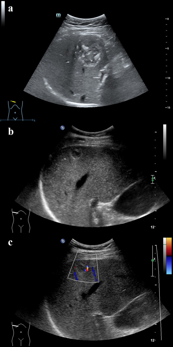
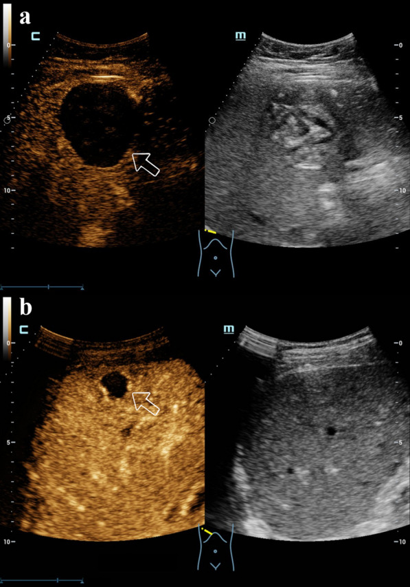
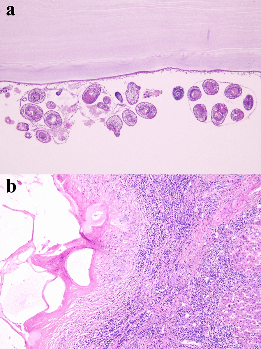
Background: Hepatic echinococcosis (HE) is a zoonotic disease caused by Echinococcus, and Echinococcus granulosus and E. multilocularis are the most common, causing cystic echinococcosis (CE) and alveolar echinococcosis (AE), respectively. Contrast-enhanced ultrasound (CEUS) is an imaging technique which has been recommended for identifying focal lesions in the liver. However, the effect of CEUS on the differentiation of hepatic echinococcosis type remains unclear.
Methods: Twenty-five patients with 46 HE lesions confirmed by histopathology in our hospital from December 2019 to May 2022 were reviewed by conventional ultrasound (US) and CEUS examinations, respectively. After US was completed, the CEUS study was performed. A bolus injection of 1.0-1.2 ml of a sulfur hexafluoride-filled microbubble contrast agent (SonoVue®) was administered. The images and clips of the lesions by US and CEUS were reviewed retrospectively. The lesions detected using US were evaluated including the location, size, morphology, margin, internal echogenicity and the internal Doppler signal. The lesions detected using CEUS were evaluated including the enhancement degree, enhancement pattern and enhancing boundary in different phases. The diagnoses of lesions by US or CEUS were respectively recorded. By taking the histopathology as the gold standard, the paired Chi-square test was performed with statistical software (IBM SPSS; IBM Corp., Armonk, NY, USA), and the results of differentiation of HE type by US and CEUS were statistically analyzed.
Results: A total of 46 lesions were involved in 25 patients, including 10 males (40.0%) and 15 females (60.0%) aged 15-55 (42.9 ± 10.3) years. By histopathology, 24 lesions of nine patients were diagnosed as CE and 22 lesions of 16 patients were diagnosed as AE. Among the 46 HE lesions, compared with histopathological examination, the accuracy rate was 65.2% and 91.3% in US and CEUS findings, respectively. Among the 24 CE lesions, 13 lesions were correctly differentiated by US, and 23 by CEUS. The difference between US and CEUS was statistically significant (Chi-square test, [Formula: see text] = 8.10, df = 23, P < 0.005). Among the total 46 HE lesions, 30 lesions were correctly differentiated by US, and 42 by CEUS. The difference between US and CEUS was statistically significant (Chi-square test, [Formula: see text] = 10.08, df = 45, P < 0.005).
Conclusions: CEUS is a more effective technique than US for differentiating the type of HE between CE and AE. It could be a reliable tool in the differentiation of HE.
Keywords: Alveolar echinococcosis; Contrast-enhanced ultrasound; Conventional ultrasound; Cystic echinococcosis; Differentiation; Hepatic echinococcosis.
© 2023. The Author(s).
Conflict of interest statement
The authors declare that no competing interests exist.
Figures
References
-
- Akhan O, Erdoğan E, Ciftci TT, Unal E, Karaağaoğlu E, Akinci D. Comparison of the long-term results of puncture, aspiration, injection and re-aspiration (PAIR) and catheterization techniques for the percutaneous treatment of CE1 and CE3a liver hydatid cysts: a prospective randomized trial. Cardiovasc Intervent Radiol. 2020;43:1034–1040. doi: 10.1007/s00270-020-02477-7. - DOI - PubMed
MeSH terms
Supplementary concepts
Grants and funding
- 2021WZK1002/Open Project of Key Laboratory for Research on Prevention and Control of Echinococcosis of National Health Commission
- 2021WZK1002/Open Project of Key Laboratory for Research on Prevention and Control of Echinococcosis of National Health Commission
- 2021WZK1002/Open Project of Key Laboratory for Research on Prevention and Control of Echinococcosis of National Health Commission
- 2021WZK1002/Open Project of Key Laboratory for Research on Prevention and Control of Echinococcosis of National Health Commission
- 2021WZK1002/Open Project of Key Laboratory for Research on Prevention and Control of Echinococcosis of National Health Commission
- 2021WZK1002/Open Project of Key Laboratory for Research on Prevention and Control of Echinococcosis of National Health Commission
- 2021WZK1002/Open Project of Key Laboratory for Research on Prevention and Control of Echinococcosis of National Health Commission
- 2021WZK1002/Open Project of Key Laboratory for Research on Prevention and Control of Echinococcosis of National Health Commission
- 2021WZK1002/Open Project of Key Laboratory for Research on Prevention and Control of Echinococcosis of National Health Commission
- 2021WZK1002/Open Project of Key Laboratory for Research on Prevention and Control of Echinococcosis of National Health Commission
- 2021WZK1002/Open Project of Key Laboratory for Research on Prevention and Control of Echinococcosis of National Health Commission
LinkOut - more resources
Full Text Sources
Miscellaneous
