Detection of microbial biofilms inside the lumen of ureteral stents: two case reports
- PMID: 37069667
- PMCID: PMC10111790
- DOI: 10.1186/s13256-023-03849-6
Detection of microbial biofilms inside the lumen of ureteral stents: two case reports
Abstract
Background: We report large biofilm structures that covered almost the entirety of the lumen and surface of double-J stents in two postrenal transplant patients, with no development of urinary tract infection. Biofilm bacteria of one patient were integrated by coccus in a net structure, whereas overlapping cells of bacilli were present in the other patient. To the best of our knowledge, this is the first time that high-quality images of the architecture of noncrystalline biofilms have been found inside double-J stents from long-term stenting in renal transplant recipients.
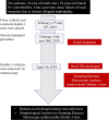
Case presentation: Two renal transplant recipients, a 34-year-old male and a 39-year-old female of Mexican-Mestizo origin, who underwent a first renal transplant and lost it due to allograft failure, had a second transplant. Two months after the surgical procedure, double-J stents were removed and analyzed using scanning electron microscopy (SEM). None of the patients had an antecedent of UTI, and none developed UTI after urinary device removal. There were no reports of injuries, encrustation, or discomfort caused by these devices.
Conclusion: The bacterial biofilm inside the J stent from long-term stenting in renal transplant recipients was mainly concentrated on unique bacteria. Biofilm structures from the outside and inside of stents do not have crystalline phases. Internal biofilms may represent a high number of bacteria in the double-J stent, in the absence of crystals.
Keywords: Biofilm; Case report; Double J stent; Renal transplant; Scanning electron microscopy (SEM).
© 2023. The Author(s).
Conflict of interest statement
The authors declare that they have no competing interests.
Figures


Similar articles
-
Observations of Bacterial Biofilm on Ureteral Stent and Studies on the Distribution of Pathogenic Bacteria and Drug Resistance.Urol Int. 2018;101(3):320-326. doi: 10.1159/000490621. Epub 2018 Sep 13. Urol Int. 2018. PMID: 30212821
-
Microbial ureteral stent colonization in renal transplant recipients: frequency and influence on the short-time functional outcome.Transpl Infect Dis. 2012 Feb;14(1):57-63. doi: 10.1111/j.1399-3062.2011.00671.x. Epub 2011 Sep 26. Transpl Infect Dis. 2012. PMID: 22093165
-
Encrustations on ureteral stents from patients without urinary tract infection reveal distinct urotypes and a low bacterial load.Microbiome. 2019 Apr 13;7(1):60. doi: 10.1186/s40168-019-0674-x. Microbiome. 2019. PMID: 30981280 Free PMC article.
-
Biofilm formation on ureteral stents - Incidence, clinical impact, and prevention.Swiss Med Wkly. 2017 Feb 3;147:w14408. doi: 10.4414/smw.2017.14408. eCollection 2017. Swiss Med Wkly. 2017. PMID: 28165539 Review.
-
Update on biofilm infections in the urinary tract.World J Urol. 2012 Feb;30(1):51-7. doi: 10.1007/s00345-011-0689-9. Epub 2011 May 18. World J Urol. 2012. PMID: 21590469 Free PMC article. Review.
Cited by
-
Urinary Tract Infections with Carbapenem-Resistant Klebsiella pneumoniae in a Urology Clinic-A Case-Control Study.Antibiotics (Basel). 2024 Jun 24;13(7):583. doi: 10.3390/antibiotics13070583. Antibiotics (Basel). 2024. PMID: 39061265 Free PMC article.
-
Analysis of Pathogens of Urinary Tract Infections Associated with Indwelling Double-J Stents and Their Susceptibility to Globularia alypum.Pharmaceutics. 2023 Oct 19;15(10):2496. doi: 10.3390/pharmaceutics15102496. Pharmaceutics. 2023. PMID: 37896256 Free PMC article.
-
How to study biofilms: technological advancements in clinical biofilm research.Front Cell Infect Microbiol. 2023 Dec 13;13:1335389. doi: 10.3389/fcimb.2023.1335389. eCollection 2023. Front Cell Infect Microbiol. 2023. PMID: 38156318 Free PMC article. Review.
-
Urological Challenges during Pregnancy: Current Status and Future Perspective on Ureteric Stent Encrustation.J Clin Med. 2024 Jul 3;13(13):3905. doi: 10.3390/jcm13133905. J Clin Med. 2024. PMID: 38999471 Free PMC article. Review.
References
-
- Friedersdorff F, Weinberger S, Biernath N, Plage H, Cash H, El-Bandar N. The ureter in the kidney transplant setting: ureteroneocystostomy surgical options, double-J stent considerations and management of related complications. Curr Urol Rep. 2020;21(1):3. doi: 10.1007/s11934-020-0956-7. - DOI - PubMed
Publication types
MeSH terms
Grants and funding
LinkOut - more resources
Full Text Sources
Medical

