

Maternal exposure to ultrafine particles enhances influenza infection during pregnancy
- PMID: 37069680
- PMCID: PMC10106898
- DOI: 10.1186/s12989-023-00521-1
Maternal exposure to ultrafine particles enhances influenza infection during pregnancy
Abstract
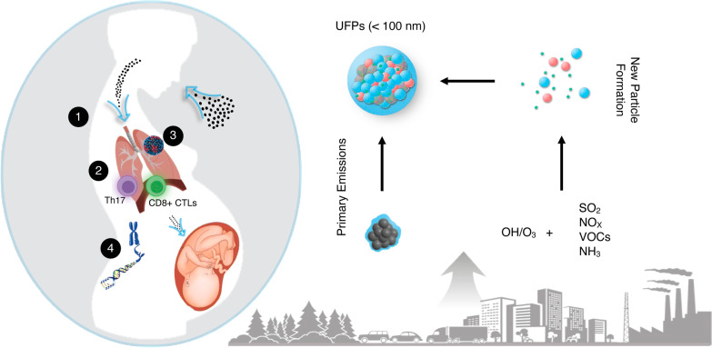
Background: Interactions between air pollution and infectious agents are increasingly recognized and critical to identify, especially to protect vulnerable populations. Pregnancy represents a vulnerable period for influenza infection and air pollution exposure, yet interactions during pregnancy remain unclear. Maternal exposure to ultrafine particles (UFPs, [Formula: see text] 100 nm diameter), a class of particulate matter ubiquitous in urban environments, elicits unique pulmonary immune responses. We hypothesized that UFP exposure during pregnancy would lead to aberrant immune responses to influenza enhancing infection severity.
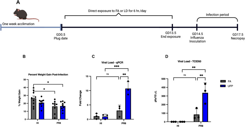
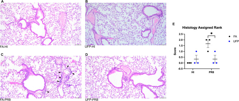
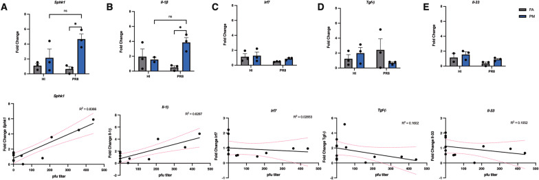
Results: Building from our well-characterized C57Bl/6N mouse model employing daily gestational UFP exposure from gestational day (GD) 0.5-13.5, we carried out a pilot study wherein pregnant dams were subsequently infected with Influenza A/Puerto Rico/8/1934 (PR8) on GD14.5. Findings indicate that PR8 infection caused decreased weight gain in filtered air (FA) and UFP-exposed groups. Co-exposure to UFPs and viral infection led to pronounced elevation in PR8 viral titer and reduced pulmonary inflammation, signifying potential suppression of innate and adaptive immune defenses. Pulmonary expression of the pro-viral factor sphingosine kinase 1 (Sphk1) and pro-inflammatory cytokine interleukin-1β (IL-1 [Formula: see text]) was significantly increased in pregnant mice exposed to UFPs and infected with PR8; expression correlated with higher viral titer.
Conclusions: Results from our model provide initial insight into how maternal UFP exposure during pregnancy enhances respiratory viral infection risk. This model is an important first step in establishing future regulatory and clinical strategies for protecting pregnant women exposed to UFPs.
Keywords: Air pollution; Infection; Influenza; Mouse model; Particulate matter; Pregnancy; Ultrafine particles.
© 2023. The Author(s).
Conflict of interest statement
Authors declare that they have no competing interests.
Figures

Similar articles
-
Prenatal Ambient Ultrafine Particle Exposure and Childhood Asthma in the Northeastern United States.Am J Respir Crit Care Med. 2021 Oct 1;204(7):788-796. doi: 10.1164/rccm.202010-3743OC. Am J Respir Crit Care Med. 2021. PMID: 34018915 Free PMC article.
-
In utero ultrafine particulate matter exposure causes offspring pulmonary immunosuppression.Proc Natl Acad Sci U S A. 2019 Feb 26;116(9):3443-3448. doi: 10.1073/pnas.1816103116. Epub 2019 Feb 11. Proc Natl Acad Sci U S A. 2019. PMID: 30808738 Free PMC article.
-
Ambient and Controlled Particle Exposures as Triggers for Acute ECG Changes.Res Rep Health Eff Inst. 2016 May;(186):5-75. Res Rep Health Eff Inst. 2016. PMID: 28661614
-
Air pollution and children's health-a review of adverse effects associated with prenatal exposure from fine to ultrafine particulate matter.Environ Health Prev Med. 2021 Jul 12;26(1):72. doi: 10.1186/s12199-021-00995-5. Environ Health Prev Med. 2021. PMID: 34253165 Free PMC article. Review.
-
Dosimetry and toxicology of inhaled ultrafine particles.Biomarkers. 2009 Jul;14 Suppl 1:67-73. doi: 10.1080/13547500902965617. Biomarkers. 2009. PMID: 19604063 Review.
Cited by
-
Particulate Matter Exposure and Viral Infections: Relevance to Highly Polluted Settings such as Ulaanbaatar, Mongolia.Curr Environ Health Rep. 2025 Apr 23;12(1):22. doi: 10.1007/s40572-025-00484-9. Curr Environ Health Rep. 2025. PMID: 40268823 Free PMC article. Review.
-
Decoding Immune Dynamics in Pregnant Women: Key Gene Expression Changes Following Influenza Vaccination.Int J Mol Sci. 2025 Apr 16;26(8):3765. doi: 10.3390/ijms26083765. Int J Mol Sci. 2025. PMID: 40332395 Free PMC article.
-
Cumulative risk assessment as the pathway to public health protection for behavioral neurotoxicity.Neurotoxicology. 2025 May;108:400-411. doi: 10.1016/j.neuro.2025.04.015. Epub 2025 May 10. Neurotoxicology. 2025. PMID: 40349850 Review.
References
-
- World Health Organization (WHO). Air pollution (WHO). 2018; https://www.who.int/airpollution/en/
-
- U.S. Environmental Protection Agency (EPA). Our Nation’s Air (US EPA). 2018; https://gispub.epa.gov/air/trendsreport/2018/
Publication types
MeSH terms
Substances
Grants and funding
LinkOut - more resources
Full Text Sources
Medical
