

Quantifying lumen diameter in coronary artery stents with high-resolution photon counting detector CT and convolutional neural network denoising
- PMID: 37069830
- PMCID: PMC10524296
- DOI: 10.1002/mp.16415
Quantifying lumen diameter in coronary artery stents with high-resolution photon counting detector CT and convolutional neural network denoising
Abstract
Background: Small coronary arteries containing stents pose a challenge in CT imaging due to metal-induced blooming artifact. High spatial resolution imaging capability is as the presence of highly attenuating materials limits noninvasive assessment of luminal patency.
Purpose: The purpose of this study was to quantify the effective lumen diameter within coronary stents using a clinical photon-counting-detector (PCD) CT in concert with a convolutional neural network (CNN) denoising algorithm, compared to an energy-integrating-detector (EID) CT system.
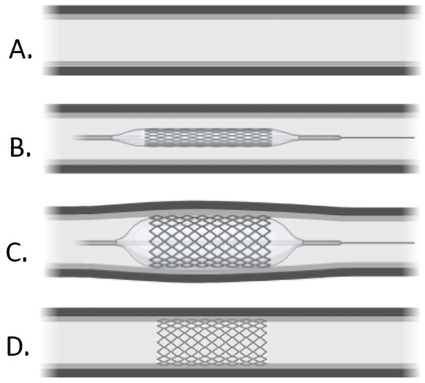
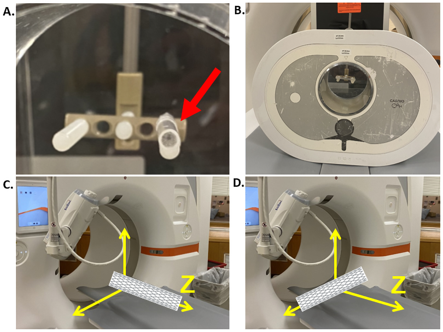
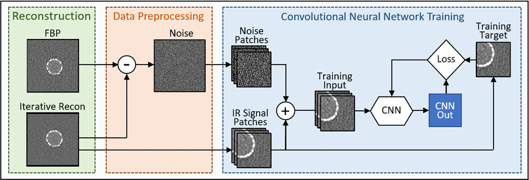
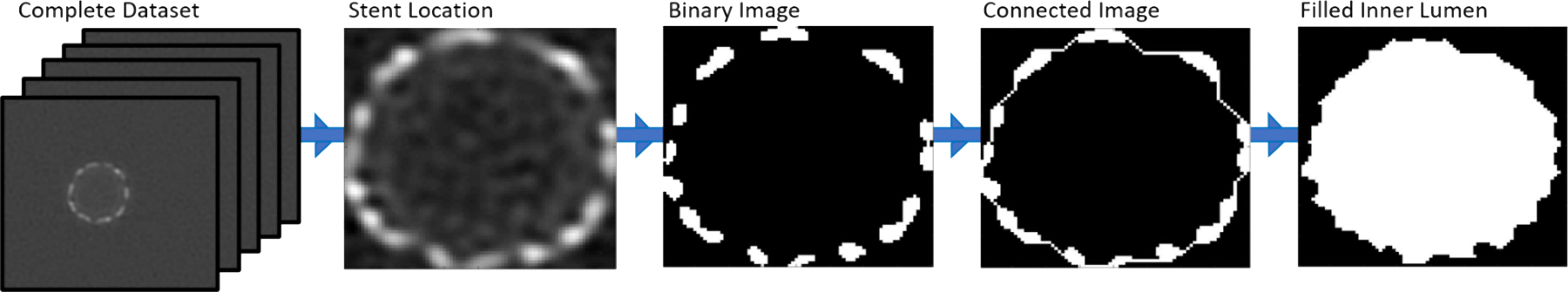
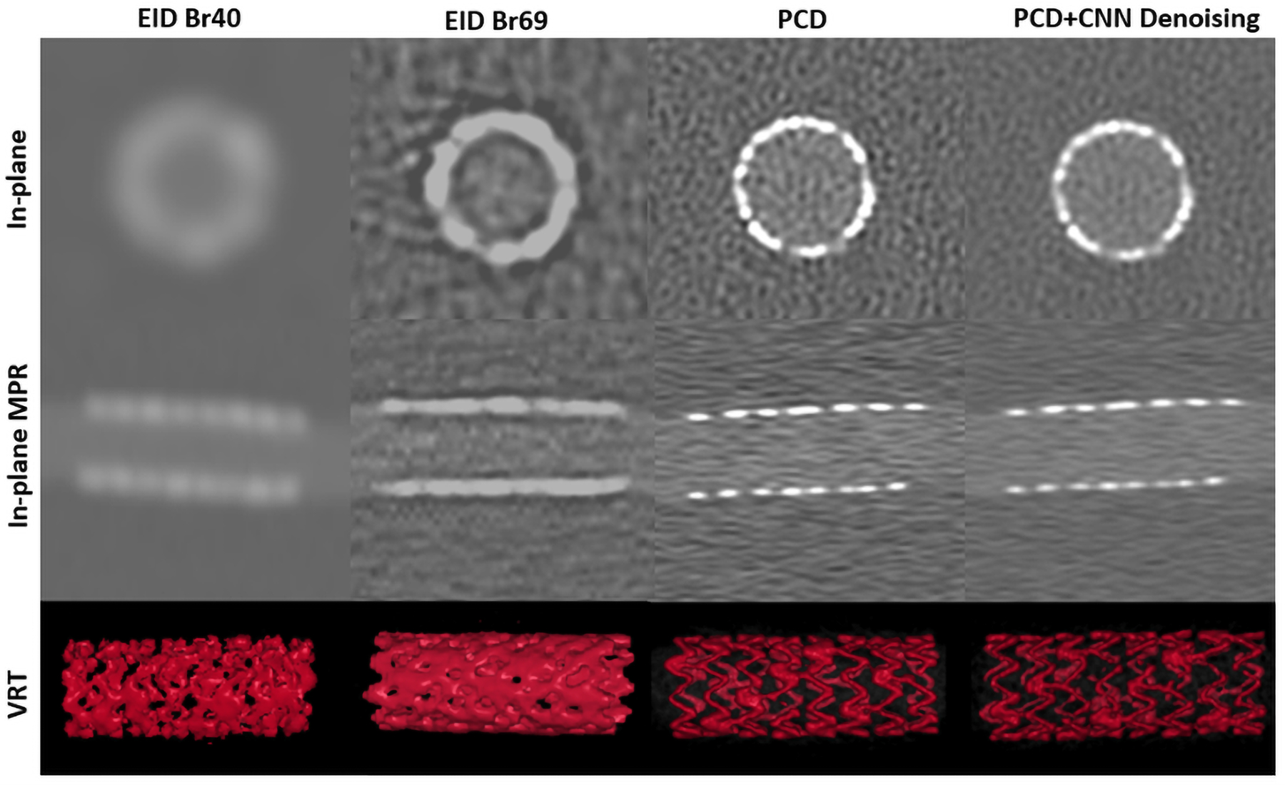
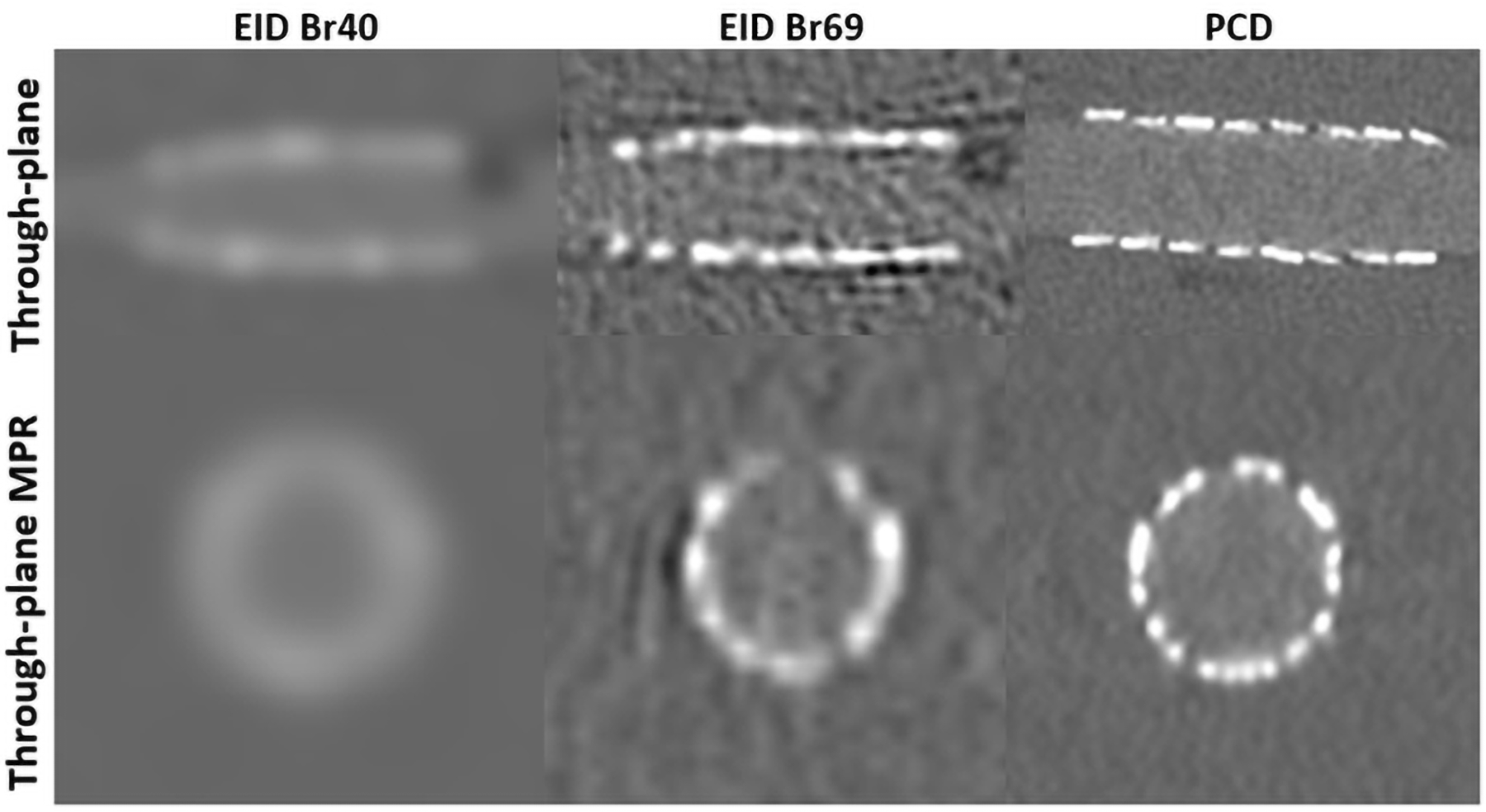
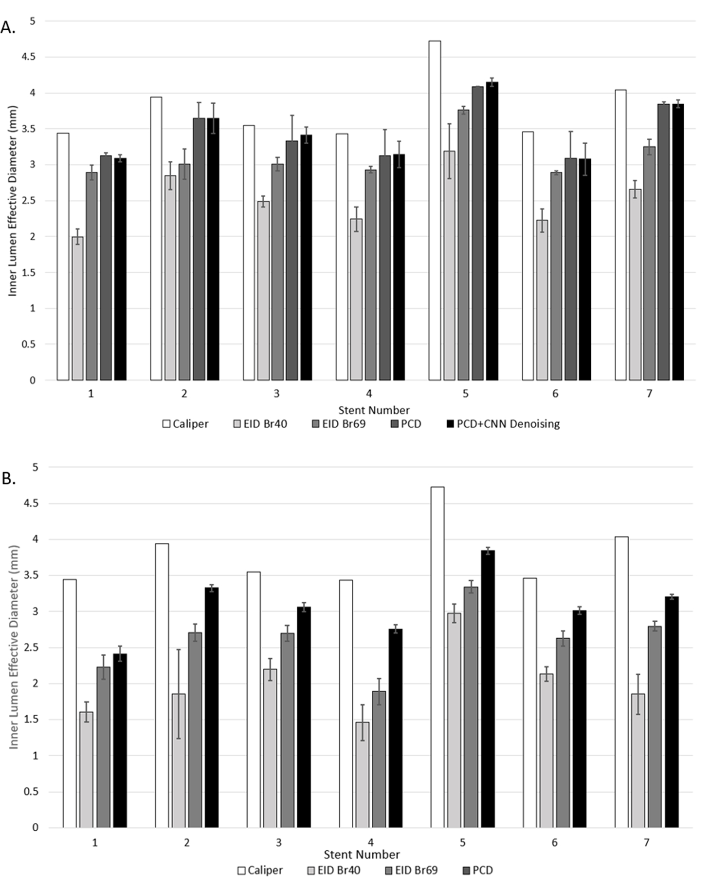
Methods: Seven coronary stents of different materials and inner diameters between 3.43 and 4.72 mm were placed in plastic tubes of diameters 3.96-4.87 mm containing 20 mg/mL of iodine solution, mimicking stented contrast-enhanced coronary arteries. Tubes were placed parallel with or perpendicular to the scanner's z-axis in an anthropomorphic phantom emulating an average-sized patient and scanned with a clinical EID-CT and PCD-CT. EID scans were performed using our standard coronary computed tomography angiography (cCTA) protocol (120 kV, 180 quality reference mAs). PCD scans were performed using the ultra-high-resolution (UHR) mode (120 × 0.2 mm collimation) at 120 kV with tube current adjusted so that CTDIvol was matched to that of EID scans. EID images were reconstructed per our routine clinical protocol (Br40, 0.6 mm thickness), and with the sharpest available kernel (Br69). PCD images were reconstructed at a thickness of 0.6 mm and a dedicated sharp kernel (Br89) which is only possible with the PCD UHR mode. To address increased image noise introduced by the Br89 kernel, an image-based CNN denoising algorithm was applied to the PCD images of stents scanned parallel to the scanner's z-axis. Stents were segmented based on full-width half maximum thresholding and morphological operations, from which effective lumen diameter was calculated and compared to reference sizes measured with a caliper.
Results: Substantial blooming artifacts were observed on EID Br40 images, resulting in larger stent struts and reduced lumen diameter (effective diameter underestimated by 41% and 47% for parallel and perpendicular orientations, respectively). Blooming artifacts were observed on EID Br69 images with 19% and 31% underestimation of lumen diameter compared to the caliper for parallel and perpendicular scans, respectively. Overall image quality was substantially improved on PCD, with higher spatial resolution and reduced blooming artifacts, resulting in the clearer delineation of stent struts. Effective lumen diameters were underestimated by 9% and 19% relative to the reference for parallel and perpendicular scans, respectively. CNN reduced image noise by about 50% on PCD images without impacting lumen quantification (<0.3% difference).
Conclusion: The PCD UHR mode improved in-stent lumen quantification for all seven stents as compared to EID images due to decreased blooming artifacts. Implementation of CNN denoising algorithms to PCD data substantially improved image quality.
Keywords: computed tomography; convolutional neural network; coronary artery stents; denoising; photon counting detector.
© 2023 American Association of Physicists in Medicine.
Conflict of interest statement
Conflict of Interest Statement
Cynthia McCollough is the recipient of a research grant to the institution from Siemens Healthcare GmbH. The other authors have no relevant conflicts of interest to disclose.
Figures
Similar articles
-
Photon-Counting Computed Tomography for Coronary Stent Imaging: In Vitro Evaluation of 28 Coronary Stents.Invest Radiol. 2021 Oct 1;56(10):653-660. doi: 10.1097/RLI.0000000000000787. Invest Radiol. 2021. PMID: 33867450
-
Photon-Counting CT: High-Resolution Imaging of Coronary Stents.Invest Radiol. 2018 Mar;53(3):143-149. doi: 10.1097/RLI.0000000000000420. Invest Radiol. 2018. PMID: 28945655
-
First In-Human Results of Computed Tomography Angiography for Coronary Stent Assessment With a Spectral Photon Counting Computed Tomography.Invest Radiol. 2022 Apr 1;57(4):212-221. doi: 10.1097/RLI.0000000000000835. Invest Radiol. 2022. PMID: 34711766 Free PMC article.
-
Coronary Stent Imaging with Photon-counting Detector CT.Radiol Cardiothorac Imaging. 2025 Jun;7(3):e240338. doi: 10.1148/ryct.240338. Radiol Cardiothorac Imaging. 2025. PMID: 40439564 Review.
-
An introduction to photon-counting detector CT (PCD CT) for radiologists.Jpn J Radiol. 2023 Mar;41(3):266-282. doi: 10.1007/s11604-022-01350-6. Epub 2022 Oct 18. Jpn J Radiol. 2023. PMID: 36255601 Free PMC article. Review.
Cited by
-
Technical principles, benefits, challenges, and applications of photon counting computed tomography in coronary imaging: a narrative review.Cardiovasc Diagn Ther. 2024 Aug 31;14(4):698-724. doi: 10.21037/cdt-24-52. Epub 2024 Jul 31. Cardiovasc Diagn Ther. 2024. PMID: 39263472 Free PMC article. Review.
-
Assessing beam hardening artifacts in coronary stent imaging using different CT acquisition parameters on photon-counting detector computed tomography.Int J Cardiovasc Imaging. 2025 Apr 10. doi: 10.1007/s10554-025-03392-z. Online ahead of print. Int J Cardiovasc Imaging. 2025. PMID: 40208433
-
Coronary artery stenosis quantification in patients with dense calcifications using ultra-high-resolution photon-counting-detector computed tomography.J Cardiovasc Comput Tomogr. 2024 Jan-Feb;18(1):56-61. doi: 10.1016/j.jcct.2023.10.009. Epub 2023 Nov 7. J Cardiovasc Comput Tomogr. 2024. PMID: 37945454 Free PMC article.
-
Evaluating small coronary stents with dual-source photon-counting computed tomography: effect of different scan modes on image quality and performance in a phantom.Diagn Interv Radiol. 2025 Jan 1;31(1):29-38. doi: 10.4274/dir.2024.242893. Epub 2024 Oct 21. Diagn Interv Radiol. 2025. PMID: 39463047 Free PMC article.
-
Spectral Photon-Counting Computed Tomography: Technical Principles and Applications in the Assessment of Cardiovascular Diseases.J Clin Med. 2024 Apr 18;13(8):2359. doi: 10.3390/jcm13082359. J Clin Med. 2024. PMID: 38673632 Free PMC article. Review.
References
-
- (CDC) CfDC. Heart Disease Facts. In: CDC; 2022.
-
- Organization WH. Cardiovascular Diseases Published 2022. Updated June 2022. Accessed July 2022.
-
- Mannil M, Hickethier T, von Spiczak J, et al. Photon-counting CT: high-resolution imaging of coronary stents. Investigative radiology 2018;53(3):143–149. - PubMed
-
- Virani SS, Alonso A, Aparicio HJ, et al. Heart disease and stroke statistics—2021 update: a report from the American Heart Association. Circulation 2021;143(8):e254–e743. - PubMed
