

Lemmel's Syndrome: A Rare Complication of Periampullary Diverticula
- PMID: 37069880
- PMCID: PMC10105574
- DOI: 10.7759/cureus.36236
Lemmel's Syndrome: A Rare Complication of Periampullary Diverticula
Abstract
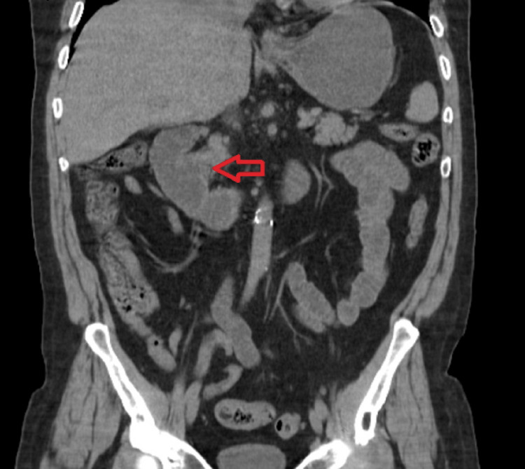
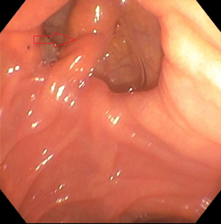
Periampullary diverticula are outpouches in the mucosa in the duodenum surrounding the ampulla of Vater. Most cases of periampullary diverticuli are asymptomatic, but complications can arise, increasing a patient's mortality. Diagnosis of periampullary diverticuli is often incidental during endoscopy or imaging studies for abdominal pain. When a patient with periampullary diverticuli is symptomatic, imaging modalities such as CT scan and MRI can help make the diagnosis, but a side-viewing endoscope provides direct visualization of the diverticuli and also allows for the potential treatment of the disease. Lemmel's syndrome is a complication of periampullary diverticuli where the diverticuli causes mechanical obstruction of the bile duct leading to obstructive jaundice without choledocholithiasis. These patients are at risk for further complications such as sepsis and perforation. Early diagnosis and treatment of these patients can help to prevent further complications from arising. We are presenting a case of Lemmel's syndrome with obstructive jaundice from a periampullary diverticuli, further complicated by cholangitis without dilation of the biliary tree.
Keywords: acute cholangitis; cholangitis; endoscopy; lemmel's syndrome; obstructive jaundice; periampullary diverticula; periampullary diverticulum.
Copyright © 2023, Battah et al.
Conflict of interest statement
The authors have declared that no competing interests exist.
Figures

Similar articles
-
Periampullary diverticulitis (Lemmel's syndrome) misdiagnosed as pancreatic head tumor: A report of two cases.Int J Surg Case Rep. 2023 May;106:108198. doi: 10.1016/j.ijscr.2023.108198. Epub 2023 Apr 20. Int J Surg Case Rep. 2023. PMID: 37087935 Free PMC article.
-
Endoscopic ultrasound of Lemmel's syndrome.Indian J Gastroenterol. 2017 Mar;36(2):155-157. doi: 10.1007/s12664-017-0744-6. Epub 2017 Mar 23. Indian J Gastroenterol. 2017. PMID: 28332175
-
Lemmel's Syndrome: Usual Presentation of an Unusual Diagnosis.Cureus. 2020 Apr 16;12(4):e7698. doi: 10.7759/cureus.7698. Cureus. 2020. PMID: 32431977 Free PMC article.
-
Recurrent ascending cholangitis with acute pancreatitis and pancreatic atrophy caused by a juxtapapillary duodenal diverticulum: A case report and literature review.Medicine (Baltimore). 2020 Jul 2;99(27):e21111. doi: 10.1097/MD.0000000000021111. Medicine (Baltimore). 2020. PMID: 32629744 Free PMC article. Review.
-
[Lemmel's syndrome caused by a giant diverticulum in the third portion of the duodenum, report of a case].Nihon Shokakibyo Gakkai Zasshi. 1995 Nov;92(11):1863-70. Nihon Shokakibyo Gakkai Zasshi. 1995. PMID: 8544356 Review. Japanese. No abstract available.
Cited by
-
Beyond the Usual Suspects: A Pictorial Case Report of Lemmel Syndrome Emphasizing the Role of Multimodality Imaging.Cureus. 2024 May 11;16(5):e60097. doi: 10.7759/cureus.60097. eCollection 2024 May. Cureus. 2024. PMID: 38860101 Free PMC article.
-
A Challenging Case of Endoscopic Retrograde Cholangiopancreatography Due to an Intradiverticular Papilla.Cureus. 2025 Jun 19;17(6):e86383. doi: 10.7759/cureus.86383. eCollection 2025 Jun. Cureus. 2025. PMID: 40688845 Free PMC article.
-
Lemmel Syndrome: A Surgical Enigma.Cureus. 2024 Jul 29;16(7):e65620. doi: 10.7759/cureus.65620. eCollection 2024 Jul. Cureus. 2024. PMID: 39205778 Free PMC article.
-
Duodenal Diverticulum Causing Lemmel Syndrome: Diagnosis by computed tomography.Sultan Qaboos Univ Med J. 2024 Nov;24(4):599-600. doi: 10.18295/squmj.11.2024.073. Epub 2024 Nov 27. Sultan Qaboos Univ Med J. 2024. PMID: 39634809 Free PMC article. No abstract available.
References
-
- Periampullary diverticula and pancreaticobiliary disease. Lobo DN, Balfour TW, Iftikhar SY, Rowlands BJ. Br J Surg. 1999;86:588–597. - PubMed
-
- Periampullary diverticula and technical success of endoscopic retrograde cholangiopancreatography. Tyagi P, Sharma P, Sharma BC, Puri AS. Surg Endosc. 2009;23:1342–1345. - PubMed
-
- Duodenal diverticulum causing intermittent-persistent cholestasis. Associated with papillitis chronica fibrosa. Manabe T, Yu GS. https://pubmed.ncbi.nlm.nih.gov/412143/ N Y State J Med. 1977;77:2132–2136. - PubMed
Publication types
LinkOut - more resources
Full Text Sources