. 2023 Jan 17;8(4):837-850.
doi: 10.1016/j.ekir.2023.01.008. eCollection 2023 Apr.

The Transcriptomics of the Human Vein Transformation After Arteriovenous Fistula Anastomosis Uncovers Layer-Specific Remodeling and Hallmarks of Maturation Failure

Affiliations

The Transcriptomics of the Human Vein Transformation After Arteriovenous Fistula Anastomosis Uncovers Layer-Specific Remodeling and Hallmarks of Maturation Failure

Laisel Martinez et al. Kidney Int Rep. .

Abstract

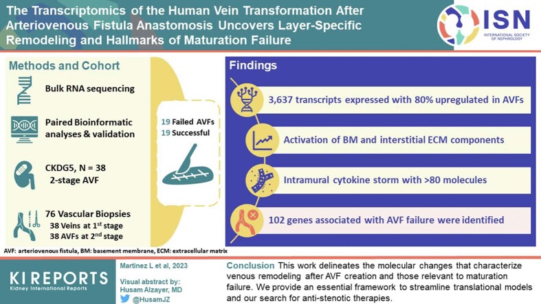
Introduction: The molecular transformation of the human preaccess vein after arteriovenous fistula (AVF) creation is poorly understood. This limits our ability to design efficacious therapies to improve maturation outcomes.

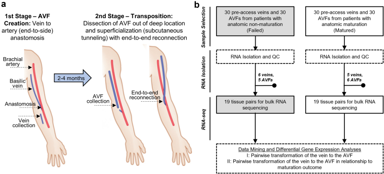
Methods: Bulk RNA sequencing (RNA-seq) followed by paired bioinformatic analyses and validation assays were performed in 76 longitudinal vascular biopsies (veins and AVFs) from 38 patients with stage 5 chronic kidney disease or end-stage kidney disease undergoing surgeries for 2-stage AVF creation (19 matured, 19 failed).

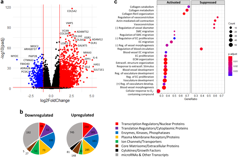
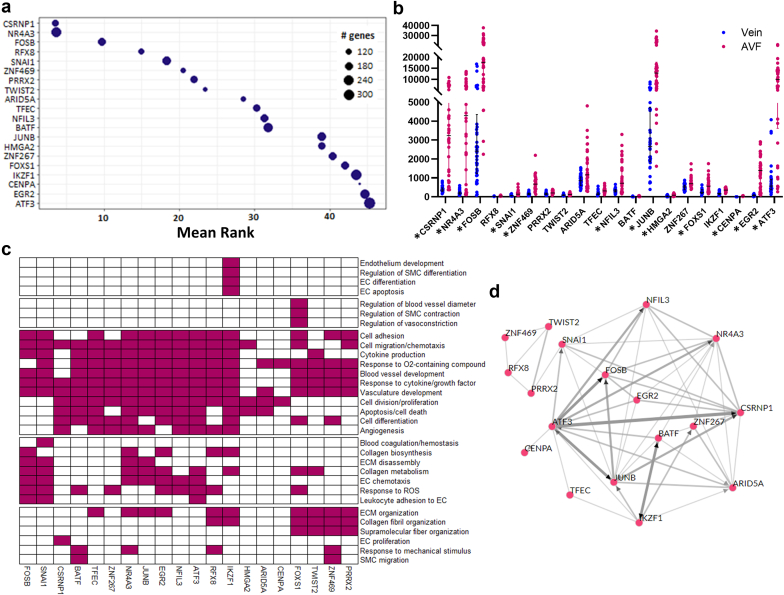
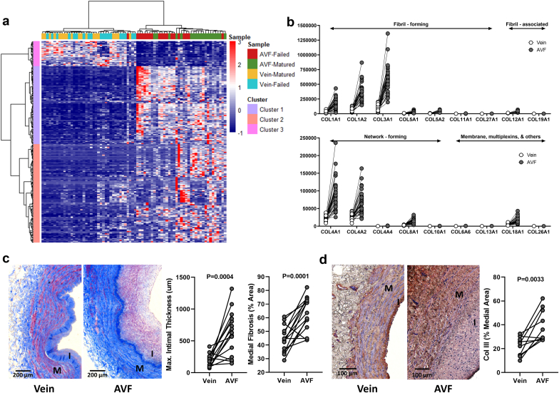
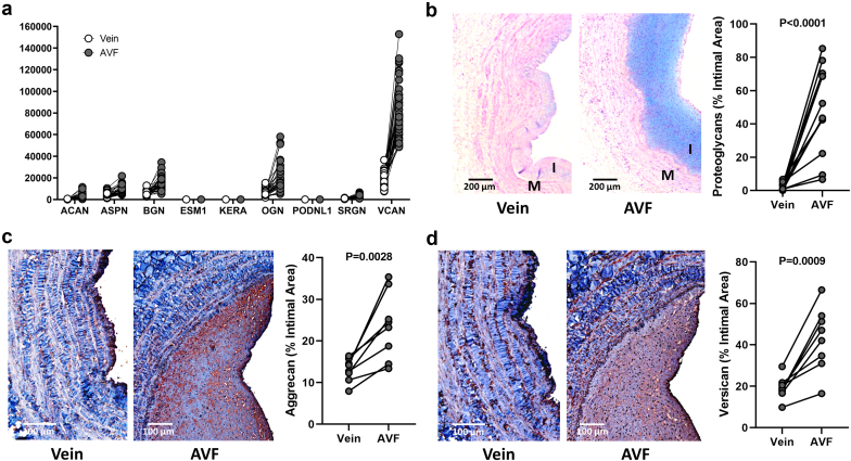
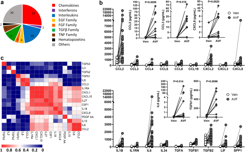
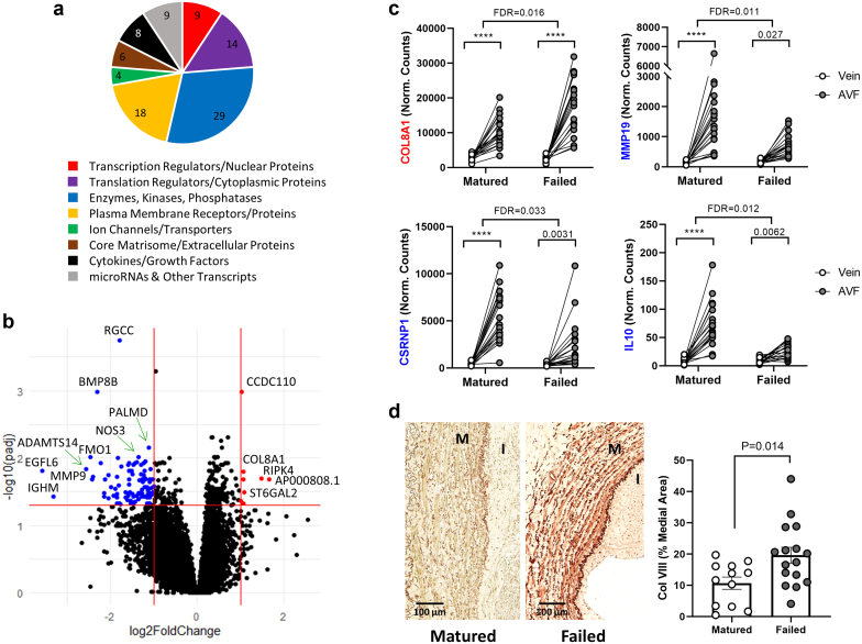
Results: A total of 3637 transcripts were differentially expressed between veins and AVFs independent of maturation outcomes, with 80% upregulated in fistulas. The postoperative transcriptome demonstrated transcriptional activation of basement membrane and interstitial extracellular matrix (ECM) components, including preexisting and novel collagens, proteoglycans, hemostasis factors, and angiogenesis regulators. A postoperative intramural cytokine storm involved >80 chemokines, interleukins, and growth factors. Postoperative changes in ECM expression were differentially distributed in the AVF wall, with proteoglycans and fibrillar collagens predominantly found in the intima and media, respectively. Interestingly, upregulated matrisome genes were enough to make a crude separation of AVFs that failed from those with successful maturation. We identified 102 differentially expressed genes (DEGs) in association with AVF maturation failure, including upregulation of network collagen VIII in medial smooth muscle cells (SMCs) and downregulation of endothelial-predominant transcripts and ECM regulators.

Conclusion: This work delineates the molecular changes that characterize venous remodeling after AVF creation and those relevant to maturation failure. We provide an essential framework to streamline translational models and our search for antistenotic therapies.

Keywords: arteriovenous fistula; extracellular matrix; maturation; transcriptomics.


Figures

None
Graphical abstract
Figure 1
Figure 1
Study design and sample collection. (a) Flow diagram of surgical procedures and sample collection. Reprinted (with minor changes) with permission from Martinez et al. (b) Total number of veins and AVFs collected for RNA isolation per maturation outcome and number of patients selected for bulk RNA sequencing after applying RNA quality control inclusion criteria (both preaccess vein and AVF sample with RNA integrity score > 5). AVF, arteriovenous fistula; QC, quality control.
Figure 2
Figure 2
DEGs and functional pathways in the vein to AVF transformation. (a) Volcano plot representation of 3637 DEGs in AVFs compared with their corresponding preaccess veins (pairwise comparisons). Changes in gene expression are presented as log2(fold change) in AVFs with respect to veins. Red dots indicate upregulated genes in AVFs (log2 fold change ≥ 1, false discovery rate < 0.05), and blue dots are genes downregulated in fistulas (log2 fold change ≤ −1, false discovery rate < 0.05). (b) Distribution of functions in 2678 DEGs with functional classifications available. (c) Pathway enrichment analyses of DEGs indicating activated and suppressed biological processes during the vein to AVF transformation. Biological processes are organized by gene ratio on the x-axis (also known as enrichment ratio), which is defined as the ratio of DEGs annotated in a term (count) to the total number of genes in this process in genome-wide annotation packages. DEG, Differentially expressed gene; EC, endothelial cell; ECM, extracellular matrix; SMC, smooth muscle cell.
Figure 3
Figure 3
Transcriptional regulation of the vein to AVF transformation. (a) TF enrichment analysis indicating the 20 best-ranked TFs in the transcriptional regulation of the vein to AVF transformation. This analysis ranks TFs based on the number of predicted targets in the input data set compared with the total number of targets experimentally identified by chromatin immunoprecipitation sequencing. (b) RNA expression in the vein and AVF of the 20 best TFs predicted in (a). The y-axis presents normalized counts, and the asterisk symbol indicates TFs that are differentially expressed during the vein to AVF transformation (log2 fold change ≥ 1, false discovery rate < 0.05). (c) Prediction of biological processes in which the above TFs participate based on pathway analyses of their corresponding gene targets. (d) Coregulatory networks of the above TFs indicating coexpression of TFs in chromatin immunoprecipitation sequencing libraries (gray lines) or direct transcriptional regulation by an indicated factor (pointed arrows). AVF, arteriovenous fistula; EC, endothelial cell; ECM, extracellular matrix; ROS, reactive oxygen species; SMC, smooth muscle cell; TF, transcription factor.
Figure 4
Figure 4
Extracellular matrix remodeling during the vein to AVF transformation. (a) Heatmap of 285 differentially expressed “matrisome” genes (absolute log2 fold change ≥ 1, false discovery rate < 0.05) in AVFs compared with their corresponding preaccess veins. Clusters 1 and 2 are upregulated in AVFs with respect to veins, whereas cluster 3 is downregulated in fistulas. (b) Expression levels and magnitude of fold change in upregulated collagen genes during the vein to AVF transformation. Y-axes indicate normalized counts. False discovery rate < 0.05 for all paired comparisons. (c) Representative trichrome stainings and quantification of morphometry in veins and AVF tissue sections. (d) Immunohistochemistry of collagen III and quantification in veins and AVF tissue sections. Values in (c) and (d) were compared using paired t-tests. AVF, arteriovenous fistula; I, intima; M, media.
Figure 5
Figure 5
Intimal remodeling during the vein to AVF transformation. (a) Expression levels and magnitude of fold change in upregulated proteoglycan genes during the vein to AVF transformation. The y-axis indicates normalized counts. False discovery rate < 0.05 for all paired comparisons. (b) Representative Alcian blue stainings and quantification in veins and AVF tissue sections. (c,d) Immunohistochemistry of aggrecan (c) and versican (d) and quantification in veins and AVF tissue sections. Values in (b) to (d) were compared using paired t-tests. AVF, arteriovenous fistula; I, intima; M, media.
Figure 6
Figure 6
Inflammation during the vein to AVF transformation. (a) Family distribution of upregulated secretable factors during the vein to AVF transformation. (b) RNA expression levels (dots) and magnitude of fold change in upregulated secretable factors. Y-axes indicate normalized counts. False discovery rate < 0.05 for all paired comparisons. Inset plots present protein quantifications (diamonds) of select factors as determined by multiplex assays. Protein levels were compared using Wilcoxon matched-pairs signed-rank tests. (c) Spearman correlation of upregulated secretable factors for which protein levels were available. AVF, arteriovenous fistula; EGF, epidermal growth factor; FGF, fibroblast growth factor; TGF, transforming growth factor; TNF, tumor necrosis factor.
Figure 7
Figure 7
Differentially expressed genes (DEGs) during the vein to AVF transformation in association with maturation failure. (a) Distribution of functions in 97 of 102 failure-associated DEGs during the vein to AVF transformation. The functional classification of 5 DEGs has not been determined. (b) Volcano plot representation of differential gene expression analysis between AVFs and their corresponding preaccess veins conditional to maturation failure. Log2(fold change) on the x-axis represents how much more (or less) a gene changed in tissue pairs that failed versus those that matured. Red dots indicate upregulated genes with failure compared with successful maturation (log2 fold change ≥ 1, false discovery rate [FDR] < 0.05), and blue dots are genes downregulated with failure (log2 fold change ≤ −1, FDR < 0.05). (c,d) Examples of DEGs during the vein to AVF transformation in association with maturation failure. COL8A1 is upregulated, whereas MMP19, CSRNP1, and IL10 are downregulated with failure. Values above brackets indicate the FDR for the corresponding comparison. ∗∗∗∗FDR < 0.001. Immunohistochemistry of collagen VIII and quantification in AVFs that matured or failed. Values were compared using an unpaired t-test. AVF, arteriovenous fistula.

Similar articles

Cited by

References

    1. Robbin M.L., Greene T., Cheung A.K., et al. Arteriovenous fistula development in the first 6 weeks after creation. Radiology. 2016;279:620–629. doi: 10.1148/radiol.2015150385. - DOI - PMC - PubMed
    1. Martinez L., Duque J.C., Tabbara M., et al. Fibrotic venous remodeling and nonmaturation of arteriovenous fistulas. J Am Soc Nephrol. 2018;29:1030–1040. doi: 10.1681/ASN.2017050559. - DOI - PMC - PubMed
    1. Lee T., Qian J.Z., Zhang Y., Thamer M., Allon M. Long-term outcomes of arteriovenous fistulas with unassisted versus assisted maturation: a retrospective National Hemodialysis Cohort Study. J Am Soc Nephrol. 2019;30:2209–2218. doi: 10.1681/ASN.2019030318. - DOI - PMC - PubMed
    1. Huijbregts H.J., Bots M.L., Wittens C.H., Schrama Y.C., Moll F.L., Blankestijn P.J. Hemodialysis arteriovenous fistula patency revisited: results of a prospective, multicenter initiative. Clin J Am Soc Nephrol. 2008;3:714–719. doi: 10.2215/CJN.02950707. - DOI - PMC - PubMed
    1. Robbin M.L., Greene T., Allon M., et al. Prediction of arteriovenous fistula clinical maturation from postoperative ultrasound measurements: findings from the hemodialysis fistula maturation study. J Am Soc Nephrol. 2018;29:2735–2744. doi: 10.1681/ASN.2017111225. - DOI - PMC - PubMed