. 2023 Feb 3;8(4):827-836.
doi: 10.1016/j.ekir.2023.01.030. eCollection 2023 Apr.

Proteomic Analysis of Complement Proteins in Glomerular Diseases

Affiliations

Proteomic Analysis of Complement Proteins in Glomerular Diseases

Sanjeev Sethi et al. Kidney Int Rep. .

Abstract

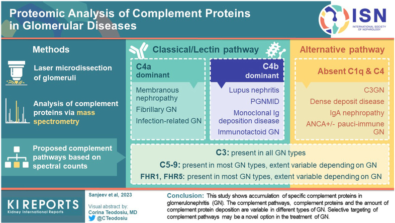
Introduction: Complement plays an important role in the pathogenesis of glomerulonephritis (GN). Even though the underlying etiology of GN might be different, complement activation with subsequent glomerular deposition of complement proteins result in glomerular injury and progression of the lesions. Routine immunofluorescence microscopy (IF) includes staining for only complement factors C3c and C1q. Therefore, with regard to evaluation of the complement pathways, routine kidney biopsy provides only limited information.

Methods: In this study, using laser microdissection of glomeruli followed by mass spectrometry, complement proteins and pathways involved in GN were analyzed.

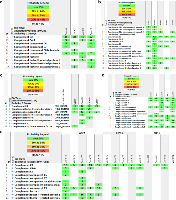
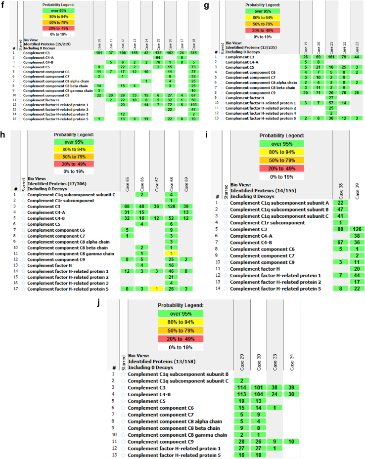
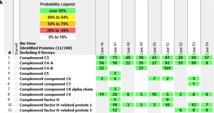
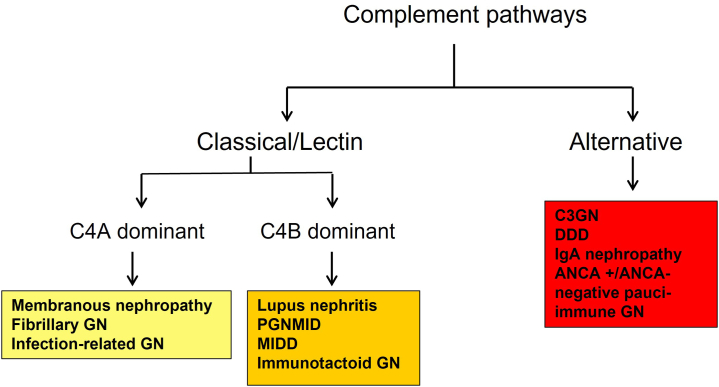
Results: We found that C3 followed by C9 are the most abundant complement proteins in GN, indicating activation of classical or lectin or alternative, and terminal pathways, either exclusively or in a combination of pathways. Furthermore, depending on the type of GN, C4A and/or C4B were also present. Therefore, membranous nephropathy (MN), fibrillary GN, and infection-related GN showed C4A dominant pathways, whereas lupus nephritis (LN), proliferative GN with monoclonal Ig deposits, monoclonal Ig deposition disease (MIDD), and immunotactoid glomerulopathy showed C4B dominant pathways. Significant deposition of complement regulatory proteins, factor H-related protein-1 (FHR-1) and factor H-related protein-5 (FHR-5), were also detected in most GN.

Conclusions: This study shows accumulation of specific complement proteins in GN. The complement pathways, complement proteins, and the amount of complement protein deposition are variable in different types of GN. Selective targeting of complement pathways may be a novel option in the treatment of GN.

Keywords: complement; glomerulonephritis; kidney; laser microdissection; mass spectrometry.


Figures

None
Graphical abstract
Figure 1
Figure 1
Proteomic Identification of complement proteins in glomerular diseases: (a) Infection related GN. (b) Lupus nephritis. (c) IgA nephropathy. (d) Fibrillary glomerulonephritis. (e) Pauci-immune ANCA-negative and ANCA-positive (PR3 and MPO) GN. (f) C3 glomerulonephritis (C3GN). (g) Dense deposit disease (DDD). (h) Proliferative glomerulonephritis with monoclonal Ig deposits (PGNMID). (i) Immunotactoid glomerulopathy. (j) Heavy chain immunoglobulin deposition disease (HCDD). (k) Membranous nephropathy. Numbers in green boxes represent spectral counts of MS/MS matches to a respective protein. Protein grouping ambiguity (red star) indicates shared amino acid sequences for certain proteins.
Figure 1
Figure 1
Proteomic Identification of complement proteins in glomerular diseases: (a) Infection related GN. (b) Lupus nephritis. (c) IgA nephropathy. (d) Fibrillary glomerulonephritis. (e) Pauci-immune ANCA-negative and ANCA-positive (PR3 and MPO) GN. (f) C3 glomerulonephritis (C3GN). (g) Dense deposit disease (DDD). (h) Proliferative glomerulonephritis with monoclonal Ig deposits (PGNMID). (i) Immunotactoid glomerulopathy. (j) Heavy chain immunoglobulin deposition disease (HCDD). (k) Membranous nephropathy. Numbers in green boxes represent spectral counts of MS/MS matches to a respective protein. Protein grouping ambiguity (red star) indicates shared amino acid sequences for certain proteins.
Figure 2
Figure 2
Proposed complement pathways in glomerulonephritis. ANCA, antineutrophil cytoplasmic antibody; DDD, dense deposit disease; MIDD, monoclonal immunoglobulin deposition disease.

References

    1. Sethi S., Haas M., Markowitz G.S., et al. Mayo Clinic/renal pathology society consensus report on pathologic classification, diagnosis, and reporting of GN. J Am Soc Nephrol. 2016;27:1278–1287. doi: 10.1681/ASN.2015060612. - DOI - PMC - PubMed
    1. Sethi S., Gamez J.D., Vrana J.A., et al. Glomeruli of Dense Deposit Disease contain components of the alternative and terminal complement pathway. Kidney Int. 2009;75:952–960. doi: 10.1038/ki.2008.657. - DOI - PMC - PubMed
    1. Vrana J.A., Gamez J.D., Madden B.J., Theis J.D., Bergen H.R., Dogan A. Classification of amyloidosis by laser microdissection and mass spectrometry based proteomic analysis in clinical biopsy specimens. Blood. 2009;114:4957–4959. doi: 10.1182/blood-2009-07-230722. - DOI - PubMed
    1. Sethi S., Vrana J.A., Theis J.D., et al. Laser microdissection and mass spectrometry-based proteomics aids the diagnosis and typing of renal amyloidosis. Kidney Int. 2012;82:226–234. doi: 10.1038/ki.2012.108. - DOI - PMC - PubMed
    1. Dasari S., Alexander M.P., Vrana J.A., et al. DnaJ heat shock protein family B Member 9 is a novel biomarker for fibrillary GN. J Am Soc Nephrol. 2018;29:51–56. doi: 10.1681/ASN.2017030306. - DOI - PMC - PubMed