. 2023 Jun 9;51(10):5210-5227.
doi: 10.1093/nar/gkad274.

A multidisciplinary approach to the identification of the protein-RNA connectome in double-stranded RNA virus capsids

Affiliations

A multidisciplinary approach to the identification of the protein-RNA connectome in double-stranded RNA virus capsids

Po-Yu Sung et al. Nucleic Acids Res. .

Abstract

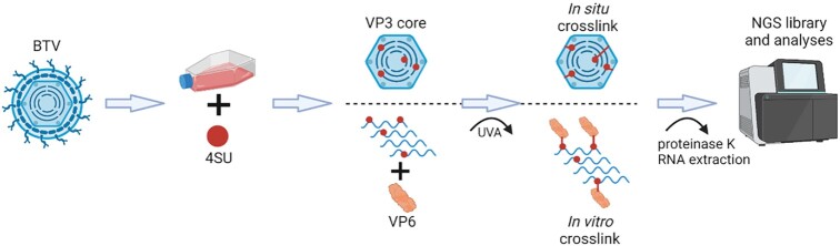
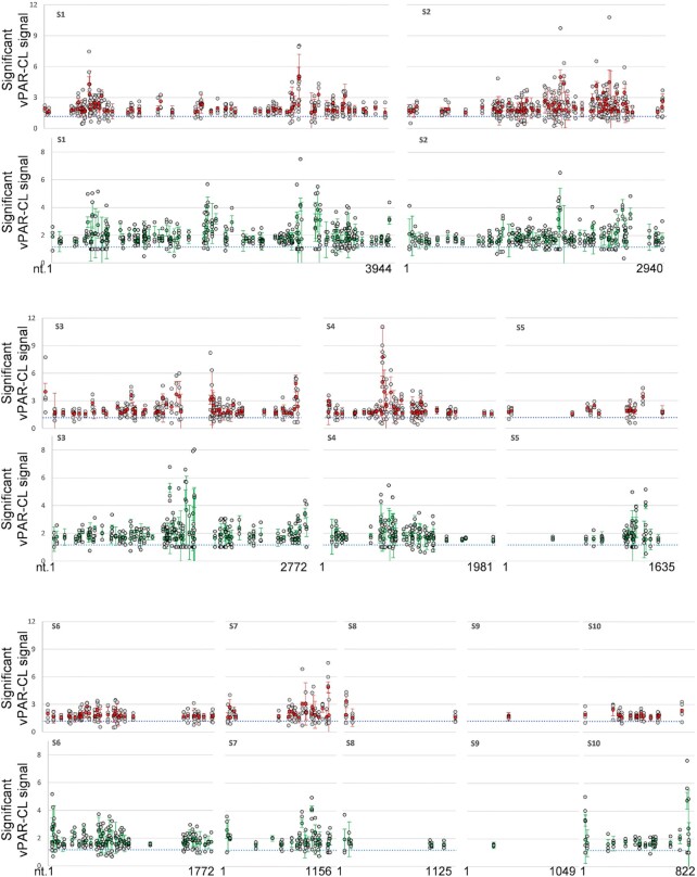
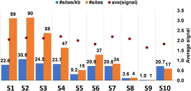
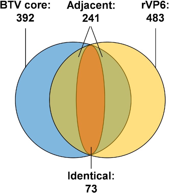
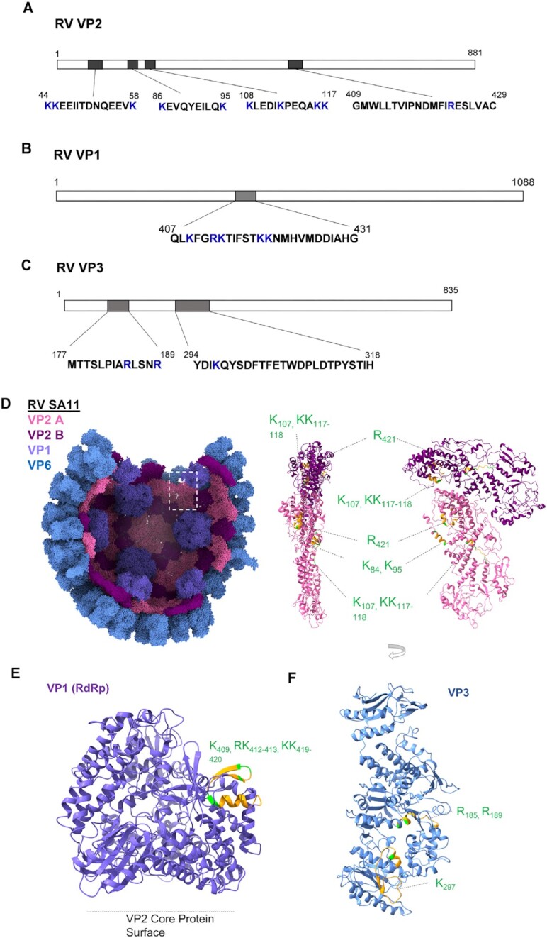
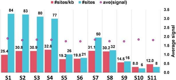
How multi-segmented double-stranded RNA (dsRNA) viruses correctly incorporate their genomes into their capsids remains unclear for many viruses, including Bluetongue virus (BTV), a Reoviridae member, with a genome of 10 segments. To address this, we used an RNA-cross-linking and peptide-fingerprinting assay (RCAP) to identify RNA binding sites of the inner capsid protein VP3, the viral polymerase VP1 and the capping enzyme VP4. Using a combination of mutagenesis, reverse genetics, recombinant proteins and in vitro assembly, we validated the importance of these regions in virus infectivity. Further, to identify which RNA segments and sequences interact with these proteins, we used viral photo-activatable ribonucleoside crosslinking (vPAR-CL) which revealed that the larger RNA segments (S1-S4) and the smallest segment (S10) have more interactions with viral proteins than the other smaller segments. Additionally, using a sequence enrichment analysis we identified an RNA motif of nine bases that is shared by the larger segments. The importance of this motif for virus replication was confirmed by mutagenesis followed by virus recovery. We further demonstrated that these approaches could be applied to a related Reoviridae member, rotavirus (RV), which has human epidemic impact, offering the possibility of novel intervention strategies for a human pathogen.


Figures

Graphical Abstract
Graphical Abstract
The molecular connectome of double-stranded RNA viruses.
Figure 1.
Figure 1.
Purified BTV cores and RCAP workflow for the analysis of BTV cores. (A) Purified BTV cores are visualised via electron microscope and SDS-PAGE. BTV proteins are indicated. (B) The schematic for RCAP workflow. Each stage is indicated.
Figure 2.
Figure 2.
Visualisation of RNA-interacting regions on VP3 and results of virus recovery from key mutations. (A) RCAP identified RNA-interacting regions and amino acids are indicated. Blue letters indicate positively charged residues. The results of virus recovery after mutating the indicated residue are indicated. (B) A partial assembly of VP7 and VP3 for BTV solved by X-ray crystallography (2btv) are illustrated (left) with VP7 coloured blue, and Chain ‘A’ and ‘B’ from the asymmetrical dimer of VP3 coloured pink and purple respectively. In the icosahedral shell of the BTV core, 60 VP3 dimers form a convex disc shape with a series of shallow grooves (35). Ribbon models for VP3 A and B are illustrated (right) to indicate RCAP identified interacting regions (orange) in each subunit. Mutated amino acid residues that yield viable virus particles are coloured green, while mutated amino acid residues that failed to recover virus are coloured red. Amino acid identities and coordinates are indicated.
Figure 3.
Figure 3.
Plagues of VP3 mutant viruses. Different VP3 mutants, Wildtype (WT) and mock infection are indicated.
Figure 4.
Figure 4.
Capsid formation and RNA packaging from different mutations on VP3-RNA interaction sites. (A) Upper panel: Different CLP samples analysed by SDS-PAGE. The VP3 and VP7 bands are indicated. Lower panel: VP3 specific immunoblotting shows the expression of mutant proteins. (B) CFA assay was performed using wild-type VP3 (WT), R274A mutant VP3, R529A mutant VP3, or without VP3 (no VP3). The BTV RNA (segments S2, S6 and S10) packaged and protected after RNase treatment was quantified and the average quantities of three repeated experiments are shown.
Figure 5.
Figure 5.
RNA-interacting regions in BTV VP1 and VP4. RCAP identified RNA-interacting regions and amino acids of BTV VP1 (A) and VP4 (B) are indicated. Blue letters indicate positively charged residues. The virus recovery results after the indicated residues mutated are indicated. (C) Ribbon model for BTV VP1 solved by cryo-EM (6pns) is illustrated (two different side views) to indicate RCAP-identified RNA-interacting regions (orange). Mutated amino acid residues that yield viable virus particles are coloured green, while mutated amino acid residues that failed to recover virus are coloured red. Amino acid identities and coordinates are indicated. (D) Ribbon model for BTV VP4 solved by X-ray crystallography (2jhc) is illustrated to indicate RCAP-identified RNA-interacting regions (orange). Mutated amino acid residues that yield viable virus particles are coloured green, while mutated amino acid residues that failed to recover virus are coloured red. Amino acid identities and coordinates are indicated.
Figure 6.
Figure 6.
vPAR-CL workflow for the analysis of genomic RNA with BTV core or BTV rVP6 protein. Each stage is indicated.
Figure 7.
Figure 7.
The average signal results from vPAR-CL for BTV RNAs. The average signals of the 10 segments of 4SU enriched BTV genomic RNA interacting with viral proteins in BTV cores (red) or rVP6 in vitro (green) are individually shown.
Figure 8.
Figure 8.
The Prominent protein interacting sites identified by vPAR-CL for BTV RNAs. Red = average signal of BTV core, green = average signal of rVP6, grey dots = individual repeat, error bar = stdev, blue dotted line = average signal of all genomic U positions.
Figure 9.
Figure 9.
The distribution of protein interacting sites in genomic RNAs within BTV cores. The numbers of protein interacting sites (#sites) and ratios of sites per kb (#sites/kb) on each segment are shown.
Figure 10.
Figure 10.
The distinct and overlapping protein interacting sites between BTV core and rVP6. Blue and yellow circles indicate the protein interacting sites identified from BTV cores and rVP6 within the whole BTV genome, respectively. The orange indicates the identical sites. Identical: co-discovery of the same sites in both BTV core and rVP6 datasets. The green and orange regions together indicate adjacent sites. Adjacent: 1 or more VP6 sites were found within 10nts from a BTV site.
Figure 11.
Figure 11.
The STREME predicted 9 base protein interacting motif for BTV. STREME analysis of vPAR-CL sites in BTV core discovered a significant RNA motif (p-value: 7.5e-3, E-value: 3e-2) with GU rich sequences.
Figure 12.
Figure 12.
RNA-interacting regions in RV VP2, VP1 and VP3. The RCAP identified RNA interacting-regions and residues in RV SA11 capsid protein VP2 (A), VP1 (B) and VP3 (C) are indicated. Positively charged residues within the regions are marked as blue. (D) A partial assembly of VP2, VP1 and VP6 RV solved by cryo-EM (6ory) are illustrated (left) with chain ‘A’ and ‘B’ from the asymmetrical dimer of VP2 coloured light pink and hot pink respectively, VP1 coloured purple, and VP6 coloured blue. Ribbon models for VP2 A and B are illustrated (right) (two side views) to indicated RCAP identified RNA-interacting regions (orange) in each subunit. (E) Ribbon model for RV VP1 solved by cryo-EM (6ory) is illustrated to indicate RCAP-identified RNA-interacting regions (orange). Positively charged residues within the regions are coloured green. (F) Ribbon model for RV VP3 solved by cryo-EM (6o6b) is illustrated to indicate RCAP-identified RNA-interacting regions (orange). Positively charged residues within the regions are coloured green. Amino acid identities and coordinates are indicated.
Figure 13.
Figure 13.
The distribution of protein interacting sites in genomic RNAs within RV DLPs. The numbers of protein interacting sites (#sites) and ratios of sites per kb (#sites/kb) on each segment are shown.

Similar articles

Cited by

References

    1. Borodavka A., Tuma R., Stockley P.G.. Evidence that viral rnas have evolved for efficient, two-stage packaging. Proc. Natl. Acad. Sci. U.S.A. 2012; 109:15769–15774. - PMC - PubMed
    1. Ford R.J., Barker A.M., Bakker S.E., Coutts R.H., Ranson N.A., Phillips S.E., Pearson A.R., Stockley P.G.. Sequence-specific, RNA-protein interactions overcome electrostatic barriers preventing assembly of satellite tobacco necrosis virus coat protein. J. Mol. Biol. 2013; 425:1050–1064. - PMC - PubMed
    1. Patel N., White S.J., Thompson R.F., Bingham R., Weiss E.U., Maskell D.P., Zlotnick A., Dykeman E., Tuma R., Twarock R.et al. .. HBV RNA pre-genome encodes specific motifs that mediate interactions with the viral core protein that promote nucleocapsid assembly. Nat. Microbiol. 2017; 2:17098. - PMC - PubMed
    1. Comas-Garcia M., Cadena-Nava R.D., Rao A.L., Knobler C.M., Gelbart W.M.. In vitro quantification of the relative packaging efficiencies of single-stranded RNA molecules by viral capsid protein. J. Virol. 2012; 86:12271–12282. - PMC - PubMed
    1. Poranen M.M., Tuma R.. Self-assembly of double-stranded RNA bacteriophages. Virus. Res. 2004; 101:93–100. - PubMed

Publication types