. 2023 Apr 3;64(4):20.
doi: 10.1167/iovs.64.4.20.

Nonmuscle Myosin IIA Regulates the Precise Alignment of Hexagonal Eye Lens Epithelial Cells During Fiber Cell Formation and Differentiation

Affiliations

Nonmuscle Myosin IIA Regulates the Precise Alignment of Hexagonal Eye Lens Epithelial Cells During Fiber Cell Formation and Differentiation

Sadia T Islam et al. Invest Ophthalmol Vis Sci. .

Abstract

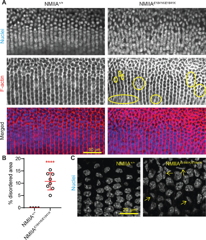
Purpose: Epithelial cells in the equatorial region of the ocular lens undergo a remarkable transition from randomly packed cells into precisely aligned and hexagon-shaped cells organized into meridional rows. We investigated the function of nonmuscle myosin IIA (encoded by Myh9) in regulating equatorial epithelial cell alignment to form meridional rows during secondary fiber cell morphogenesis.

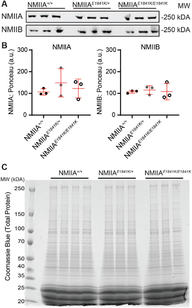
Methods: We used genetic knock-in mice to study a common human Myh9 mutation, E1841K, in the rod domain. The E1841K mutation disrupts bipolar filament assembly. Lens shape, clarity, and stiffness were evaluated, and Western blots were used to determine the level of normal and mutant myosins. Cryosections and lens whole mounts were stained and imaged by confocal microscopy to investigate cell shape and organization.

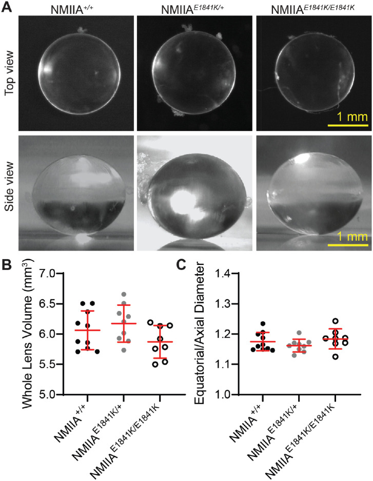
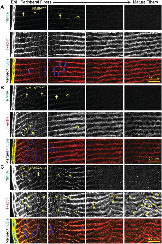
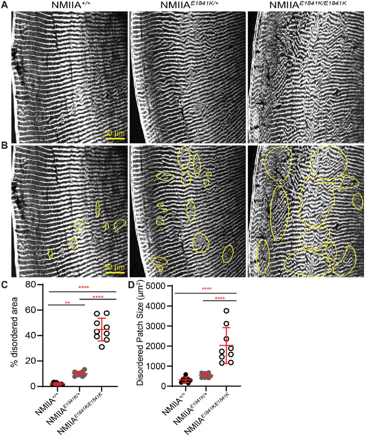
Results: We observed no obvious changes in lens size, shape, and biomechanical properties (stiffness and resilience) between the control and nonmuscle myosin IIA-E1841K mutant mice at 2 months of age. Surprisingly, we found misalignment and disorder of fiber cells in heterozygous and homozygous mutant lenses. Further analysis revealed misshapen equatorial epithelial cells that cause disorientation of the meridional rows before fiber cell differentiation in homozygous mutant lenses.

Conclusions: Our data indicate that nonmuscle myosin IIA bipolar filament assembly is required for the precise alignment of the meridional rows at the lens equator and that the organization of lens fiber cells depends on the proper patterning of meridional row epithelial cells. These data also suggest that lens fiber cell organization and a hexagonal shape are not required for normal lens size, shape transparency, or biomechanical properties.


Conflict of interest statement

Disclosure: S.T. Islam, None; C. Cheng, None; J. Parreno, None; V.M. Fowler, None

Figures

Figure 1.
Figure 1.
Diagrams of mouse lens anatomy and NMIIA structure. (A) A cartoon of a sagittal view of the mouse lens. The ocular lens is composed of two types of cells, epithelial cells (colored) and fiber cells (gray). The anterior epithelial cells are quiescent (blue), whereas the equatorial epithelial cells (orange) have proliferative activity and can migrate further down to the equator to differentiate into meridional row epithelial cells (green). The meridional row epithelial cells further differentiate into secondary fiber cells (grey). (B) A cartoon of the en face view of the lens equator shows that the irregularly shaped and randomly packed equatorial epithelial cells (orange) become precisely aligned, hexagon shaped and arranged in a honeycomb pattern (green). The green cells are arranged into meridional rows, which further elongate and differentiate into secondary fiber cells (gray). (C) NMIIA molecules are hexamers containing two heavy chains (purple), each consisting of an N-terminal motor domain with actin-activated ATPase activity, a flexible neck, and a rod domain. The essential light chain (ELC) (green) and regulatory light chain (RLC) (orange) bind to the heavy chain in the neck region. NMIIA activity is increased or decreased by RLC phosphorylation or dephosphorylation, respectively. The E1841K mutation (pink) in the rod domain is one of the most common MYH9-RD mutations in humans. Cartoons not drawn to scale.
Figure 2.
Figure 2.
The NMIIA E1841K mutation has no effect on whole lens size and shape. (A) (Upper panel) Top-view images of freshly dissected 2-month-old NMIIA+/+, NMIIAE1841K/+, and NMIIAE1841K/E1841K lenses. All lenses were transparent without obvious opacities. Lower panel, side view images of lenses. Scale bars, 1 mm. (B) Whole lens volume and (C) aspect ratio (equatorial to axial diameter ratio) from 2-month-old control and mutant mice show no significant differences in whole lens size and shape. Plots reflect mean ± SD of 8–10 lenses from 4–5 biological replicates per genotype.
Figure 3.
Figure 3.
The NMIIA-E1841K mutation does not affect the global expression of NMIIA/B isoforms in the lens. (A) Western blots of NMIIA and NMIIB heavy chains in whole lens lysates from 6- to 8-week-old NMIIA+/+, NMIIAE1841K/+, and NMIIAE1841K/E1841K mice. (B) Relative NMIIA/B heavy chain expression in NMIIA+/+, NMIIAE1841K/+, and NMIIAE1841K/E1841K whole lenses. NMIIA and NMIIB heavy chain protein levels were normalized to total protein level (Ponceau S staining), which shows no significant changes in protein expression level in the NMIIA-E1841K mutant lenses compared with control lenses. Plots reflect the mean ± SD of n = 3 biological replicates per genotype. *P < 0.05. (C) Coomassie blue staining of total lens extracts from 6- to 8-week-old NMIIA+/+, NMIIAE1841K/+, and NMIIAE1841K/E1841K mice. No noticeable changes in total protein levels were observed between control and mutant lens lysates. Three or four biological replicates were tested for each genotype.
Figure 4.
Figure 4.
NMIIA is predominantly localized in lens epithelial cells of NMIIA+/+, NMIIAE1841K/+, and NMIIAE1841K/E1841K lenses. Immunostaining of frozen sections in the cross-orientation for (A) NMIIA+/+, (B) NMIIAE1841K/+, and (C) NMIIAE1841K/E1841K lenses for NMIIA (green), F-actin (red), and cell nuclei (blue). Images are equatorial sections, with sequential panels showing from left to right, the lens epithelium (Epi) and peripheral fiber cells (leftmost panel) inwards to the mature fiber cells (rightmost panel, approximately 260 µm deep). The first and second leftmost panels are 0 to approximately 65 µm deep and approximately 65 µm to approximately 130 µm deep, respectively. The mature fiber cells are seen in the third panel from the left (approximately 130 to approximately 195 µm deep) and rightmost panel (approximately 195 to approximately 260 µm deep). Although NMIIA is mostly localized to the lens epithelium, faint NMIIA puncta are present along the short vertices of the fiber cells (arrows). F-actin staining shows misaligned fiber cells in NMIIAE1841K/+ and NMIIAE1841K/E1841K lens sections. Asterisks indicate regions of disorder. Scale bar, 20 µm.
Figure 5.
Figure 5.
NMIIAE1841K/E1841K lenses display large areas of fiber cell disorganization. (A, B) Equatorial cryosections of NMIIA+/+, NMIIAE1841K/+, and NMIIAE1841K/E1841K lenses were immunolabeled with rhodamine-phalloidin (F-actin). (A) Cryosections without outlined disordered regions, whereas (B) shows regions of disorder outlined in yellow. Both NMIIAE1841K/+ and NMIIAE1841K/E1841K lenses displayed more areas of fiber cell disorder as compared with NMIIA+/+ lenses. Scale bar, 50 µm. (C) The percent disordered area was significantly greater in the NMIIAE1841K/+ and NMIIAE1841K/E1841K lenses compared with NMIIA+/+ lenses. (D) Disordered patch sizes were significantly higher in NMIIAE1841K/E1841K lenses compared with NMIIA+/+ and NMIIAE1841K/+ lenses, whereas the patch size is not significantly different between NMIIA+/+ and NMIIAE1841K/+ lenses. Plots reflect the mean ± SD of n = 9 independent immunostained sections from three different mice per genotype. **P < 0.01; ***P < 0.001; ****P < 0.0001.
Figure 6.
Figure 6.
Peripheral fiber cells are disordered in the NMIIAE1841K/E1841K lenses. Whole fixed lenses were labeled with rhodamine-phalloidin (F-actin) (red), WGA (cell membrane, green), and nuclei (blue). (A) A single optical section of the peripheral fiber cells in the region of the lens fulcrum is shown in the XY plane. The fulcrum can be identified by a change in cell morphology and bright phalloidin staining (red dashed line). The fulcrum is irregular and discontinuous (short red dashed line) in the homozygous mutant lens, and peripheral fiber cells are not aligned in parallel rows in NMIIAE1841K/E1841K lenses in contrast to the NMIIA+/+ lenses. Scale bars, 20 µm. (B) Images of a single optical section of peripheral fiber cells approximately 5.0 to 5.5 µm inwards from the fulcrum. The F-actin and WGA staining reveal irregularly spaced and misaligned fiber cells in the NMIIAE1841K/E1841K lens, while precisely aligned and regularly spaced fiber cell membranes are observed in the NMIIA+/+ lens. Scale bars, 50 µm.
Figure 7.
Figure 7.
Equatorial epithelial cells in meridional rows are misaligned in NMIIAE1841K/E1841K lenses. (A) Whole mounts of fixed lenses from NMIIA+/+ and NMIIAE1841K/E1841K mice labeled for nuclei (top) and F-actin (middle). Merged images (bottom) with nuclei in blue and F-actin in red. A single optical section in the XY plane displays the middle region of the meridional row cells at low magnification. Meridional row cells are hexagonal and precisely aligned, with nuclei arranged in parallel rows in the NMIIA+/+ lens, whereas meridional rows are misaligned and branching (yellow circles), with disordered and misaligned nuclei in the NMIIAE1841K/E1841K lens. Scale bar, 50 µm. (B) Percent disordered area, plotted as the mean ± SD of four NMIIA+/+ and eight NMIIAE1841K/E1841K lenses from at least four to five different mice. Each dot represents an individual lens, and there is a statistically significant increase in disordered area in homozygous mutant lenses compared with the control lenses. ****P < 0.0001. (C) High magnification view of meridional row cell nuclei in NMIIA+/+ and NMIIAE1841K/E1841K lenses. A single optical section in the XY plane displays the nuclei at the mid-region of meridional row cells (i.e., middle of the lateral membrane with respect to apical–basal cell domains). The mid-region of the meridional row cells was identified based on the maximum diameter of the nuclei. The nuclei of the NMIIAE1841K/E1841K lens seem to be misaligned, out of plane, and abnormally shaped, in contrast with the aligned and regular pattern of nuclei in the NMIIA+/+ lens. Scale bar, 20 µm.
Figure 8.
Figure 8.
Cells in meridional rows exhibit aberrant cell shapes and irregular packing in NMIIAE1841K/E1841K lenses. (A) F-actin staining in single optical sections at the basal region of meridional row cells in NMIIA+/+ and NMIIAE1841K/E1841K lenses, shown in XY plane. F-actin is enriched along all six sides of meridional row epithelial cells. Scale bar, 20 µm. Yellow boxes, regions enlarged in (B). (B) High magnification of a region from A (yellow boxes), showing an individual cell (C) (central cell) surrounded by six neighboring cells (numbered) in the NMIIA+/+ lens, but surrounded by seven neighboring cells in the NMIIAE1841K/E1841K lens. Scale bar, 10 µm. (C) Frequency distribution (%) of the number of adjacent cells for 200 cells from 5 different lenses. Almost all cells in NMIIA+/+ lenses have six nearest neighbors, whereas cells in NMIIAE1841K/E1841K lenses have variable numbers of nearest neighbors (4–8). (D) Percentage of hexagonal adjacent cells in NMIIA+/+ and NMIIAE1841K/E1841K lenses. The plot represents the mean ± SD of five different lens images from at least four different mice. *P < 0.05.

Similar articles

Cited by

References

    1. Bassnett S. On the mechanism of organelle degradation in the vertebrate lens. Exp Eye Res. 2009; 88(2): 133–139, doi:10.1016/j.exer.2008.08.017. - DOI - PMC - PubMed
    1. Lovicu F, Robinson M. Development of the ocular lens. Cambridge, UK: Cambridge University Press; 2004.
    1. Bassnett S, Šikić H.. The lens growth process. Prog Retin Eye Res. 2017; 60: 181–200, doi:10.1016/j.preteyeres.2017.04.001. - DOI - PMC - PubMed
    1. Šikić H, Shi Y, Lubura S, Bassnett S.. A full lifespan model of vertebrate lens growth. R Soc Open Sci. 2017; 4(1): 160695, doi:10.1098/rsos.160695. - DOI - PMC - PubMed
    1. Lovicu F, Robinson M. Development of the ocular lens. Cambridge, UK: Cambridge University Press; 2004.

Publication types

Substances