. 2023 May;307(4):e222045.
doi: 10.1148/radiol.222045. Epub 2023 Apr 18.

Middle Meningeal Artery Embolization for Chronic Subdural Hematoma: Predictors of Clinical and Radiographic Failure from 636 Embolizations

Affiliations

Middle Meningeal Artery Embolization for Chronic Subdural Hematoma: Predictors of Clinical and Radiographic Failure from 636 Embolizations

Mohamed M Salem et al. Radiology. 2023 May.

Abstract

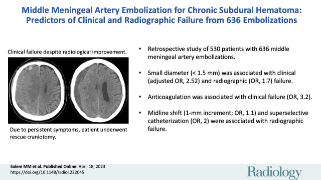
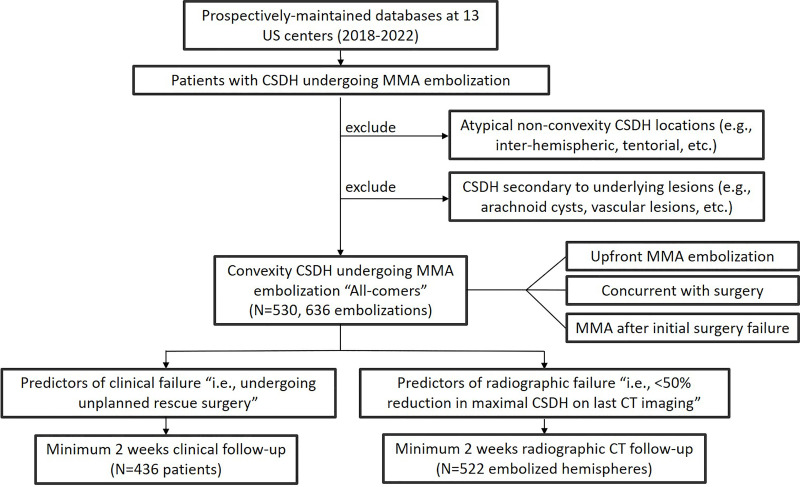
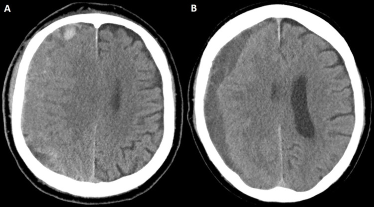
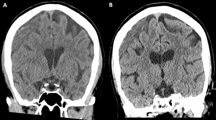
Background Knowledge regarding predictors of clinical and radiographic failures of middle meningeal artery (MMA) embolization (MMAE) treatment for chronic subdural hematoma (CSDH) is limited. Purpose To identify predictors of MMAE treatment failure for CSDH. Materials and Methods In this retrospective study, consecutive patients who underwent MMAE for CSDH from February 2018 to April 2022 at 13 U.S. centers were included. Clinical failure was defined as hematoma reaccumulation and/or neurologic deterioration requiring rescue surgery. Radiographic failure was defined as a maximal hematoma thickness reduction less than 50% at last imaging (minimum 2 weeks of head CT follow-up). Multivariable logistic regression models were constructed to identify independent failure predictors, controlling for age, sex, concurrent surgical evacuation, midline shift, hematoma thickness, and pretreatment baseline antiplatelet and anticoagulation therapy. Results Overall, 530 patients (mean age, 71.9 years ± 12.8 [SD]; 386 men; 106 with bilateral lesions) underwent 636 MMAE procedures. At presentation, the median CSDH thickness was 15 mm and 31.3% (166 of 530) and 21.7% (115 of 530) of patients were receiving antiplatelet and anticoagulation medications, respectively. Clinical failure occurred in 36 of 530 patients (6.8%, over a median follow-up of 4.1 months) and radiographic failure occurred in 26.3% (137 of 522) of procedures. At multivariable analysis, independent predictors of clinical failure were pretreatment anticoagulation therapy (odds ratio [OR], 3.23; P = .007) and an MMA diameter less than 1.5 mm (OR, 2.52; P = .027), while liquid embolic agents were associated with nonfailure (OR, 0.32; P = .011). For radiographic failure, female sex (OR, 0.36; P = .001), concurrent surgical evacuation (OR, 0.43; P = .009), and a longer imaging follow-up time were associated with nonfailure. Conversely, MMA diameter less than 1.5 mm (OR, 1.7; P = .044), midline shift (OR, 1.1; P = .02), and superselective MMA catheterization (without targeting the main MMA trunk) (OR, 2; P = .029) were associated with radiographic failure. Sensitivity analyses retained these associations. Conclusion Multiple independent predictors of failure of MMAE treatment for chronic subdural hematomas were identified, with small diameter (<1.5 mm) being the only factor independently associated with both clinical and radiographic failures. © RSNA, 2023 Supplemental material is available for this article. See also the editorial by Chaudhary and Gemmete in this issue.


Conflict of interest statement

Disclosures of conflicts of interest: M.M.S. No relevant relationships. O.K. No relevant relationships. A.N.H. No relevant relationships. A.A.B. No relevant relationships. M.K. No relevant relationships. C.B. No relevant relationships. J.C.H. No relevant relationships. A.A.M. No relevant relationships. G.C. No relevant relationships. J.M.D. Institutional grants or contracts from National Institutes of Health (NIH), NSF SBIR, University at Buffalo Center for Advanced Technology, Buffalo Translational Consortium, Cummings Foundation, NVIDIA, and Google; royalties from RIST Neurovascular; lecture payment from Medtronic; advisory board for NIH Strokenet; leadership role, Cerebrovascular section of the Congress of Neurological Surgeons; stockholder, QAS.ai, RIST Neurovascular, Cerebrotech, Synchron, and Hyperion. S.N. Lecture payment from Cerenovus; board of directors, Society of NeuroInterventional Surgery. C.M.C. No relevant relationships. H.A.R. No relevant relationships. J.M.M. No relevant relationships. A.M.S. Grants or contracts from Medtronic, Stryker, Penumbra, Avail, and RapidAI; consulting fees from Stryker, Penumbra, RapidAI, and Terumo; medical advisory board, Brain Aneurysm Foundation; stockholder, Avail Med. A.A.K. Consulting fees from Medtronic; payment for expert testimony from Procopio US Attorney; advisory board, Route 92 and Medtronic; leadership role, Congress of Neurological Surgeons and American College of Surgeons; stockholder, Ospitek, Synaptive, and Proximie. B.M.H. No relevant relationships. R.H. Grants or contracts from NIH, Interline Endowment, Microvention, Stryker, CNX, and Balt; consulting fees from Medtronic, Balt, Stryker, Q’Apel Medical, Codman Neuro (J&J), Cerenovus, Microvention, Imperative Care, Phenox, and Rapid Medical; advisory board, MiVI, eLum, Three Rivers, Shape Medical, and Corindus; associate editor of the endovascular section for Neurosurgery Journal; stockholder, InNeuroCo, Cerebrotech, eLum, Endostream, Three Rivers Medical, Scientia, RisT, Blink TBI, and Corindus. O.T. No relevant relationships. E.I.L. Consulting fees from Clarion, GLG Consulting, Guidepoint Global, Imperative Care, Medtronic, StimMed, Misionix, and Mosiac; lecture payments from Clarion, GLG Consulting, Guidepoint Global, Imperative Care, Medtronic, StimMed, Misionix, and Mosiac; expert testimony payment from Renders Medical; patents planned, issued, or pending for Ultrasonic surgical blade; advisory board for Stryker, NeXtGen Biologics, MEDX, Cognition Medical, Endostream Medical, and IRRAS; stockholder, NeXtGen Biologics, RAPID Medical, Claret Medical, Cognition Medical, Imperative Care, Rebound Therapeutics, StimMed, and Three Rivers Medical; chief medical officer for Haniva Technology. R.G. Consulting fees from Medtronic Neurovascular, Balt Neurovascular, and Cerenovus. M.J.L No relevant relationships. A.H.S. Grant from NIH; consulting fees from Amnis Therapeutics, Apellis Pharmaceuticals, Boston Scientific, Canon Medical Systems, Cardinal Health 200, Cerebrotech Medical Systems, Cerenovus, Cerevatech Medical, Cordis, Corindus, Endostream Medical, Imperative Care, InspireMD, Integra, IRRAS AB, Medtronic, MicroVention, Minnetronix Neuro, Peijia Medical, Penumbra, Piraeus Medical, Q’Apel Medical, Rapid Medical, Serenity Medical, Silk Road Medical, StimMed, Stryker Neurovascular, Three Rivers Medical, VasSol, and Viz.ai; stockholder, Adona Medical, Amnis Therapeutics, Bend IT Technologies, BlinkTBI, Borvo Medical, Cerebrotech Medical Systems, Cerevatech Medical, Cognition Medical, Collavidence, CVAID, E8, Endostream Medical, Galaxy Therapeutics, Imperative Care, InspireMD, Instylla, International Medical Distribution Partners, Launch NY, Neurolutions, NeuroRadial Technologies, NeuroTechnology Investors, Neurovascular Diagnostics, Peijia Medical, PerFlow Medical, Piraeus Medical, Q’Apel Medical, QAS.ai, Radical Catheter Technologies, Rebound Therapeutics, RIST Neurovascular, Sense Diagnostics, Serenity Medical, Silk Road Medical, Sim & Cure, SongBird Therapy, Spinnaker Medical, StimMed, Synchron, Three Rivers Medical, Truvic Medical, Tulavi Therapeutics, Vastrax, VICIS, Viseon, and Whisper Medical; payments related to Cerenovus EXCELLENT and ARISE II Trial; Medtronic SWIFT PRIME, VANTAGE, EMBOLISE, and SWIFT DIRECT Trials; MicroVention FRED Trial & CONFIDENCE Study; MUSC POSITIVE Trial; Penumbra 3D Separator Trial, COMPASS Trial, INVEST Trial, MIVI Neuroscience EVAQ Trial; Rapid Medical SUCCESS Trial; and InspireMD C-GUARDIANS IDE Pivotal Trial. P.K. Grants from NIH (1U18EB029353-01), Medtronic (ERP-2019-12070), Siemens (CON30434), and Joe Niekro Foundation (CON30914); consulting fees from Stryker Neurovascular, Imperative Care, Cerenovus, and Microvention; editorial board, Journal of Neurointerventional Surgery. C.S.O. No relevant relationships. B.A.G. Consulting fees from Medtronic and Microvention. A.J.T. Consulting fees from Stryker, Medtronic, and Cerevasc. B.T.J. No relevant relationships. J.K.B. Consulting fees from Q`Apel Medical, Stryker, Medtronic, Cerenovous, and Microvention.

Figures

None
Graphical abstract
Flowchart shows patient inclusion and exclusion. CSDH = chronic
subdural hematoma, MMA = middle meningeal artery.
Figure 1:
Flowchart shows patient inclusion and exclusion. CSDH = chronic subdural hematoma, MMA = middle meningeal artery.
Temporal depiction of chronic subdural hematoma (CSDH) response after
middle meningeal artery (MMA) embolization. Line graph shows the percentage
of CSDHs that achieved radiographic success (≥50% reduction in
maximal hematoma thickness), stratified according to preprocedural CSDH
maximal thickness (group 1, <10 mm; group 2, 10 mm to <15 mm;
group 3, 15 mm to <20 mm, and group 4, ≥20 mm) over
prespecified imaging follow-up intervals, including 2–4 weeks
(14–28 days), greater than 4 weeks to 6 weeks (29–42 days), 6
weeks to 90 days (43–90 days), and greater than 90 days. The data on
the right indicate the number of cases achieving the end point (numerator)
from the total cases (denominator) for each CSDH group over each time
interval.
Figure 2:
Temporal depiction of chronic subdural hematoma (CSDH) response after middle meningeal artery (MMA) embolization. Line graph shows the percentage of CSDHs that achieved radiographic success (≥50% reduction in maximal hematoma thickness), stratified according to preprocedural CSDH maximal thickness (group 1, <10 mm; group 2, 10 mm to <15 mm; group 3, 15 mm to <20 mm, and group 4, ≥20 mm) over prespecified imaging follow-up intervals, including 2–4 weeks (14–28 days), greater than 4 weeks to 6 weeks (29–42 days), 6 weeks to 90 days (43–90 days), and greater than 90 days. The data on the right indicate the number of cases achieving the end point (numerator) from the total cases (denominator) for each CSDH group over each time interval.
Illustrative cases of each preprocedural CSDH maximal thickness group.
(A) Noncontrast head CT image (left) shows a maximal hematoma thickness of 9
mm (group 1, <10 mm) in a 77-year-old man who underwent middle
meningeal artery embolization (MMAE) concurrently with left-sided craniotomy
for hematoma evacuation. Follow-up noncontrast head CT image (right) at 4
months shows complete resolution of the hematoma. (B) Noncontrast head CT
image (left) shows a maximal hematoma thickness of 11.6 mm (group 2, 10 mm
to <15 mm) in an 80-year-old man who underwent MMAE following
recurrence after a prior left-sided burr hole procedure (3 months earlier).
Follow-up noncontrast head CT image (right) at 9 weeks shows complete
resolution of the CSDH. (C) Noncontrast head CT image (left) shows a maximal
hematoma thickness of 16 mm (group 3, 15 mm to <20 mm) in a
68-year-old man who underwent MMAE following recurrence after prior
left-sided craniotomy. Follow-up noncontrast head CT image (right) at 6
weeks shows a minimal hematoma thickness of 5 mm and resolution of symptoms.
(D) Noncontrast head CT image (left) shows a maximal hematoma thickness of
25 mm (group 4, ≥20 mm) in a 94-year-old man who underwent MMAE
following recurrence after prior left-sided craniotomy (2 months earlier).
Follow-up noncontrast head CT image (right) at 8 weeks shows near resolution
of the right-sided hematoma.
Figure 3:
Illustrative cases of each preprocedural CSDH maximal thickness group. (A) Noncontrast head CT image (left) shows a maximal hematoma thickness of 9 mm (group 1, <10 mm) in a 77-year-old man who underwent middle meningeal artery embolization (MMAE) concurrently with left-sided craniotomy for hematoma evacuation. Follow-up noncontrast head CT image (right) at 4 months shows complete resolution of the hematoma. (B) Noncontrast head CT image (left) shows a maximal hematoma thickness of 11.6 mm (group 2, 10 mm to <15 mm) in an 80-year-old man who underwent MMAE following recurrence after a prior left-sided burr hole procedure (3 months earlier). Follow-up noncontrast head CT image (right) at 9 weeks shows complete resolution of the CSDH. (C) Noncontrast head CT image (left) shows a maximal hematoma thickness of 16 mm (group 3, 15 mm to <20 mm) in a 68-year-old man who underwent MMAE following recurrence after prior left-sided craniotomy. Follow-up noncontrast head CT image (right) at 6 weeks shows a minimal hematoma thickness of 5 mm and resolution of symptoms. (D) Noncontrast head CT image (left) shows a maximal hematoma thickness of 25 mm (group 4, ≥20 mm) in a 94-year-old man who underwent MMAE following recurrence after prior left-sided craniotomy (2 months earlier). Follow-up noncontrast head CT image (right) at 8 weeks shows near resolution of the right-sided hematoma.
Case illustration of clinical failure. (A) Preprocedural head CT image
in a 74-year-old man with an extensive cardiovascular history (receiving
aspirin and clopidogrel) and stage IV lung cancer shows a right-sided
isodense subacute subdural hematoma. The patient was initially managed
conservatively and eventually offered middle meningeal artery embolization
because of persistent headaches, which was uneventful. The patient
subsequently returned from rehabilitation after 4 weeks. (B) Head CT image
in the same patient shows changes in hematoma density and decreased size.
Despite these radiologic changes, the patient underwent subsequent rescue
craniotomy because of progression of headaches.
Figure 4:
Case illustration of clinical failure. (A) Preprocedural head CT image in a 74-year-old man with an extensive cardiovascular history (receiving aspirin and clopidogrel) and stage IV lung cancer shows a right-sided isodense subacute subdural hematoma. The patient was initially managed conservatively and eventually offered middle meningeal artery embolization because of persistent headaches, which was uneventful. The patient subsequently returned from rehabilitation after 4 weeks. (B) Head CT image in the same patient shows changes in hematoma density and decreased size. Despite these radiologic changes, the patient underwent subsequent rescue craniotomy because of progression of headaches.
Case illustration of radiographic failure. (A) Head CT image in a
63-year-old woman shows a large right-sided chronic subdural hematoma
(maximal thickness of 19 mm), which was discovered after the patient fell.
Because of progressive headaches, the patient underwent a middle meningeal
artery embolization procedure, which was uneventful. (B) Head CT image
obtained at the 10-week follow-up (last available) shows minimal reduction
of hematoma thickness, and the patient reported little improvement of
headaches.
Figure 5:
Case illustration of radiographic failure. (A) Head CT image in a 63-year-old woman shows a large right-sided chronic subdural hematoma (maximal thickness of 19 mm), which was discovered after the patient fell. Because of progressive headaches, the patient underwent a middle meningeal artery embolization procedure, which was uneventful. (B) Head CT image obtained at the 10-week follow-up (last available) shows minimal reduction of hematoma thickness, and the patient reported little improvement of headaches.

Comment in

References

    1. Ducruet AF , Grobelny BT , Zacharia BE , et al. . The surgical management of chronic subdural hematoma . Neurosurg Rev 2012. ; 35 ( 2 ): 155 – 169 ; discussion 169 . - PubMed
    1. Miranda LB , Braxton E , Hobbs J , Quigley MR . Chronic subdural hematoma in the elderly: not a benign disease . J Neurosurg 2011. ; 114 ( 1 ): 72 – 76 . - PubMed
    1. Ivamoto HS , Lemos HP Jr , Atallah AN . Surgical treatments for chronic subdural hematomas: a comprehensive systematic review . World Neurosurg 2016. ; 86 : 399 – 418 . - PubMed
    1. Liu W , Bakker NA , Groen RJ . Chronic subdural hematoma: a systematic review and meta-analysis of surgical procedures . J Neurosurg 2014. ; 121 ( 3 ): 665 – 673 . - PubMed
    1. Ironside N , Nguyen C , Do Q , et al. . Middle meningeal artery embolization for chronic subdural hematoma: a systematic review and meta-analysis . J Neurointerv Surg 2021. ; 13 ( 10 ): 951 – 957 . - PubMed

LinkOut - more resources