

The IQGAP scaffolds: Critical nodes bridging receptor activation to cellular signaling
- PMID: 37071417
- PMCID: PMC10120595
- DOI: 10.1083/jcb.202205062
The IQGAP scaffolds: Critical nodes bridging receptor activation to cellular signaling
Abstract
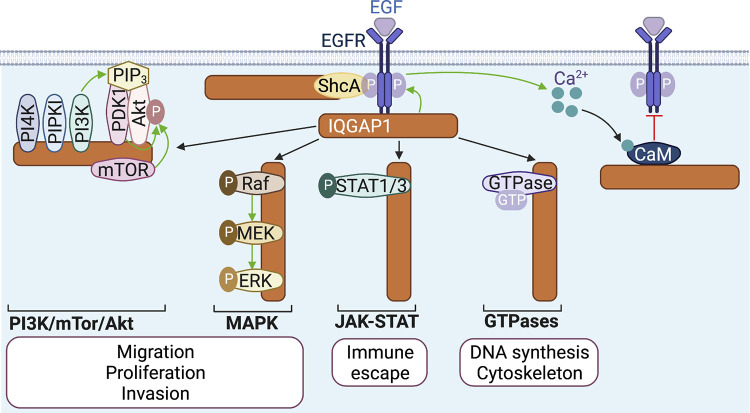
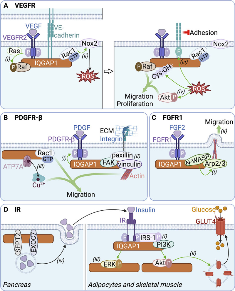
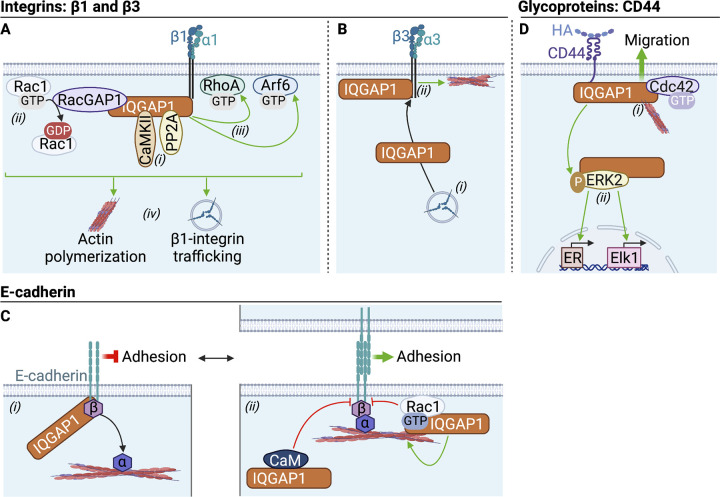
The scaffold protein IQGAP1 assembles multiprotein signaling complexes to influence biological functions. Cell surface receptors, particularly receptor tyrosine kinases and G-protein coupled receptors, are common IQGAP1 binding partners. Interactions with IQGAP1 modulate receptor expression, activation, and/or trafficking. Moreover, IQGAP1 couples extracellular stimuli to intracellular outcomes via scaffolding of signaling proteins downstream of activated receptors, including mitogen-activated protein kinases, constituents of the phosphatidylinositol 3-kinase pathway, small GTPases, and β-arrestins. Reciprocally, some receptors influence IQGAP1 expression, subcellular localization, binding properties, and post-translational modifications. Importantly, the receptor:IQGAP1 crosstalk has pathological implications ranging from diabetes and macular degeneration to carcinogenesis. Here, we describe the interactions of IQGAP1 with receptors, summarize how they modulate signaling, and discuss their contribution to pathology. We also address the emerging functions in receptor signaling of IQGAP2 and IQGAP3, the other human IQGAP proteins. Overall, this review emphasizes the fundamental roles of IQGAPs in coupling activated receptors to cellular homeostasis.
This is a work of the U.S. Government and is not subject to copyright protection in the United States. Foreign copyrights may apply.
Conflict of interest statement
Disclosures: The authors declare no competing interests exist.
Figures

Similar articles
-
IQGAPs choreograph cellular signaling from the membrane to the nucleus.Trends Cell Biol. 2015 Mar;25(3):171-84. doi: 10.1016/j.tcb.2014.12.005. Epub 2015 Jan 21. Trends Cell Biol. 2015. PMID: 25618329 Free PMC article. Review.
-
Identification of the interactome of the DP1 receptor for Prostaglandin D2: Regulation of DP1 receptor signaling and trafficking by IQGAP1.Biochim Biophys Acta Gen Subj. 2021 Nov;1865(11):129969. doi: 10.1016/j.bbagen.2021.129969. Epub 2021 Aug 2. Biochim Biophys Acta Gen Subj. 2021. PMID: 34352343
-
IQGAPs in cancer: a family of scaffold proteins underlying tumorigenesis.FEBS Lett. 2009 Jun 18;583(12):1817-24. doi: 10.1016/j.febslet.2009.05.007. Epub 2009 May 9. FEBS Lett. 2009. PMID: 19433088 Free PMC article. Review.
-
IQGAP1 and its binding proteins control diverse biological functions.Cell Signal. 2012 Apr;24(4):826-34. doi: 10.1016/j.cellsig.2011.12.005. Epub 2011 Dec 11. Cell Signal. 2012. PMID: 22182509 Free PMC article. Review.
-
IQGAP1 gene silencing induces apoptosis and decreases the invasive capacity of human hepatocellular carcinoma cells.Tumour Biol. 2016 Oct;37(10):13927-13939. doi: 10.1007/s13277-016-5283-8. Epub 2016 Aug 3. Tumour Biol. 2016. PMID: 27488117
Cited by
-
IQGAP1 participates in bone marrow-derived macrophage recruitment and involves in liver inflammation/fibrosis.J Mol Med (Berl). 2025 Jul 19. doi: 10.1007/s00109-025-02573-6. Online ahead of print. J Mol Med (Berl). 2025. PMID: 40682669
-
Coordination of actin plus-end dynamics by IQGAP1, formin, and capping protein.bioRxiv [Preprint]. 2024 Apr 1:2023.05.04.539490. doi: 10.1101/2023.05.04.539490. bioRxiv. 2024. Update in: J Cell Biol. 2024 Sep 2;223(9):e202305065. doi: 10.1083/jcb.202305065. PMID: 37205555 Free PMC article. Updated. Preprint.
-
ATG9A facilitates the closure of mammalian autophagosomes.J Cell Biol. 2025 Feb 3;224(2):e202404047. doi: 10.1083/jcb.202404047. Epub 2025 Jan 2. J Cell Biol. 2025. PMID: 39745851 Free PMC article.
-
Cytoskeletal mechanisms regulating attaching/effacing bacteria interactions with host cells: It takes a village to build the pedestal.Bioessays. 2024 Nov;46(11):e2400160. doi: 10.1002/bies.202400160. Epub 2024 Sep 20. Bioessays. 2024. PMID: 39301984 Review.
-
The Personalized Inherited Signature Predisposing to Non-Small-Cell Lung Cancer in Non-Smokers.Cancers (Basel). 2024 Aug 20;16(16):2887. doi: 10.3390/cancers16162887. Cancers (Basel). 2024. PMID: 39199663 Free PMC article.
References
-
- Alemayehu, M., Dragan M., Pape C., Siddiqui I., Sacks D.B., Di Guglielmo G.M., Babwah A.V., and Bhattacharya M.. 2013. β-Arrestin2 regulates lysophosphatidic acid-induced human breast tumor cell migration and invasion via Rap1 and IQGAP1. PLoS One. 8:e56174. 10.1371/journal.pone.0056174 - DOI - PMC - PubMed
-
- Ashino, T., Kohno T., Sudhahar V., Ash D., Ushio-Fukai M., and Fukai T.. 2018. Copper transporter ATP7A interacts with IQGAP1, a Rac1 binding scaffolding protein: Role in PDGF-induced VSMC migration and vascular remodeling. Am. J. Physiol. Cell Physiol. 315:C850–C862. 10.1152/ajpcell.00230.2018 - DOI - PMC - PubMed
Publication types
MeSH terms
Substances
LinkOut - more resources
Full Text Sources
Miscellaneous
