. 2023 May 22;8(10):e167042.
doi: 10.1172/jci.insight.167042.

Interferon-dependent signaling is critical for viral clearance in airway neutrophils

Affiliations

Interferon-dependent signaling is critical for viral clearance in airway neutrophils

Camilla Margaroli et al. JCI Insight. .

Abstract

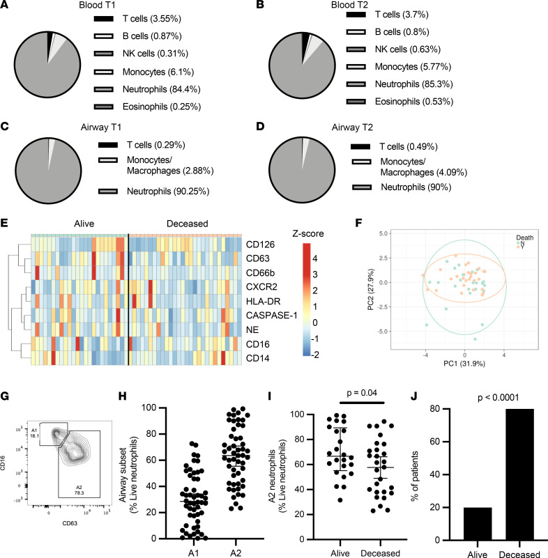
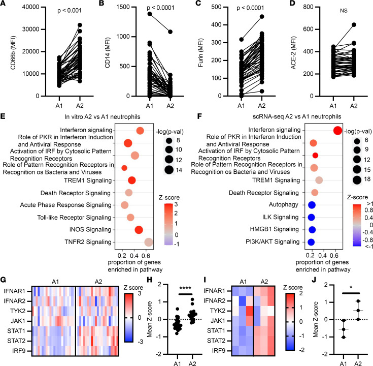
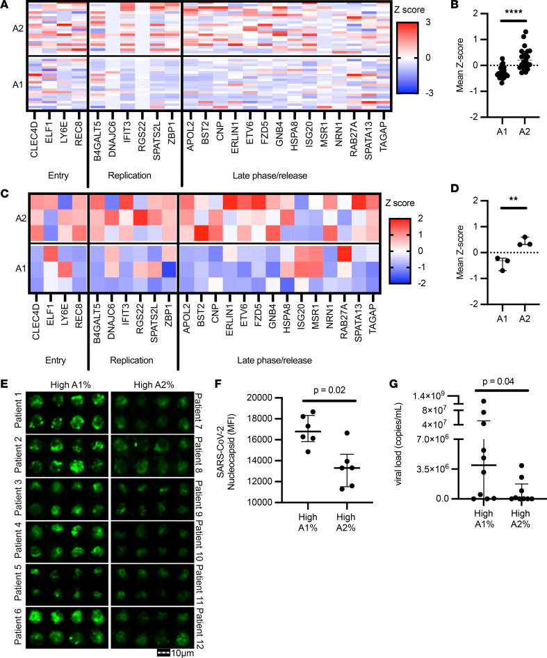
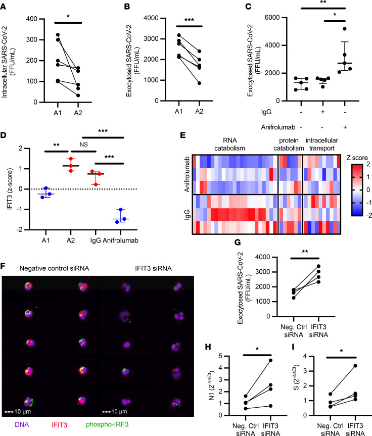
Neutrophilic inflammation characterizes several respiratory viral infections, including COVID-19-related acute respiratory distress syndrome, although its contribution to disease pathogenesis remains poorly understood. Blood and airway immune cells from 52 patients with severe COVID-19 were phenotyped by flow cytometry. Samples and clinical data were collected at 2 separate time points to assess changes during ICU stay. Blockade of type I interferon and interferon-induced protein with tetratricopeptide repeats 3 (IFIT3) signaling was performed in vitro to determine their contribution to viral clearance in A2 neutrophils. We identified 2 neutrophil subpopulations (A1 and A2) in the airway compartment, where loss of the A2 subset correlated with increased viral burden and reduced 30-day survival. A2 neutrophils exhibited a discrete antiviral response with an increased interferon signature. Blockade of type I interferon attenuated viral clearance in A2 neutrophils and downregulated IFIT3 and key catabolic genes, demonstrating direct antiviral neutrophil function. Knockdown of IFIT3 in A2 neutrophils led to loss of IRF3 phosphorylation, with consequent reduced viral catabolism, providing the first discrete mechanism to our knowledge of type I interferon signaling in neutrophils. The identification of this neutrophil phenotype and its association with severe COVID-19 outcomes emphasizes its likely importance in other respiratory viral infections and potential for new therapeutic approaches in viral illness.

Keywords: Cellular immune response; Immunology; Innate immunity; Neutrophils.


Figures

Figure 1
Figure 1. Airway neutrophil subsets associate with survival.
Blood and airway immune cell frequencies (live and CD45+) and profiles were determined by flow cytometry. COVID-19 patients displayed blood neutrophilia (A) upon ICU admission (T1, n = 52) (normal neutrophil frequencies: 40%–65%). (B) These profiles were maintained at time point 2 (T2, n = 28). (C and D) Airway immune cell frequencies in mBAL fluid displayed marked neutrophil infiltration, which was maintained through both time points. (E) No significant difference was observed between surviving and deceased patients for individual surface markers, or as a combined profile by principal component analysis (F). (G and H) Presence of specific neutrophil subsets, including airway neutrophil profiles matching the A1 and A2 populations. (I) A2 neutrophil frequency at time of admission discriminated 30-day mortality (alive = 28, deceased = 24). (J) Low frequencies of the A2 population correlated with mortality (alive = 3, deceased = 10). Results in G and H are shown as median and interquartile range. Statistical analysis was performed using an unpaired, 2-tailed t test upon normality testing (I) and Fisher’s exact test for unpaired analysis (J).
Figure 2
Figure 2. A2 neutrophils show antiviral gene signatures.
(AD) Airway neutrophils were profiled by flow cytometry at time point 1 (n = 52). A1 and A2 neutrophil expression of surface CD66b, CD14, furin, and ACE-2. MFI, median fluorescence intensity. (E) Pathway analysis for genes enriched in A2 neutrophils generated in vitro (n = 3 donors). (F) Pathway analysis for genes enriched in A2 vs. A1 BAL neutrophils from single-cell RNA sequencing (scRNA-seq; n = 21 patients). (G and H) Type I interferon pathway gene expression for A2 vs. A1 BAL neutrophils from scRNA-seq, with mean z scores (n = 21 patients). (I and J) Type I interferon pathway gene expression for ex vivo–generated A2 vs. A1 neutrophil mean z scores (n = 3 per group). Data are shown as median and interquartile range. Statistical analysis was performed using Wilcoxon’s matched-pair signed-rank test for paired analysis (AD) and Wilcoxon’s rank-sum test for unpaired analysis (H and J). *P < 0.05, ****P < 0.0001.
Figure 3
Figure 3. A2 neutrophils show differential anti–SARS-CoV-2 responses.
(A and B) Expression analysis of genes implicated in SARS-CoV-2 intracellular antiviral response, with mean z scores. Data were obtained from sc-RNA-seq and each column represents 1 patient (n = 21 per group). (C and D) A1 and A2 neutrophils generated using an in vitro transmigration model showed differential gene expression for SARS-CoV-2 intracellular antiviral response. (E) Airway neutrophils from a subset of patients with high A1 or high A2 frequencies (n = 6 per group) were stained for SARS-CoV-2 nucleocapsid (green) and acquired by image cytometry (see Supplemental Figure 3). Scale bar: 10 μm. (F) Patients with high A1 percentage showed increased presence of intracellular SARS-CoV-2 in airway neutrophils. (G) Patients with high A1 percentage showed increased presence of extracellular SARS-CoV-2 in the mBAL supernatant (n = 19 patients). Results are shown as median and interquartile range. Statistical analysis was performed using Wilcoxon’s rank-sum test for unpaired analysis. **P < 0.01, ****P < 0.0001.
Figure 4
Figure 4. IFIT3 signaling modulates viral clearance in A2 neutrophils.
(A) A1 and A2 neutrophils incubated with SARS-CoV-2 (MOI = 1) (n = 6 neutrophil donors). (B) A1 and A2 neutrophils show differential exocytosis of infectious SARS-CoV-2 (n = 6 neutrophil donors). (C) Type I interferon blockade with anifrolumab increased exocytosis of infectious SARS-CoV-2 in A2 neutrophils (n = 5 neutrophil donors) compared with IgG control or media alone. (D) IFIT3 expression by RNA-seq. (E) Expression of genes in the macromolecular catabolic processes (GO: 0009057). (F) Image cytometry analysis of airway neutrophils for IFIT3 (red) and phospho-IRF3 (green) expression. Nuclei were stained with DAPI (purple). Scale bars: 10 μm. (G) IFIT3 knockdown increased exocytosis of infectious SARS-CoV-2 in A2 neutrophils (n = 4 neutrophil donors). (H and I) IFIT3 knockdown modulates viral RNA catabolism (N1 and S RNA) in A2 neutrophils (n = 4 neutrophil donors). FFU, foci-forming units. Data are shown as median and interquartile range. Statistical analysis was performed using Wilcoxon’s matched-pair signed-rank test (A, B, and GI) or 1-way ANOVA with Tukey’s test for multiple comparisons (C and D). *P < 0.05; **P < 0.01; ***P < 0.001. NS, not significant.

Update of

Similar articles

Cited by

References

    1. GBD 2016 Lower Respiratory Infections Collaborators Estimates of the global, regional, and national morbidity, mortality, and aetiologies of lower respiratory infections in 195 countries, 1990-2016: a systematic analysis for the Global Burden of Disease Study 2016. Lancet Infect Dis. 2018;18(11):1191–1210. doi: 10.1016/S1473-3099(18)30310-4. - DOI - PMC - PubMed
    1. Hariri L, Hardin CC. Covid-19, angiogenesis, and ARDS endotypes. N Engl J Med. 2020;383(2):182–183. doi: 10.1056/NEJMe2018629. - DOI - PubMed
    1. Johansson C, Kirsebom FCM. Neutrophils in respiratory viral infections. Mucosal Immunol. 2021;14(4):815–827. doi: 10.1038/s41385-021-00397-4. - DOI - PMC - PubMed
    1. Liu J, et al. Neutrophil-to-lymphocyte ratio predicts critical illness patients with 2019 coronavirus disease in the early stage. J Transl Med. 2020;18(1):206. doi: 10.1186/s12967-020-02374-0. - DOI - PMC - PubMed
    1. Mathew D, et al. Deep immune profiling of COVID-19 patients reveals distinct immunotypes with therapeutic implications. Science. 2020;369(6508):eabc8511. doi: 10.1126/science.abc8511. - DOI - PMC - PubMed

Publication types