. 2023 Jan-Dec:15:17590914231169140.
doi: 10.1177/17590914231169140.

Activation of the PACAP/PAC1 Signaling Pathway Accelerates the Repair of Impaired Spatial Memory Caused by an Ultradian Light Cycle

Affiliations

Activation of the PACAP/PAC1 Signaling Pathway Accelerates the Repair of Impaired Spatial Memory Caused by an Ultradian Light Cycle

Dejiao Xu et al. ASN Neuro. 2023 Jan-Dec.

Abstract

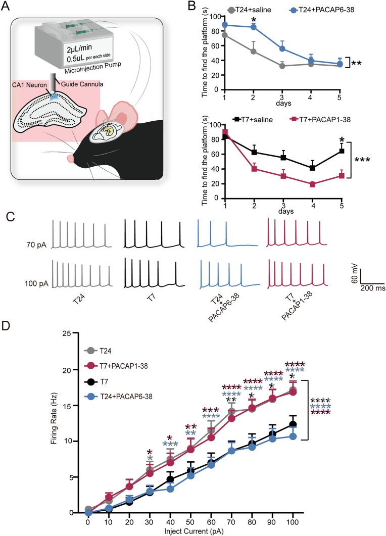
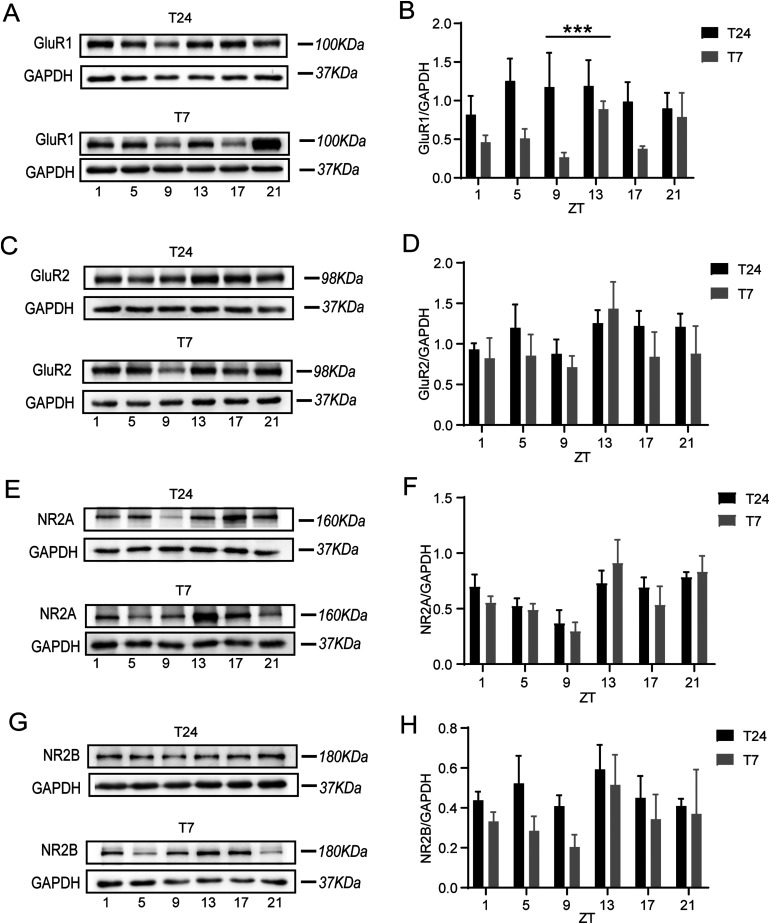
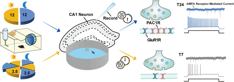
The mechanism of light-induced spatial memory deficits, as well as whether rhythmic expression of the pituitary adenylyl cyclase-activating polypeptides (PACAP)-PAC1 pathway influenced by light is related to this process, remains unclear. Here, we aimed to investigate the role of the PACAP-PAC1 pathway in light-mediated spatial memory deficits. Animals were first housed under a T24 cycle (12 h light:12 h dark), and then light conditions were transformed to a T7 cycle (3.5 h light:3.5 h dark) for at least 4 weeks. The spatial memory function was assessed using the Morris water maze (MWM). In line with behavioral studies, rhythmic expression of the PAC1 receptor and glutamate receptors in the hippocampal CA1 region was assessed by western blotting, and electrophysiology experiments were performed to determine the influence of the PACAP-PAC1 pathway on neuronal excitability and synaptic signaling transmission. Spatial memory was deficient after mice were exposed to the T7 light cycle. Rhythmic expression of the PAC1 receptor was dramatically decreased, and the excitability of CA1 pyramidal cells was decreased in T7 cycle-housed mice. Compensation with PACAP1-38, a PAC1 receptor agonist, helped T7 cycle-housed mouse CA1 pyramidal cells recover neuronal excitability to normal levels, and cannulas injected with PACAP1-38 shortened the time to find the platform in MWM. Importantly, the T7 cycle decreased the frequency of AMPA receptor-mediated excitatory postsynaptic currents. In conclusion, the PACAP-PAC1 pathway is an important protective factor modulating light-induced spatial memory function deficits, affecting CA1 pyramidal cell excitability and excitatory synaptic signaling transmission.

Keywords: CA1 pyramidal cell; Morris water maze; PACAP-PAC1 pathway; excitability; light; spatial memory.


Conflict of interest statement

The author(s) declared no potential conflicts of interest with respect to the research, authorship, and/or publication of this article.

Figures

Figure 1.
Figure 1.
The ultradian light cycle induces learning deficits in mice. A, Wheel-running activity of mice housed under the T24/T7 cycle. B, Associated periodograms during at least 2 weeks in T24 cycle, followed by at least 3 weeks in T7 cycle. C, Representative trajectories of mice from each experimental group in the probe trial and working trial. D, Using the MWM, we observed significant deficits during learning and test trials in mice exposed to the T7 (gray, N = 12 mice) cycle versus T24-housed (blue, N = 13 mice) mice (two-way ANOVA (F(1,115) = 30.85, ****P < 0.0001) with Sikak's multiple comparisons test: *P(day2) = 0.031, **P(day3) = 0.0029, **P(day4) = 0.0016). E, Representative immunoblots showing the expression of PAC1 in the CA1 region from T24 and T7 mice (PAC1: pituitary adenylate cyclase-activating polypeptide, PACAP receptor subtype 1). F, Relative quantification of PAC1 expression. Significant differences in the rhythmicity of PAC1 levels were observed in mice exposed to the T7 (gray, N = 4 mice) cycle versus T24-housed (black, N = 4 mice) mice. The data were analyzed using two-way ANOVA (F(1,36) = 56.60, ****P < 0.0001) with Bonferroni's multiple comparisons test (*P < 0.05, **P < 0.01; *P(ZT5) = 0.0218; *P(ZT9) = 0.0017; *P(ZT13) = 0.0019; *P(ZT17) = 0.0022) n = 4 mice)). ZT, Zeitgeber time.
Figure 2.
Figure 2.
The PACAP-PAC1 pathway is implicated in ultradian light cycle-induced learning deficits in mice. A, Representative bilateral implanted cannula in mice and bilateral microinjected drugs into the hippocampal CA1 region. B, Using the MWM, (top) deficits during learning in T24-housed mice microinjected with PACAP6-38 (blue, N = 8 mice) versus T24-housed mice microinjected with saline (gray, N = 7 mice) (two-way ANOVA (F(1,65) = 7.935, **P = .0064) are shown with Bonferroni's multiple comparisons test: *P(day2) = 0.0462). (Bottom) Recover during learning in T7-housed mice microinjected with PACAP1-38 (red, N = 8 mice) versus T7-housed mice microinjected with saline (black, N = 8 mice) (two-way ANOVA (F(1,70) = 14.96, ***P = 0.0002) with Bonferroni's multiple comparisons test: *P(day5)  = 0.02) (PACAP6-38: antagonist of PAC1 receptor; PACAP1-38: agonist of PAC1 receptor). C, Representative current steps of changing amplitude applied during whole-cell recordings from CA1 pyramidal cells in the presence of T24 (gray), T7 (black), T24 + PACAP6-38 (blue), and T7 + PACAP1-38 (red) (PACAP1-38: PAC1 receptor agonist, PACAP6-38: PAC1 receptor antagonist). Horizontal bars represent 200 ms, and vertical bars represent 60 mV. D, Action potential firing rate of CA1 pyramidal neurons (n = 12 cells; N = 4 mice). Compared to T24-housed (gray) mice, the frequency was decreased in the cells of T7-housed (black) mouse brain slices (two-way ANOVA (F(1,242) = 50.06, ****P < 0.0001) and Sidak's multiple comparisons test: **P(70pA) = 0.0075; *P(80pA) = 0.0299; *P(90pA) = 0.0299; *P(100pA) = 0.0299), and the cells were puffed with PACAP6-38 in the T24 mouse (blue) brain slices (two-way ANOVA (F(1,121) = 166.6, ****P < 0.0001) and Sidak's multiple comparisons test. *P(30pA) = 0.0298; ***P(40pA) = 0.0005; **P(50pA) = 0.0017; ****P(60pA) < 0.0001; ****P(70pA) < 0.0001; ****P(80pA) < 0.0001; ****P(90pA) < 0.0001; ****P(100pA) < 0.0001). The frequency increased in the cells puffed with PACAP1-38 in T7 (red) mouse brain slices(two-way ANOVA (F(1,121) = 165.8, ****P < 0.0001 and Sidak's multiple comparisons test * P(30pA)  = 0.0132; *P(40pA)  = 0.0474; **P(50pA)  = 0.0032; ***P(60pA)  = 0.0003; ****P(70pA) < 0.0001; ****P(80pA) < 0.0001; ****P(90pA) < 0.0001; ****P(100pA) < 0.0001).Compared to T24-housed (gray) mice, T7 + PACAP1-38 (red) has no significant difference (two-way ANOVA and Sidak's multiple comparisons test; P > 0.05). Compared to T7-housed (gray) mice, T24 + PACAP6-38 (blue) has no significant difference (two-way ANOVA and Sidak's multiple comparisons test; P > 0.05).
Figure 3.
Figure 3.
The ultradian light cycle disrupts the rhythmic expression of AMPA receptors and NMDA receptors in the CA1 region. A, C, E, G, Representative immunoblots showing expression of GluR1, GluR2, NR2A, and NR2B in CA1 from T24-housed (black, N = 4) and T7-housed (gray, N = 4) mice (GluR1: α-amino-3-hydroxy-5-methyl-4-isoxazolepropionic acid (AMPA) receptor subunit 1; GluR2: AMPA receptor subunit 2; NR2A: N-methyl-D-aspartate (NMDA) receptor subunits NR2A; NR2B: NMDB receptor subunits NR2B). B,T24 vs T7 has a different expression of GluR1(The data were analyzed using two-way ANOVA (F(1,36) = 13.15, *** P < 0.0009). D, No differences in the rhythmicity of GluR2 levels were observed between groups two-way ANOVA: P > 0.05). F, No differences in the rhythmicity of NR2A levels were observed between groups two-way ANOVA: P > 0.05). H, No differences in the rhythmicity of NR2B levels were observed between groups two-way ANOVA: P > 0.05).
Figure 4.
Figure 4.
The ultradian light cycle disrupts the rhythmic expression of p-PKA, P-PKC, CaMK I, and p-ERK in the CA1 region. A, C, E, G Representative immunoblots showing expression of p-PKA, p-PKC, CaMK II, and p-ERK in CA1 from T24-housed (black, N = 5 mice) and T7-housed (gray, N = 5 mice) mice (p-PKA: phospho-protein kinase A; p-PKC: phospho-proteinkinase C; CaMK Ⅱ : Ca2 + /calmodulin-dependent protein kinase, p-ERK:phospho-ERK). B, No differences in the rhythmicity of p-PKA levels were observed between groups two-way ANOVA: P > 0.05). D, No differences in the rhythmicity of p-PKC levels were observed between groups two-way ANOVA: P > 0.05). F, No differences in the rhythmicity of CaMK Ⅱ levels were observed between groups two-way ANOVA: P > 0.05). H. T24 versus T7 has a different expression of p-ERK (The data were analyzed using two-way ANOVA (F(1,36) = 5.079, * P < 0.0304) with Bonferroni's multiple comparisons test (** P < 0.01, P(ZT13) = 0.0092).
Figure 5.
Figure 5.
The PACAP-PAC1 pathway modulates the AMPA current in memory-deficit mice. A, Representative AMPA current trace on CA1 pyramidal cells. Horizontal bars represent 1 s, and vertical bars represent 20 pA. B, A cumulative probability plot of the AMPA-EPSC interevent interval (IEI) and frequency of AMPA-EPSCs in T24 (blue), T7 (gray), and T24 + PACAP6-38 (red). Compared to T24-housed (n = 11 cells, N = 4 mice) mice, the frequency was decreased in the cells of T7-housed (n = 11 cells, N = 4 mice) mouse brain slices (unpaired Student's t test: * P = 0.0321), and the cells were puffed with PACAP6-38 in the T24 mouse brain slices (n = 11 cells, N = 4 mice) (paired Student's t test: * P = 0.0109). There was no significant change between T7-housed mice and T24 + PACAP6-38 mice (unpaired Student's t test: P > 0.05). C, There was no significant change in the amplitude of the AMPA current (unpaired Student's t test: P > 0.05). D, Representative NMDA current trace in CA1 pyramidal cells. Horizontal bars represent 1 s, and vertical bars represent 20 pA. E, A cumulative probability plot of the NMDA-EPSC interevent interval (IEI) and frequency of NMDA-EPSCs at T24 (blue) and T7 (gray). Compared to T24-housed (n = 11 cells, N = 4 mice) mice, the frequency was no different in the cells of T7-housed (n = 11 cells, N = 4 mice) mouse brain slices (unpaired Student's t test: P > 0.05). F, There was no significant change in the amplitude of the NMDA current (unpaired Student's t test: P > 0.05).
Figure 6.
Figure 6.
Schematic diagram summarizing the role of the PACAP-PAC1 pathway in light-mediated spatial memory deficits. Aberrant light stimulation can lead to alterations in cognitive functions. Two different light conditions, a regular T24 cycle (12 h light: 12 h dark) and an ultradian T7 light cycle (3.5 h light: 3.5 h dark), were used in this study. In Morris water maze (MWM), a significantly prolonged time to find the platform was observed in mice exposed to the T7 cycle versus T24-housed mice. Rhythmic expressions of PAC1 receptor were decreased when housed under the T7 cycle compared to the T24 cycle. Patch clamp recording results showed that the excitability of CA1 pyramidal cells, as well as the frequency of AMPAR-mediated excitatory postsynaptic currents in mice housed under the T7 cycle, were decreased, which was related to the decrease in PAC1 receptor and GluR1 expression. The PACAP-PAC1 pathway is an essential protective factor in modulating light-induced spatial memory deficits.

Similar articles

Cited by

References

    1. Ago, Y., Hiramatsu, N., Ishihama, T., Hazama, K., Hayata-Takano, A., Shibasaki, Y., Shintani, N., Hashimoto, H., Kawasaki, T., Onoe, H., Chaki, S., Nakazato, A., Baba, A., Takuma, K., & Matsuda, T. (2013). The selective metabotropic glutamate 2/3 receptor agonist MGS0028 reverses psychomotor abnormalities and recognition memory deficits in mice lacking the pituitary adenylate cyclase-activating polypeptide. Behav Pharmacol, 24(1), 74–77. 10.1097/FBP.0b013e32835cf3e5 - DOI - PubMed
    1. Ajpru S., McArthur A. J., Piggins H. D., Sugden D. (2002). Identification of PAC1 receptor isoform mRNAs by real-time PCR in rat suprachiasmatic nucleus. Brain Res Mol Brain Res, 105(1--2), 29–37. 10.1016/S0169-328X(02)00387-X - DOI - PubMed
    1. Arimura A., Somogyvári-Vigh A., Miyata A., Mizuno K., Coy D. H., Kitada C. (1991). Tissue distribution of PACAP as determined by RIA: Highly abundant in the rat brain and testes. Endocrinology, 129(5), 2787–2789. 10.1210/endo-129-5-2787 - DOI - PubMed
    1. Asok A., Leroy F., Rayman J. B., Kandel E. R. (2019). Molecular mechanisms of the memory trace. Trends Neurosci, 42(1), 14–22. 10.1016/j.tins.2018.10.005 - DOI - PMC - PubMed
    1. Bannerman, D. M., Sprengel, R., Sanderson, D. J., McHugh, S. B., Rawlins, J. N., Monyer, H., & Seeburg, P. H. (2014). Hippocampal synaptic plasticity, spatial memory and anxiety. Nature Reviews Neuroscience, 15(3), 181–192. 10.1038/nrn3677 - DOI - PubMed

Publication types

MeSH terms

Substances

LinkOut - more resources