Abl kinase-mediated FUS Tyr526 phosphorylation alters nucleocytoplasmic FUS localization in FTLD-FUS
- PMID: 37071594
- PMCID: PMC10545532
- DOI: 10.1093/brain/awad130
Abl kinase-mediated FUS Tyr526 phosphorylation alters nucleocytoplasmic FUS localization in FTLD-FUS
Abstract
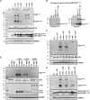
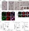
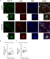
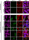
Nuclear to cytoplasmic mislocalization and aggregation of multiple RNA-binding proteins (RBPs), including FUS, are the main neuropathological features of the majority of cases of amyotrophic lateral sclerosis (ALS) and frontotemporal lobular degeneration (FTLD). In ALS-FUS, these aggregates arise from disease-associated mutations in FUS, whereas in FTLD-FUS, the cytoplasmic inclusions do not contain mutant FUS, suggesting different molecular mechanisms of FUS pathogenesis in FTLD that remain to be investigated. We have previously shown that phosphorylation of the C-terminal Tyr526 of FUS results in increased cytoplasmic retention of FUS due to impaired binding to the nuclear import receptor TNPO1. Inspired by the above notions, in the current study we developed a novel antibody against the C-terminally phosphorylated Tyr526 FUS (FUSp-Y526) that is specifically capable of recognizing phosphorylated cytoplasmic FUS, which is poorly recognized by other commercially available FUS antibodies. Using this FUSp-Y526 antibody, we demonstrated a FUS phosphorylation-specific effect on the cytoplasmic distribution of soluble and insoluble FUSp-Y526 in various cells and confirmed the involvement of the Src kinase family in Tyr526 FUS phosphorylation. In addition, we found that FUSp-Y526 expression pattern correlates with active pSrc/pAbl kinases in specific brain regions of mice, indicating preferential involvement of cAbl in the cytoplasmic mislocalization of FUSp-Y526 in cortical neurons. Finally, the pattern of immunoreactivity of active cAbl kinase and FUSp-Y526 revealed altered cytoplasmic distribution of FUSp-Y526 in cortical neurons of post-mortem frontal cortex tissue from FTLD patients compared with controls. The overlap of FUSp-Y526 and FUS signals was found preferentially in small diffuse inclusions and was absent in mature aggregates, suggesting possible involvement of FUSp-Y526 in the formation of early toxic FUS aggregates in the cytoplasm that are largely undetected by commercially available FUS antibodies. Given the overlapping patterns of cAbl activity and FUSp-Y526 distribution in cortical neurons, and cAbl induced sequestration of FUSp-Y526 into G3BP1 positive granules in stressed cells, we propose that cAbl kinase is actively involved in mediating cytoplasmic mislocalization and promoting toxic aggregation of wild-type FUS in the brains of FTLD patients, as a novel putative underlying mechanism of FTLD-FUS pathophysiology and progression.
Keywords: FTLD; FUS; c-Abl; c-Src; cytoplasmic aggregates; phosphorylation.
© The Author(s) 2023. Published by Oxford University Press on behalf of the Guarantors of Brain.
Conflict of interest statement
The authors report no competing interests.
Figures


Similar articles
-
FUS is phosphorylated by DNA-PK and accumulates in the cytoplasm after DNA damage.J Neurosci. 2014 Jun 4;34(23):7802-13. doi: 10.1523/JNEUROSCI.0172-14.2014. J Neurosci. 2014. PMID: 24899704 Free PMC article.
-
Transportin 1 colocalization with Fused in Sarcoma (FUS) inclusions is not characteristic for amyotrophic lateral sclerosis-FUS confirming disrupted nuclear import of mutant FUS and distinguishing it from frontotemporal lobar degeneration with FUS inclusions.Neuropathol Appl Neurobiol. 2013 Aug;39(5):553-61. doi: 10.1111/j.1365-2990.2012.01300.x. Neuropathol Appl Neurobiol. 2013. PMID: 22934812
-
FET proteins TAF15 and EWS are selective markers that distinguish FTLD with FUS pathology from amyotrophic lateral sclerosis with FUS mutations.Brain. 2011 Sep;134(Pt 9):2595-609. doi: 10.1093/brain/awr201. Epub 2011 Aug 19. Brain. 2011. PMID: 21856723 Free PMC article.
-
Conjoint pathologic cascades mediated by ALS/FTLD-U linked RNA-binding proteins TDP-43 and FUS.Neurology. 2011 Oct 25;77(17):1636-43. doi: 10.1212/WNL.0b013e3182343365. Epub 2011 Sep 28. Neurology. 2011. PMID: 21956718 Free PMC article. Review.
-
Frontotemporal lobar degeneration and amyotrophic lateral sclerosis: molecular similarities and differences.Rev Neurol (Paris). 2013 Oct;169(10):793-8. doi: 10.1016/j.neurol.2013.07.019. Epub 2013 Sep 5. Rev Neurol (Paris). 2013. PMID: 24011641 Review.
Cited by
-
Regulation of HNRNP family by post-translational modifications in cancer.Cell Death Discov. 2024 Oct 4;10(1):427. doi: 10.1038/s41420-024-02198-7. Cell Death Discov. 2024. PMID: 39366930 Free PMC article. Review.
-
c-Abl/TFEB Pathway Activation as a Common Pathogenic Mechanism in Lysosomal Storage Diseases: Therapeutic Potential of c-Abl Inhibitors.Antioxidants (Basel). 2025 May 20;14(5):611. doi: 10.3390/antiox14050611. Antioxidants (Basel). 2025. PMID: 40427492 Free PMC article.
-
Pathological Involvement of Protein Phase Separation and Aggregation in Neurodegenerative Diseases.Int J Mol Sci. 2024 Sep 23;25(18):10187. doi: 10.3390/ijms251810187. Int J Mol Sci. 2024. PMID: 39337671 Free PMC article. Review.
-
SUMO2/3 conjugation of TDP-43 protects against aggregation.Sci Adv. 2025 Feb 21;11(8):eadq2475. doi: 10.1126/sciadv.adq2475. Epub 2025 Feb 21. Sci Adv. 2025. PMID: 39982984 Free PMC article.
-
SRC Kinase Isoforms Regulate mRNA Splicing during Neural Development.J Neurosci. 2025 Aug 20;45(34):e1705242025. doi: 10.1523/JNEUROSCI.1705-24.2025. J Neurosci. 2025. PMID: 40750357 Free PMC article.
References
-
- Kwiatkowski TJJ, Bosco DA, Leclerc AL, et al. . Mutations in the FUS/TLS gene on chromosome 16 cause familial amyotrophic lateral sclerosis. Science. 2009;323:1205–1208. - PubMed
-
- Cairns NJ, Ghoshal N. FUS: a new actor on the frontotemporal lobar degeneration stage. Neurology. 2010;74:354–356. - PubMed
-
- Prpar Mihevc S, Darovic S, Kovanda A, Bajc Česnik A, Župunski V, Rogelj B. Nuclear trafficking in amyotrophic lateral sclerosis and frontotemporal lobar degeneration. Brain. 2017;140:13–26. - PubMed
Publication types
MeSH terms
Substances
LinkOut - more resources
Full Text Sources
Other Literature Sources
Medical
Research Materials
Miscellaneous

