. 2023 Apr 25;120(17):e2220565120.
doi: 10.1073/pnas.2220565120. Epub 2023 Apr 18.

DNA hydrogels for bone regeneration

Affiliations

DNA hydrogels for bone regeneration

Dimitra Athanasiadou et al. Proc Natl Acad Sci U S A. .

Abstract

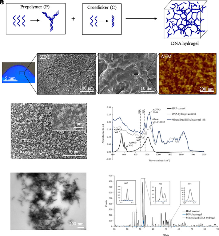
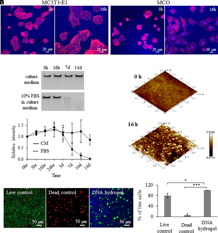
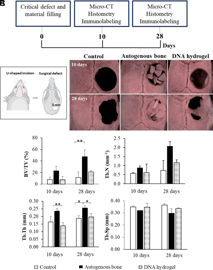
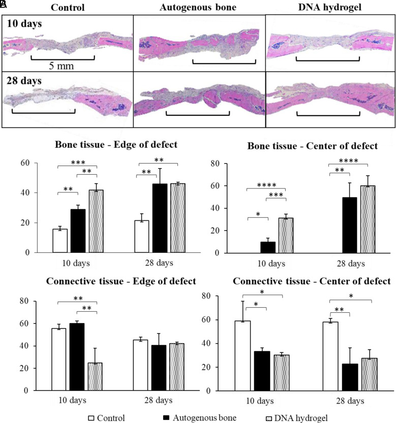
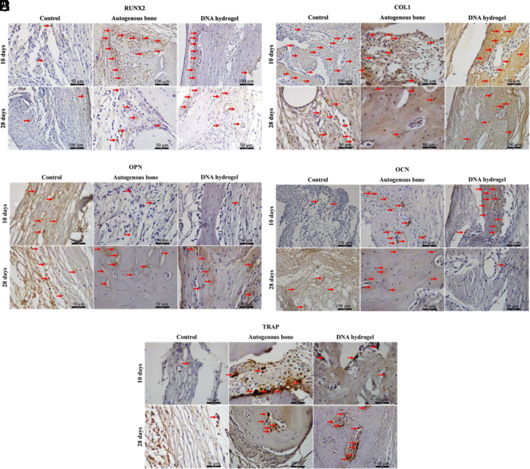
DNA-based biomaterials have been proposed for tissue engineering approaches due to their predictable assembly into complex morphologies and ease of functionalization. For bone tissue regeneration, the ability to bind Ca2+ and promote hydroxyapatite (HAP) growth along the DNA backbone combined with their degradation and release of extracellular phosphate, a known promoter of osteogenic differentiation, make DNA-based biomaterials unlike other currently used materials. However, their use as biodegradable scaffolds for bone repair remains scarce. Here, we describe the design and synthesis of DNA hydrogels, gels composed of DNA that swell in water, their interactions in vitro with the osteogenic cell lines MC3T3-E1 and mouse calvarial osteoblast, and their promotion of new bone formation in rat calvarial wounds. We found that DNA hydrogels can be readily synthesized at room temperature, and they promote HAP growth in vitro, as characterized by Fourier transform infrared spectroscopy, X-ray diffraction, scanning electron microscopy, atomic force microscopy, and transmission electron microscopy. Osteogenic cells remain viable when seeded on DNA hydrogels in vitro, as characterized by fluorescence microscopy. In vivo, DNA hydrogels promote the formation of new bone in rat calvarial critical size defects, as characterized by micro-computed tomography and histology. This study uses DNA hydrogels as a potential therapeutic biomaterial for regenerating lost bone.

Keywords: DNA hydrogels; DNA nanotechnology; biomineralization; bone regeneration.


Conflict of interest statement

The authors declare no competing interest.

Figures

Fig. 1.
Fig. 1.
DNA hydrogel synthesis and characterization. (A) Schematic representation of DNA hydrogel formation after mixing DNA prepolymer (P) with the DNA cross-linker (C). (B) Picture of the DNA hydrogel under UV irradiation (Left), and SEM along with AFM imaging showing a fibrous porous morphology at microscale and nanoscale, respectively. (C) SEM imaging of DNA hydrogels after 16 h of mineralization. (D) Infrared spectra of mineralized DNA hydrogels confirming that the observed mineral phase is HAP. (E) TEM imaging of DNA hydrogels after 16 h of mineralization. (F) XRD spectra of DNA hydrogels after 16 h mineralization confirming that the observed mineral phase is HAP.
Fig. 2.
Fig. 2.
DNA hydrogel interaction and degradation with osteogenic cells. Fluorescence microscopy images of (A) MC3T3-E1 and (B) MCO cell interaction on DNA hydrogels up to 16 h. (C) (Top), Denaturing gel electrophoresis of DNA hydrogel incubated in cell culture medium or in cell culture medium with 10% FBS for up to 14 d. (Bottom), Degradation kinetics of DNA hydrogel in the presence of cell culture medium with and without 10% FBS. (D) AFM images of DNA hydrogels after 0- and 16-h incubation in cell culture medium with 10% FBS. (E) MCO cell viability assay in the presence of DNA hydrogel. Significant difference is indicated by brackets (*P < 0.05 and ***P < 0.001). Values were compared by the repeated measures ANOVA test.
Fig. 3.
Fig. 3.
In vivo rat calvarial regeneration using DNA hydrogels. (A) Timeline of the performed analysis. (B) Experimental design according to the procedures performed in the study and micro-CT characterization of the studied groups. (C) Measured parameters by micro-CT including BV/TV (%), trabecular number (Tb.N; mm−1), trabecular thickness (Tb.Th; mm), and trabecular separation (Tb. Sp; mm). Significant difference is indicated by brackets (*P < 0.05 and **P < 0.01). Values were compared by a one-way ANOVA test.
Fig. 4.
Fig. 4.
Histological analysis of rat calvarial regeneration using DNA hydrogels. (A) Histological images of treated and untreated rat calvarial defects at 10 and 28 d after surgery. (B) Bone and connective tissue measurements among the studied groups. Significant difference is indicated by brackets (*P < 0.05, **P < 0.01, ***P < 0.001, and ****P < 0.0001). Values were compared by a one-way ANOVA test.
Fig. 5.
Fig. 5.
Immunolabeling analysis during DNA hydrogel calvarial healing. Immunohistochemical detection of osteogenic differentiation markers. (A) Runt-related transcription factor 2 (RUNX2), (B) collagen type I (Col1), (C) osteopontin (OPN), (D) osteocalcin (OCN), and (E) tartrate-resistant acid phosphatase (TRAP) among the studied groups at 10 and 28 d after surgery.

References

    1. Seeman N. C., Sleiman H. F., DNA nanotechnology. Nat. Rev. Mater. 3, 17068 (2018).
    1. Hong F., Zhang F., Liu Y., Yan H., Origami D. N. A., Scaffolds for creating higher order structures. Chem. Rev. 117, 12584–12640 (2017). - PubMed
    1. Wang D., Liu P., Luo D., Putting DNA to work as generic polymeric materials. Angew. Chem. Int. Ed. 61, e202110666 (2022). - PubMed
    1. Carneiro K. M. M., Avakyan N., Sleiman H. F., Long-range assembly of DNA into nanofibers and highly ordered networks. Wires Nanomed. Nanobi. 5, 266–285 (2013). - PubMed
    1. Sethi S., Sugiyama H., Endo M., Biomimetic DNA nanotechnology to understand and control cellular responses. ChemBioChem 23, e202100446 (2022). - PubMed

Publication types