Cerebral white matter rarefaction has both neurodegenerative and vascular causes and may primarily be a distal axonopathy
- PMID: 37071794
- PMCID: PMC10209646
- DOI: 10.1093/jnen/nlad026
Cerebral white matter rarefaction has both neurodegenerative and vascular causes and may primarily be a distal axonopathy
Abstract
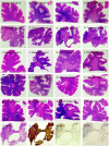
Cerebral white matter rarefaction (CWMR) was considered by Binswanger and Alzheimer to be due to cerebral arteriolosclerosis. Renewed attention came with CT and MR brain imaging, and neuropathological studies finding a high rate of CWMR in Alzheimer disease (AD). The relative contributions of cerebrovascular disease and AD to CWMR are still uncertain. In 1181 autopsies by the Arizona Study of Aging and Neurodegenerative Disorders (AZSAND), large-format brain sections were used to grade CWMR and determine its vascular and neurodegenerative correlates. Almost all neurodegenerative diseases had more severe CWMR than the normal control group. Multivariable logistic regression models indicated that Braak neurofibrillary stage was the strongest predictor of CWMR, with additional independently significant predictors including age, cortical and diencephalic lacunar and microinfarcts, body mass index, and female sex. It appears that while AD and cerebrovascular pathology may be additive in causing CWMR, both may be solely capable of this. The typical periventricular pattern suggests that CWMR is primarily a distal axonopathy caused by dysfunction of the cell bodies of long-association corticocortical projection neurons. A consequence of these findings is that CWMR should not be viewed simply as "small vessel disease" or as a pathognomonic indicator of vascular cognitive impairment or vascular dementia.
Keywords: Alzheimer disease; Hyperintensity; Lacunar infarct; Leukoaraiosis; Microscopic infarct; Small vessel disease; Vascular cognitive impairment.
© The Author(s) 2023. Published by Oxford University Press on behalf of American Association of Neuropathologists, Inc. All rights reserved. For permissions, please email: journals.permissions@oup.com.
Conflict of interest statement
Dr Beach has had recent unrelated personal paid consultancies with Aprinoia Therapeutics, Vivid Genomics, and Acadia Pharmaceuticals.
Figures


Similar articles
-
The Contribution of Cerebral Vascular Neuropathology to Mild Stage of Alzheimer's Dementia Using the NACC Database.Curr Alzheimer Res. 2020;17(13):1167-1176. doi: 10.2174/1567205018666210212160902. Curr Alzheimer Res. 2020. PMID: 33583381 Free PMC article.
-
Concomitant pathologies among a spectrum of parkinsonian disorders.Parkinsonism Relat Disord. 2014 May;20(5):525-9. doi: 10.1016/j.parkreldis.2014.02.012. Epub 2014 Feb 22. Parkinsonism Relat Disord. 2014. PMID: 24637124 Free PMC article.
-
Neuropathologic basis of white matter hyperintensity accumulation with advanced age.Neurology. 2013 Sep 10;81(11):977-83. doi: 10.1212/WNL.0b013e3182a43e45. Epub 2013 Aug 9. Neurology. 2013. PMID: 23935177 Free PMC article.
-
White matter degeneration in vascular and other ageing-related dementias.J Neurochem. 2018 Mar;144(5):617-633. doi: 10.1111/jnc.14271. Epub 2018 Jan 9. J Neurochem. 2018. PMID: 29210074 Review.
-
Overlap between pathology of Alzheimer disease and vascular dementia.Alzheimer Dis Assoc Disord. 1999 Oct-Dec;13 Suppl 3:S115-23. doi: 10.1097/00002093-199912003-00017. Alzheimer Dis Assoc Disord. 1999. PMID: 10609690 Review.
Cited by
-
Gray matter microstructure from in-vivo diffusion MRI reflects post-mortem neuropathology severity and clinical progression of Alzheimer's disease.medRxiv [Preprint]. 2025 Jun 4:2025.05.30.25328630. doi: 10.1101/2025.05.30.25328630. medRxiv. 2025. PMID: 40502575 Free PMC article. Preprint.
-
Alcohol Use Disorder and Dementia: A Review.Alcohol Res. 2024 May 23;44(1):03. doi: 10.35946/arcr.v44.1.03. eCollection 2024. Alcohol Res. 2024. PMID: 38812709 Free PMC article. Review.
-
Comparison of histological procedures and antigenicity of human post-mortem brains fixed with solutions used in gross anatomy laboratories.Front Neuroanat. 2024 Apr 10;18:1372953. doi: 10.3389/fnana.2024.1372953. eCollection 2024. Front Neuroanat. 2024. PMID: 38659652 Free PMC article.
-
Revised criteria for diagnosis and staging of Alzheimer's disease: Alzheimer's Association Workgroup.Alzheimers Dement. 2024 Aug;20(8):5143-5169. doi: 10.1002/alz.13859. Epub 2024 Jun 27. Alzheimers Dement. 2024. PMID: 38934362 Free PMC article.
-
White matter hyperintensity patterns: associations with comorbidities, amyloid, and cognition.Alzheimers Res Ther. 2024 Apr 1;16(1):67. doi: 10.1186/s13195-024-01435-6. Alzheimers Res Ther. 2024. PMID: 38561806 Free PMC article.
References
-
- Alzheimer A. Arteriosclerotic brain atrophy (H. Forstl, R. Levy, Trans). Int J Geriat Psychiatry 1991;6:129–30 (Original work published 1895)
-
- Alzheimer A. A contribution concerning the pathological anatomy of mental disturbances in old age (H. Forstl, B. Beats, Trans). Alz Dis Assoc Dis 1991;5:69–70 (Original work published 1899) - PubMed
-
- Beach TG. The history of Alzheimer’s disease: Three debates. J Hist Med Allied Sci 1987;42:327–49 - PubMed
-
- Olszewski J; Subcortical Arteriosclerotic Encephalopathy. Review of the literature on the so-called Binswanger's disease and presentation of two cases. World Neurol 1962;3:359–75 - PubMed
-
- Babikian V, Ropper AH.. Binswanger's disease: A review. Stroke 1987;18:2–12 - PubMed
Publication types
MeSH terms
Grants and funding
LinkOut - more resources
Full Text Sources
Medical

