. 2023 Aug;69(2):210-219.
doi: 10.1165/rcmb.2022-0373OC.

Local Complement Contributes to Pathogenic Activation of Lung Endothelial Cells in SARS-CoV-2 Infection

Affiliations

Local Complement Contributes to Pathogenic Activation of Lung Endothelial Cells in SARS-CoV-2 Infection

Hui Zhang et al. Am J Respir Cell Mol Biol. 2023 Aug.

Abstract

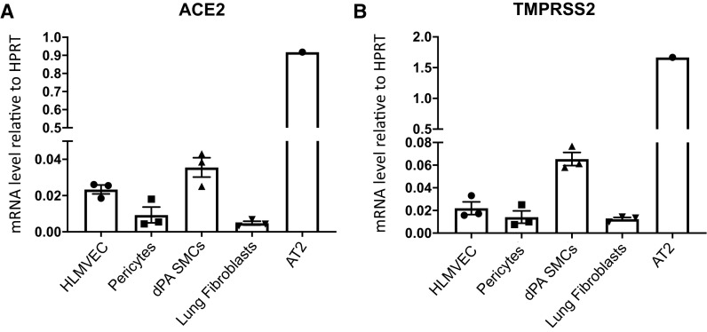
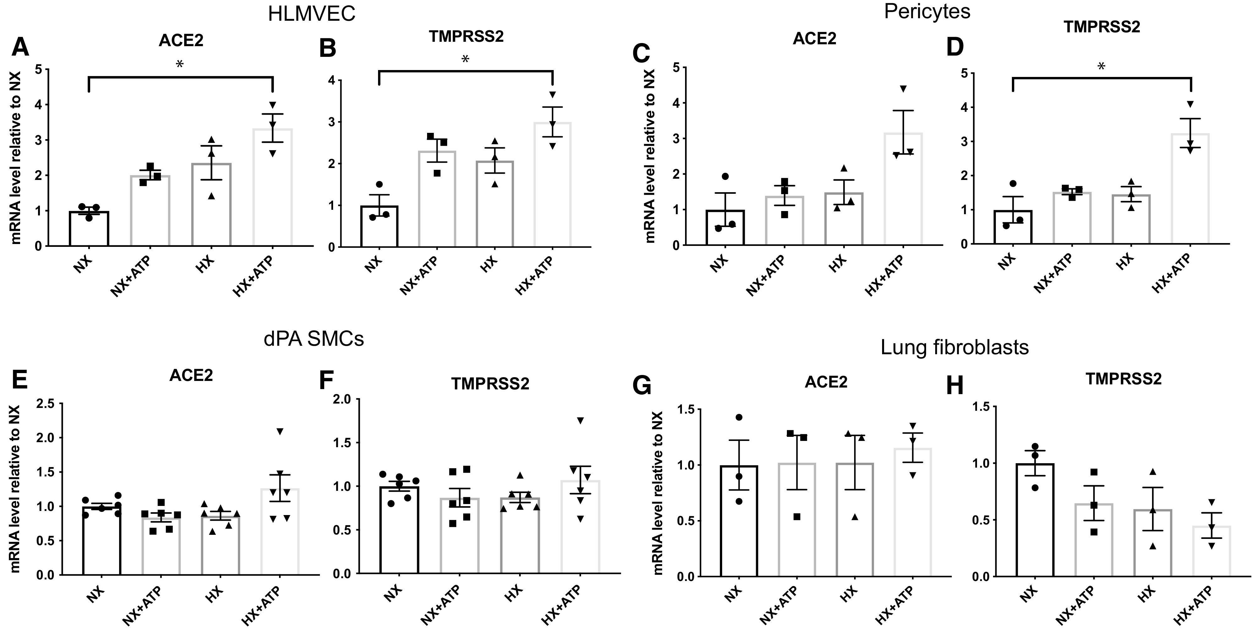
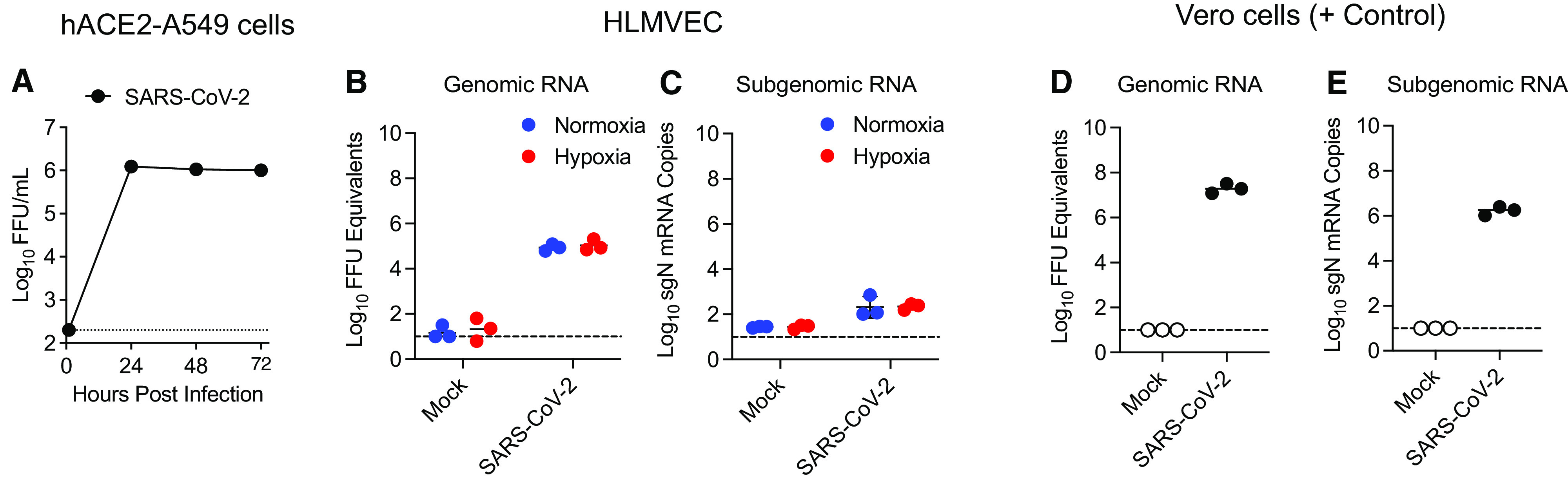
Endothelial dysfunction and inflammation contribute to the vascular pathology of coronavirus disease (COVID-19). However, emerging evidence does not support direct infection of endothelial or other vascular wall cells, and thus inflammation may be better explained as a secondary response to epithelial cell infection. In this study, we sought to determine whether lung endothelial or other resident vascular cells are susceptible to productive severe acute respiratory syndrome coronavirus 2 (SARS-CoV-2) infection and how local complement activation contributes to endothelial dysfunction and inflammation in response to hypoxia and SARS-CoV-2-infected lung alveolar epithelial cells. We found that ACE2 (angiotensin-converting enzyme 2) and TMPRSS2 (transmembrane serine protease 2) mRNA expression in lung vascular cells, including primary human lung microvascular endothelial cells (HLMVECs), pericytes, smooth muscle cells, and fibroblasts, was 20- to 90-fold lower compared with primary human alveolar epithelial type II cells. Consistently, we found that HLMVECs and other resident vascular cells were not susceptible to productive SARS-CoV-2 infection under either normoxic or hypoxic conditions. However, viral uptake without replication (abortive infection) was observed in HLMVECs when exposed to conditioned medium from SARS-CoV-2-infected human ACE2 stably transfected A549 epithelial cells. Furthermore, we demonstrated that exposure of HLMVECs to conditioned medium from SARS-CoV-2-infected human ACE2 stably transfected A549 epithelial cells and hypoxia resulted in upregulation of inflammatory factors such as ICAM-1 (intercellular adhesion molecule 1), VCAM-1 (vascular cell adhesion molecule 1), and IL-6 (interleukin 6) as well as complement components such as C3 (complement C3), C3AR1 (complement C3a receptor 1), C1QA (complement C1q A chain), and CFB (complement factor B). Taken together, our data support a model in which lung endothelial and vascular dysfunction during COVID-19 involves the activation of complement and inflammatory signaling and does not involve productive viral infection of endothelial cells.

Keywords: coronavirus disease (COVID-19); extracellular ATP; hypoxia; inflammation; lung microvascular endothelial cells.


Figures

Figure 1.
Figure 1.
ACE2 (angiotensin-converting enzyme 2) and TMPRSS2 (transmembrane serine protease 2) are differentially expressed among different lung vascular cell types. (A and B) Total RNA was isolated from human HLMVECs, pericytes, dPA SMCs, lung fibroblasts, and AT2 cells (cultured in normal medium), and mRNA concentrations of ACE2 (A) and TMPRSS2 (B) were determined using qRT-PCR analysis. Data are presented as mean ± SEM. AT2 = alveolar epithelial type II; dPA = distal pulmonary artery; HLMVEC = human lung microvascular endothelial cell; HPRT = hypoxanthine phosphoribosyl transferase; SMC = smooth muscle cell.
Figure 2.
Figure 2.
Hypoxia (HX) and extracellular ATP upregulate ACE2 and TMPRSS2 mRNA concentrations in HLMVECs but do not affect ACE2 and TMPRSS2 mRNA concentrations in SMCs and lung fibroblasts. Cells were cultured in normal medium and exposed to HX (3% O2) or extracellular ATP (100 μM) for 24 hours or remained untreated, and mRNA concentrations of ACE2 and TMPRSS2 were determined using qRT-PCR analysis. (A and B) ACE2 (A) and TMPRSS2 (B) concentrations in HLMVECs. (C and D) ACE2 (C) and TMPRSS2 (D) concentrations in pericytes. (E and F) ACE2 (E) and TMPRSS2 (F) concentrations in dPA SMCs. (G and H) ACE2 (G) and TMPRSS2 (H) concentrations in lung fibroblasts. Data are presented as mean ± SEM. *P < 0.05. NX = normoxia.
Figure 3.
Figure 3.
SARS-CoV-2 does not productively infect primary HLMVECs. (A) Measure of infectious virus in HLMVEC supernatants by FFA. (B) mNeonGreen reporter assay in infected HLMVECs. (C) Measure of infectious virus in susceptible Vero E6 cell supernatants by FFA. (D) mNeonGreen reporter assay in infected Vero E6 cells. See Methods for details. FFA = focus formation assay; FFU = fluorescence-forming units; hpi = hours postinfection; MOI = multiplicity of infection; MVEC = microvascular endothelial cell; SARS-CoV-2 = severe acute respiratory syndrome coronavirus 2; SSC = side scatter.
Figure 4.
Figure 4.
SARS-CoV-2 genomic RNA can be detected in HLMVECs after SARS-CoV-2 exposure. (A) Human ACE2 stably transfected A549 (hACE2-A549) cells were inoculated with SARS-CoV-2 (MOI = 0.5 FFU/cell). At 0 (input), 24, 48, and 72 hours postinoculation, infectious virus in cell culture supernatants was quantified using FFA. (B and C): HLMVECs were cultured under normoxic or hypoxic (3% O2) conditions for 24 hours. Cells were then stimulated for 24 hours with conditioned medium from mock-infected or SARS-CoV-2–infected hACE2-A549 cells (MOI = 1.5 FFU/cell). At 24 hours after conditioned medium treatment, viral genomic RNA (B) and N (nucleocapsid) subgenomic (sgN) mRNA (C) were quantified using qRT-PCR. (D and E) Susceptible and permissive Vero E6 cells were inoculated with SARS-CoV-2-mNeonGreen at an MOI of 0.001 FFU/cell in normoxia. At 24 hours postinoculation, viral genomic RNA (D) and sgN mRNA (E) were quantified using qRT-PCR.
Figure 5.
Figure 5.
SARS-CoV-2 and hypoxia stimulate inflammatory responses in HLMVECs. HLMVEC were preexposed to normoxic or hypoxic (3% O2) conditions for 24 hours, followed by the addition of conditioned medium from mock-infected or SARS-CoV-2–infected hACE2-A549 cells for 24 hours. (A) Total RNA was isolated, and the mRNA concentrations of indicated inflammatory markers were determined using qRT-PCR analysis. Data are presented as mean ± SEM. *P < 0.05 and **P < 0.01. (B) Total cell lysates were collected and analyzed for the expression of indicated markers using western blot analysis, and the representative blots are presented. (C) Densitometry of the blots in B is presented (technical replicates). ICAM-1 = intercellular adhesion molecule 1.
Figure 6.
Figure 6.
Effects of the conditioned medium of SARS-CoV-2–infected hACE2-A549 cells and hypoxia on the expression of complement proteins in HLMVECs. HLMVECs were preexposed to normoxic or hypoxic (3% O2) conditions for 24 hours, followed by the addition of conditioned medium from mock-infected or SARS-CoV-2–infected hACE2-A549 cells for 24 hours. (A) Total RNA was isolated, and mRNA concentrations of C3 (complement C3), C3AR1 (complement C3a receptor 1), C1QA (complement C1q A chain), and CFB (complement factor B) were determined using qRT-PCR analysis. Data are presented as mean ± SEM. *P < 0.05, **P < 0.01, ***P < 0.001, ****P < 0.0001, and #P = 0.0508 (borderline significant). (B) Total cell lysates were collected and analyzed for the expression of indicated complement components using western blot analysis, and the representative blots are presented. (C) Densitometry of the blots in B is presented (technical replicates).

References

    1. Teuwen LA, Geldhof V, Pasut A, Carmeliet P. COVID-19: the vasculature unleashed. Nat Rev Immunol . 2020;20:389–391. - PMC - PubMed
    1. Guzik TJ, Mohiddin SA, Dimarco A, Patel V, Savvatis K, Marelli-Berg FM, et al. COVID-19 and the cardiovascular system: implications for risk assessment, diagnosis, and treatment options. Cardiovasc Res . 2020;116:1666–1687. - PMC - PubMed
    1. Ackermann M, Verleden SE, Kuehnel M, Haverich A, Welte T, Laenger F, et al. Pulmonary vascular endothelialitis, thrombosis, and angiogenesis in COVID-19. N Engl J Med . 2020;383:120–128. - PMC - PubMed
    1. Gadanec LK, McSweeney KR, Qaradakhi T, Ali B, Zulli A, Apostolopoulos V. Can SARS-CoV-2 virus use multiple receptors to enter host cells? Int J Mol Sci . 2021;22:992. - PMC - PubMed
    1. Attaway AH, Scheraga RG, Bhimraj A, Biehl M, Hatipoğlu U. Severe COVID-19 pneumonia: pathogenesis and clinical management. BMJ . 2021;372:n436. - PubMed

Publication types

MeSH terms

Substances