Review
. 2023 Sep 29;10(1):77-98.
doi: 10.1146/annurev-virology-100220-112120. Epub 2023 Apr 18.

Viral Evolution Shaped by Host Proteostasis Networks

Affiliations
Review

Viral Evolution Shaped by Host Proteostasis Networks

Jimin Yoon et al. Annu Rev Virol. .

Abstract

Understanding the factors that shape viral evolution is critical for developing effective antiviral strategies, accurately predicting viral evolution, and preventing pandemics. One fundamental determinant of viral evolution is the interplay between viral protein biophysics and the host machineries that regulate protein folding and quality control. Most adaptive mutations in viruses are biophysically deleterious, resulting in a viral protein product with folding defects. In cells, protein folding is assisted by a dynamic system of chaperones and quality control processes known as the proteostasis network. Host proteostasis networks can determine the fates of viral proteins with biophysical defects, either by assisting with folding or by targeting them for degradation. In this review, we discuss and analyze new discoveries revealing that host proteostasis factors can profoundly shape the sequence space accessible to evolving viral proteins. We also discuss the many opportunities for research progress proffered by the proteostasis perspective on viral evolution and adaptation.

Keywords: chaperone; drug and immune system resistance; protein folding biophysics; quality control; stress response; viral adaptation.


Figures

Figure 1
Figure 1
Examples of protein folding defects. (a) Kinetic defects can increase the energy required to attain the native state or reduce the energy needed to attain a misfolded/unfolded state. For example, in this figure, the protein favors a misfolded conformation and/or is trapped in its unfolded (U) conformation owing to a kinetic defect. (b) Thermodynamic defects can increase the energy of the native folded (F) state. They can also decrease the energy of a misfolded/unfolded state, making the native state relatively less stable. (c) A variant that is more aggregation prone might have a partially misfolded (M) state, a low kinetic barrier that can lead preferentially to an aggregated state (A).
Figure 2
Figure 2
Organization of the cellular proteostasis network. (a) Proteins must fold into their native conformations to function but may instead misfold or aggregate. Chaperones can promote protein folding and/or prevent and correct misfolding and aggregation. Proteins with unresolved defects are usually identified and targeted for proteasomal or lysosomal degradation by quality control factors. (b) Proteostasis networks are dynamically regulated via transcriptional stress response pathways. In metazoan cells, these pathways are specific to different subcellular compartments. Abbreviations: ATF4, activating transcription factor 4; ATF6, activating transcription factor 6; ATF6f, activating transcription factor 6 fragment; BiP, binding immunoglobulin protein; HSF1, heat shock factor 1; IRE1, inositol requiring enzyme-1; PERK, PRK-like endoplasmic reticulum kinase; XBP1s, X-box binding protein 1 spliced.
Figure 3
Figure 3
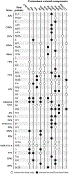
Select examples of viral proteins whose proper folding likely depends on host proteostasis network components. White circles indicate the existence of data showing that the viral protein and the host proteostasis network component interact (e.g., coimmunoprecipitation). Black circles indicate that, in addition to interaction data, there are functional data further supporting that the protein folding depends on the proteostasis network component (e.g., the steady-state level of the viral protein decreases upon chaperone inhibition). Related references are provided in the Supplemental Material. Abbreviations: AdV, adenovirus; BiP, binding immunoglobulin protein; CHIKV, chikungunya virus; CNX, Calnexin; CRT, calreticulin; CSFV, classical swine fever virus; DENV, dengue virus; EBOV, Ebola virus; Grp94, glucose-regulated protein 94; HBV, hepatitis B virus; HCV, hepatitis C virus; HIV, human immunodeficiency virus; Hsp, heat shock protein; JEV, Japanese encephalitis virus; MuV, mumps virus; NoV, norovirus; PDI, protein disulfide isomerase; PPI, peptidyl prolyl isomerase; PRV, pseudorabies virus; RABV, rabies virus; RSV, respiratory syncytial virus; SARS-CoV-2, severe acute respiratory syndrome coronavirus 2; SeV, Sendai virus; SV40, simian virus 40; ZIKV, Zika virus.
Figure 4
Figure 4
Proteostasis networks can influence the sequence space accessible to client proteins and thereby shape their evolution (25). (left) Viruses can sustain adaptive mutations (x-axis) that influence their relative fitness (gradient scale). However, if a mutation results in a protein variant with severe folding defects (y-axis), the virus harboring that protein variant is not viable (marked by an X in the trajectory), regardless of the potential fitness benefits the mutation would otherwise confer. The extent to which a viral protein can successfully fold thus determines the sequence space accessible to any given viral protein (shaded area of the graph). (right) The protein folding landscape can change depending on the compositions and activities of the proteostasis networks. (a) Hypothetical accessible sequence space at basal levels of chaperones and quality control factors. (b) When chaperones are upregulated, more protein variants can fold into their native conformations and the accessible sequence space expands. (c) When quality control factors are increased, more protein variants are targeted for degradation and the accessible sequence space constricts.
Figure 5
Figure 5
Methods for studying the evolution of viral proteins in the context of proteostasis perturbation in a laboratory environment include (a) serial passaging and (b) DMS. (c) There are advantages and disadvantages to both methods, depending on the type of information one wishes to obtain from the experiment. Abbreviations: DMS, deep mutational scanning; WT, wild-type.

References

    1. Elena SF, Lenski RE. 2003. Evolution experiments with microorganisms: the dynamics and genetic bases of adaptation. Nat. Rev. Genet 4:457–69 - PubMed
    1. Sanjuán R, Nebot MR, Chirico N, Mansky LM, Belshaw R. 2010. Viral mutation rates. J. Virol 84:9733–48 - PMC - PubMed
    1. Peck KM, Lauring AS. 2018. Complexities of viral mutation rates. J. Virol 92:e01031–17 - PMC - PubMed
    1. Worobey M, Holmes EC. 1999. Evolutionary aspects of recombination in RNA viruses. J. Gen. Virol 80(Part 10):2535–43 - PubMed
    1. Lowen AC. 2017. Constraints, drivers, and implications of influenza A virus reassortment. Annu. Rev. Virol 4:105–21 - PubMed

Publication types

LinkOut - more resources