. 2023 May;228(3):152384.
doi: 10.1016/j.imbio.2023.152384. Epub 2023 Apr 11.

Higher proinflammatory responses possibly contributing to suppressed cytotoxicity in patients with COVID-19 associated mucormycosis

Affiliations

Higher proinflammatory responses possibly contributing to suppressed cytotoxicity in patients with COVID-19 associated mucormycosis

Ashwini Shete et al. Immunobiology. 2023 May.

Abstract

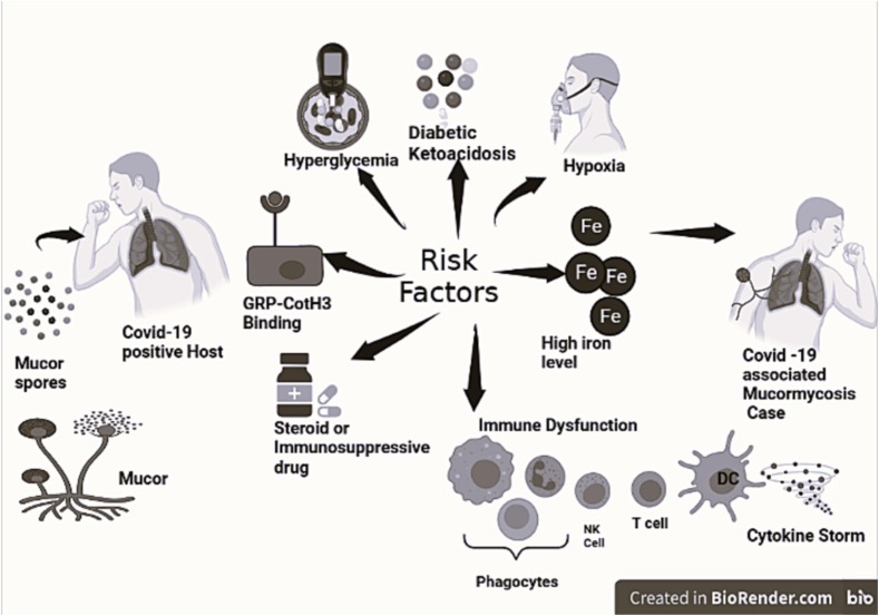
Introduction: COVID-19 Associated Mucormycosis (CAM), an opportunistic fungal infection, surged during the second wave of SARS Cov-2 pandemic. Since immune responses play an important role in controlling this infection in immunocompetent hosts, it is required to understand immune perturbations associated with this condition for devising immunotherapeutic strategies for its control. We conducted a study to determine different immune parameters altered in CAM cases as compared to COVID-19 patients without CAM.

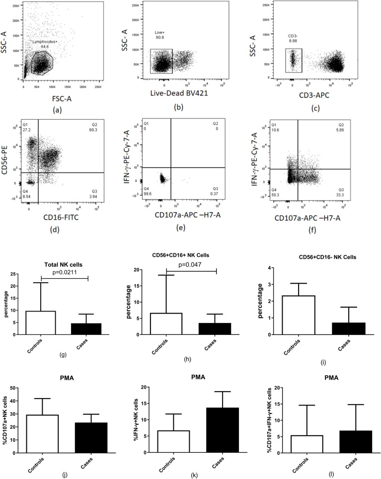
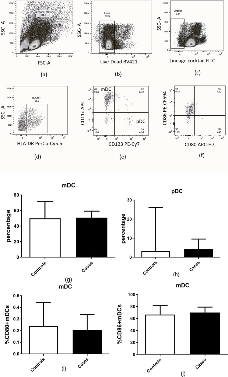
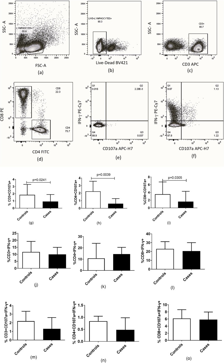
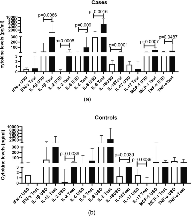
Methodology: Cytokine levels in serum samples of CAM cases (n = 29) and COVID-19 patients without CAM (n = 20) were determined using luminex assay. Flow cytometric assays were carried out in 20 CAM cases and 10 controls for determination of frequency of NK cells, DCs, phagocytes, T cells and their functionalities. The cytokine levels were analyzed for their association with each other as well as with T cell functionality. The immune parameters were also analyzed with respect to the known risk factors such as diabetes mellitus and steroid treatment.

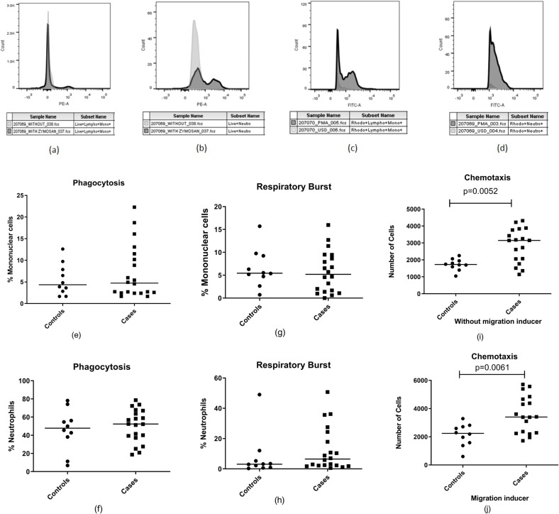
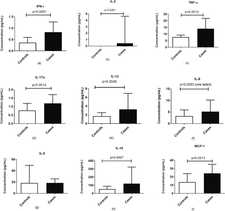
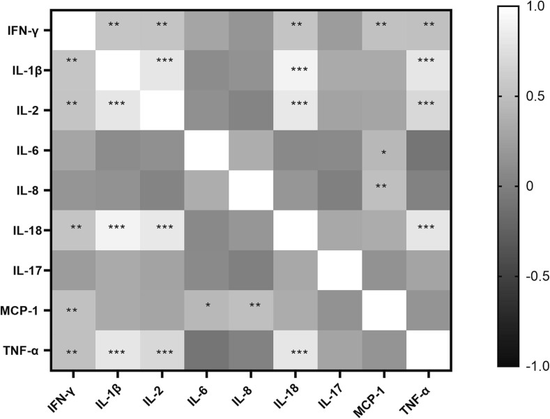
Results: Significant reduction in frequencies of total and CD56 + CD16 + NK cells (cytotoxic subset) was noted in CAM cases. Degranulation responses indicative of cytotoxicity of T cell were significantly hampered in CAM cases as compared to the controls. Conversely, phagocytic functions showed no difference in CAM cases versus their controls except for migratory potential which was found to be enhanced in CAM cases. Levels of proinflammatory cytokines such as IFN-γ, IL-2, TNF-α, IL-17, IL-1β, IL-18 and MCP-1 were significantly elevated in cases as compared to the control with IFN-γ and IL-18 levels correlating negatively with CD4 T cell cytotoxicity. Steroid administration was associated with higher frequency of CD56 + CD16- NK cells (cytokine producing subset) and higher MCP-1 levels. Whereas diabetic participants had higher phagocytic and chemotactic potential and had higher levels of IL-6, IL-17 and MCP-1.

Conclusion: CAM cases differed from the controls in terms of higher titers of proinflammatory cytokines, reduced frequency of total and cytotoxic CD56 + CD16 + NK cell. They also had reduced T cell cytotoxicity correlating inversely with IFN-γ and IL-18 levels, possibly indicating induction of negative feedback mechanisms while diabetes mellitus or steroid administration did not affect the responses negatively.

Keywords: COVID-19 Associated Mucormycosis (CAM); NK cells; Proinflammatory cytokines; T cell cytotoxicity.


Conflict of interest statement

Declaration of Competing Interest The authors declare that they have no known competing financial interests or personal relationships that could have appeared to influence the work reported in this paper.

Figures

Fig. 1
Fig. 1
Risk factors playing role in COVID-19 associated mucormycosis (CAM): Host factors in COVID-19 patients contributing to their increased susceptibility to development of mucormycosis are displayed in the figure.
Fig. 2
Fig. 2
Estimation of NK cell percentages in patients with COVID-19 and mucormycosis (n = 20) compared to patients with COVID-19 (n = 10). (a-f) Representative flow cytometry plots showing gating strategy used for analysis of NK cells are presented. Lymphocytes identified by forward and side scatter were further gated for identification of the NK cells using the markers displayed in the figure. (g) Percentages of total natural killer (NK) cell (h) Percentages of CD56 + CD16 + NK cells (i) Percentage of CD56 + CD16- NK cells, (j-i) NK cell functionality after stimulation with PMA: (j) Percentage of CD 107a + NK cells (k) Percentage of IFN-γ + NK cells (l) Percentage of CD 107a and IFN-γ + NK cells are shown in the figure. Histograms and error bars represent median ± IQR of the percentages.
Fig. 3
Fig. 3
Estimation of mDCs and pDCs in patients with COVID-19 and mucormycosis (n = 20) compared to patients with COVID-19 (n = 10). (a-f) Representative flow cytometry plots showing gating strategy used for analysis of dendritic cells are presented. Markers used for identification of dendritic cells are displayed in the figure. (g) Percentage of mDCs.(h) Percentage of pDCs (i) Percentage of CD80 + mDCs (j) Percentage of CD86 + mDCs are shown in the figure. Histograms and error bars represent median ± IQR of the percentages.
Fig. 4
Fig. 4
Estimation of phagocytic cell functionality in cases (n = 20) and control (n = 10) groups. (a,b) Histogram showing zymosan particle phagocytosis by monocytes (a) and neutrophils (b), (c,d) Histogram showing respiratory burst activity demonstrated by monocytes (c) and neutrophils (d) as indicated by rhodamine 123 fluoroscence, (e,f) Dot plots showing Phagocytic cell activity of mononuclear cells and neutrophils (g,h) Dot plots showing Respiratory burst activity of mononuclear cells and neutrophils. (i,j) Dot plots showing number of cells exhibiting chemotactic migration without inducer (P = 0.0068) and with inducer (P = 0.0078). Horizontal lines in the dot plots represent median values.
Fig. 5
Fig. 5
Estimation of acquired immune response in the patients with COVID-19 associated mucormycosis (n = 20) compared to patients with COVID −19 (n = 10). (a–f) Representative flow cytometry plots showing gating strategy used for analysis of CD4 + and CD8 + T cells are presented. Lymphocytes identified by forward and side scatter were further gated for identification of the T cells using the markers displayed in the figure. (g–i) Percentage of CD107a expressing CD3+, CD4 + and CD8 +. (j–l) Percentage of IFN-γ expressing CD3+, CD4 + and CD8 +. (m–o) Percentage of CD107a and IFN-γ in unison expressing CD3+, CD4 + and CD8 after stimulation with PMA and Ionomycin are shown in the figure. Histograms and error bars represent median ± IQR of the percentages.
Fig. 6
Fig. 6
Concentration of cytokines in serum samples of the patients with COVID-19 and mucormycosis (n = 29) compared to patients with COVID-19 (n = 20). Histograms represent the levels expressed as median ± IQR of (a) IFN-γ, (b) IL-2, (c) TNF-α, (d) IL-17a, (e) IL-1b, (f) IL-6, (g) IL-8, (h) IL-18 and (i) MCP-1.
Fig. 7
Fig. 7
Concentration of cytokines in supernatants of PBMCs stimulated with CotH3 peptide. Histograms represent the levels expressed as median ± IQR. Levels of the cytokines mentioned on X-axis in unstimulated (USD) versus peptide stimulated (Test) cells of cases (a) and controls (b) are shown in the figure.
Fig. 8
Fig. 8
Heatmap plotted to show correlation coefficients (r values) as per Spearman correlation analysis of serum levels of different cytokines with each other. Scale for r values is given in the figure. * indicate significant p values for correlation between the cytokines. * - p < 0.05, **- p < 0.01, ***- p < 0.001.
Fig. 9
Fig. 9
Comparison of Immune functionalities in participants with and without diabetes mellitus and steroid treatment irrespective of presence or absence of mucormycosis. Histograms represent the levels expressed as median ± IQR.

References

    1. Abassi Z., Higazi A.A.R., Kinaneh S., Armaly Z., Skorecki K., Heyman S.N. ACE2, COVID-19 infection, inflammation, and coagulopathy: missing pieces in the puzzle. Front. Physiol. 2020;11 - PMC - PubMed
    1. Akiba H., Motoki Y., Satoh M., Iwatsuki K., Kaneko F. Recalcitrant trichophytic granuloma associated with NK-cell deficiency in a SLE patient treated with corticosteroid. Eur. J. Dermatol. 2001;11:58–62. - PubMed
    1. Ali M.A.M., Spinler S.A. COVID-19 and thrombosis: from bench to bedside. Trends Cardiovasc. Med. 2021;31:143–160. - PMC - PubMed
    1. Al-Kuraishy H.M., Al-Gareeb A.I., Al-Niemi M.S., Alexiou A., Batiha G.E. Calprotectin: the link between acute lung injury and gastrointestinal injury in Covid-19: Ban or Boon. Curr. Protein Pept. Sci. 2022;23:310–320. - PubMed
    1. Al-Kuraishy H.M., Al-Gareeb A.I., Alkazmi L., Habotta O.A., Batiha G.E. High-mobility group box 1 (HMGB1) in COVID-19: extrapolation of dangerous liaisons. Inflammopharmacology. 2022;30:811–820. - PMC - PubMed

Publication types