

Developing an experimental-computational workflow to study the biomechanics of the human conventional aqueous outflow pathway
- PMID: 37072067
- PMCID: PMC10226761
- DOI: 10.1016/j.actbio.2023.04.008
Developing an experimental-computational workflow to study the biomechanics of the human conventional aqueous outflow pathway
Abstract
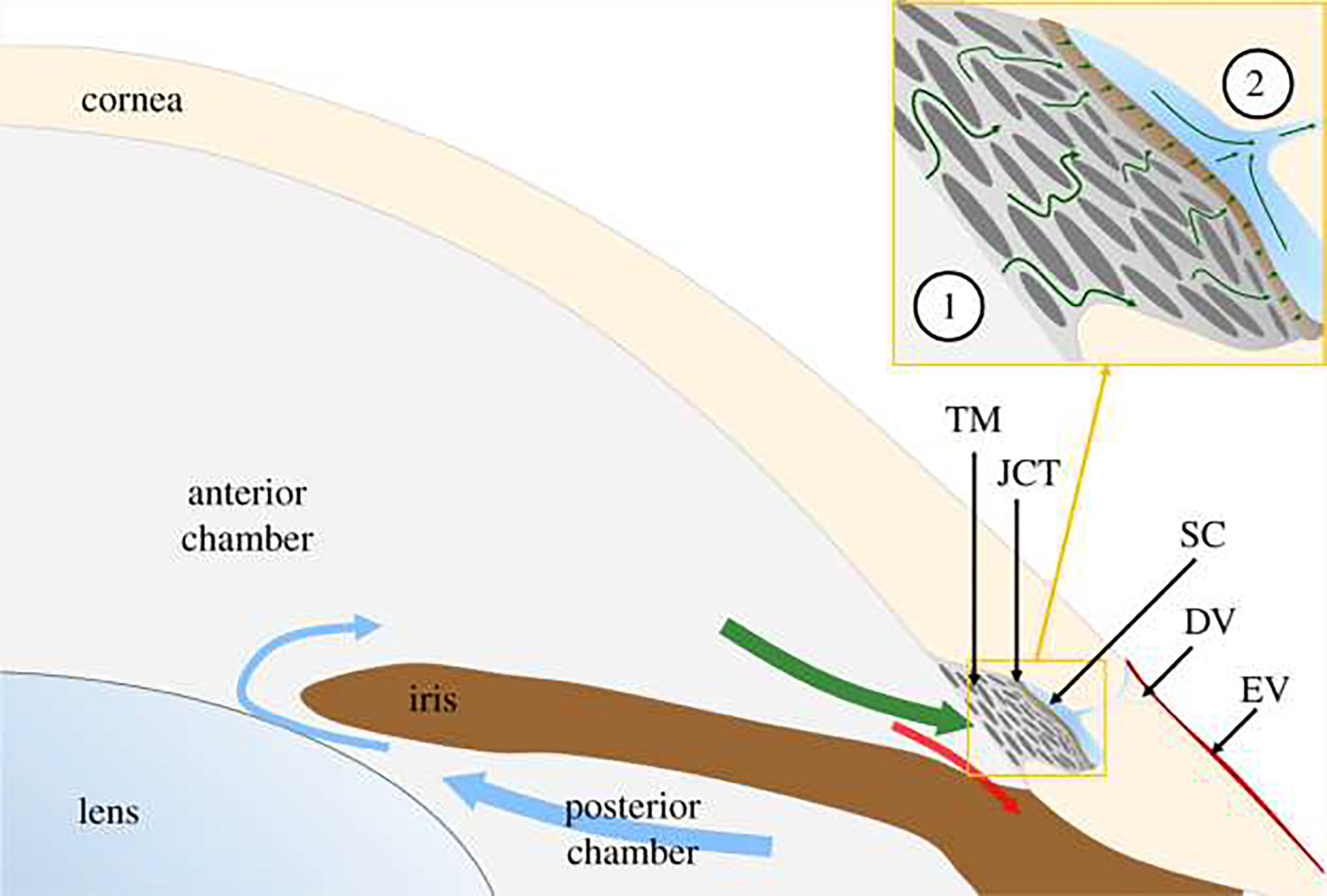
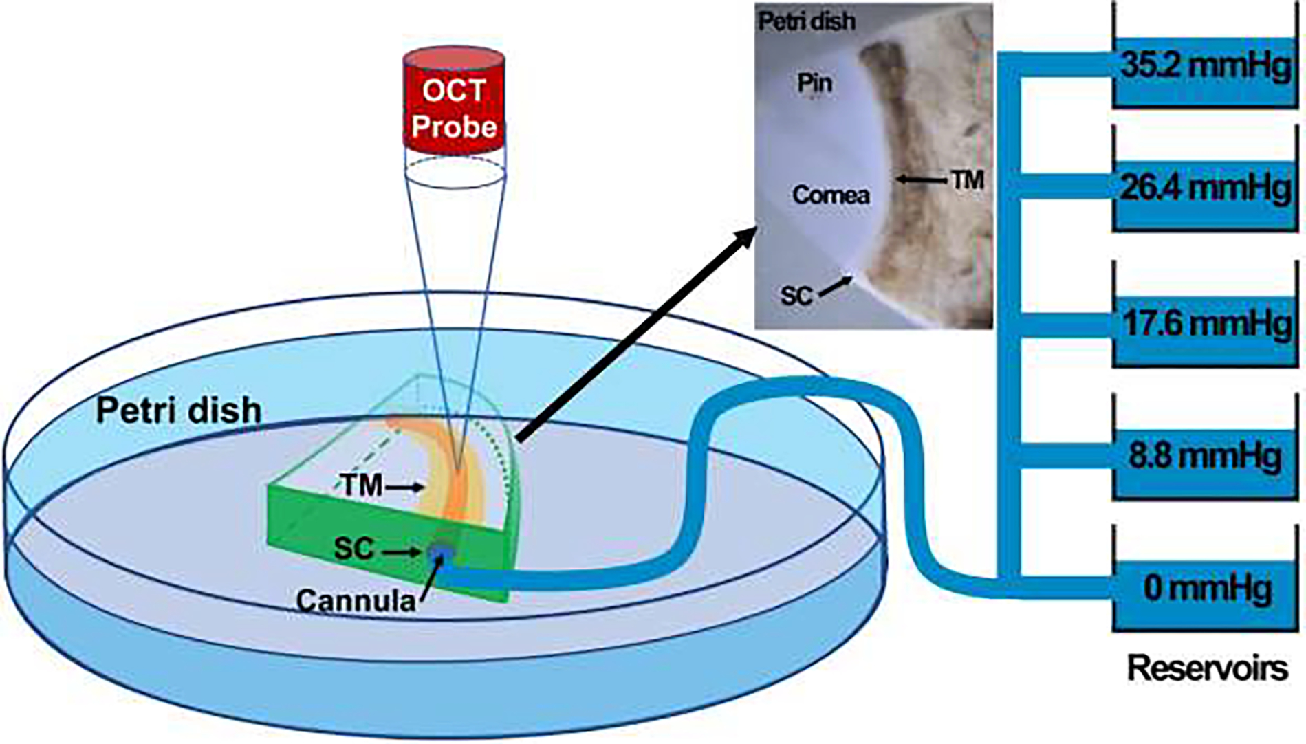
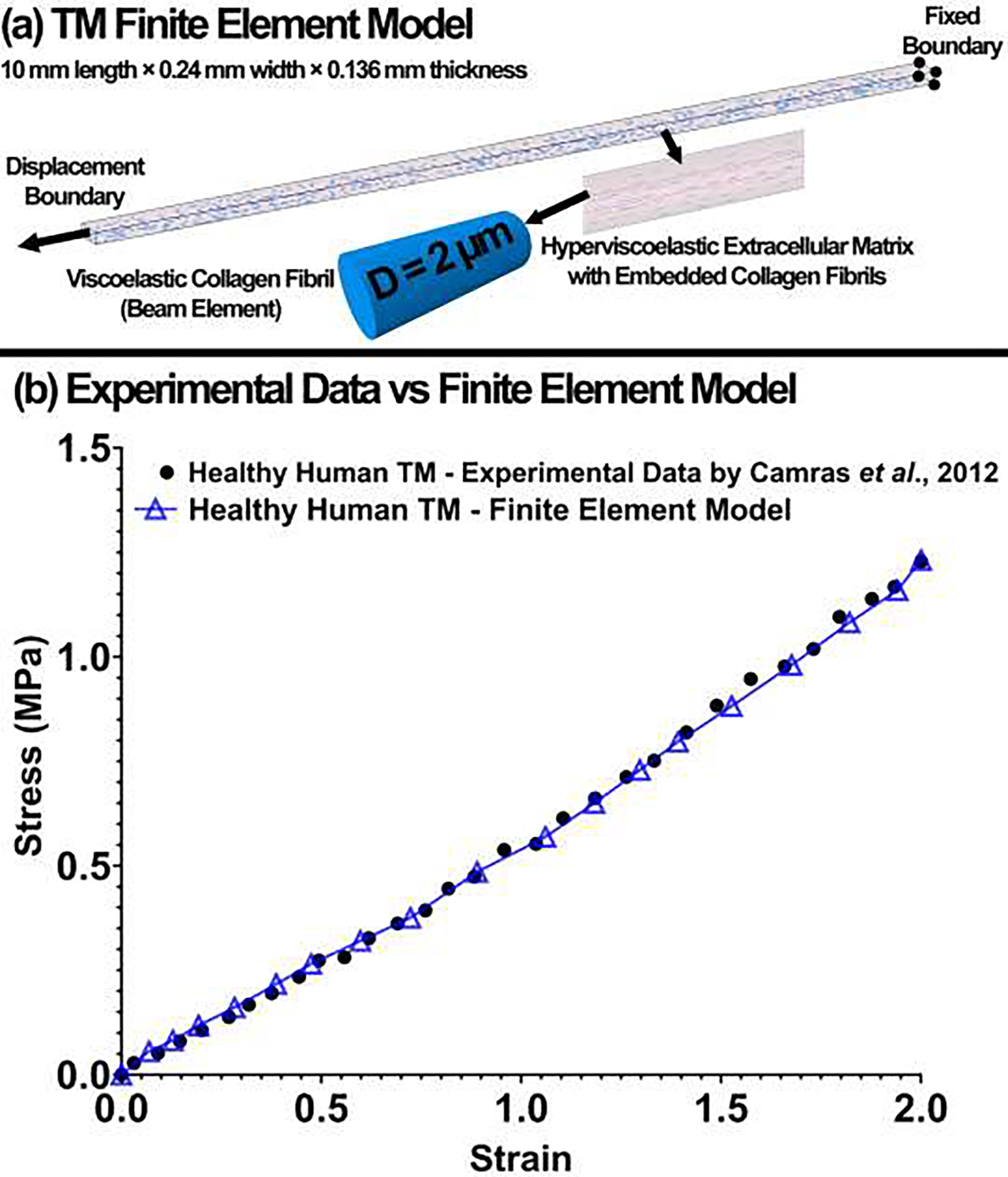
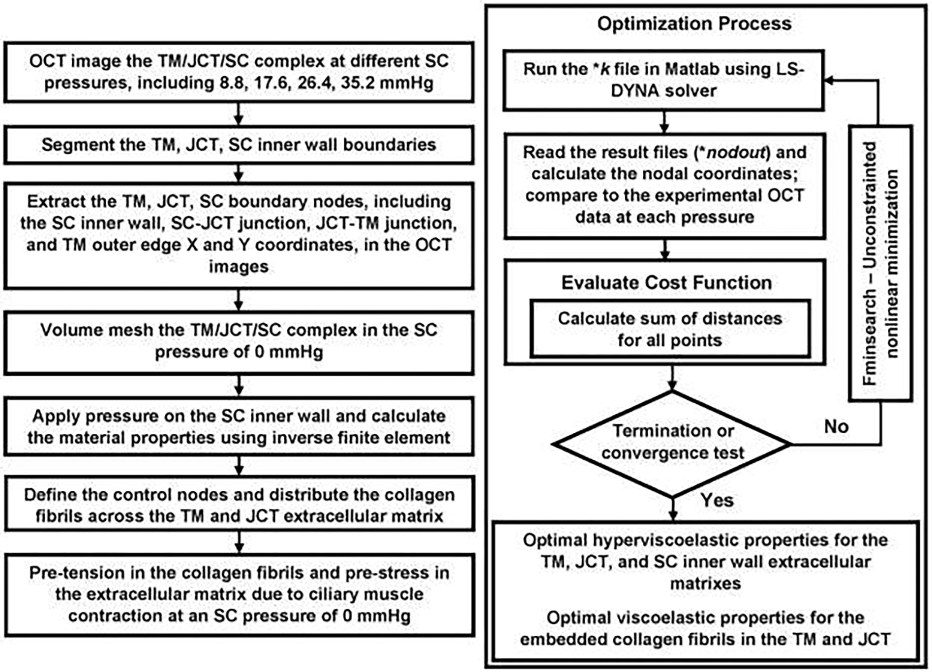
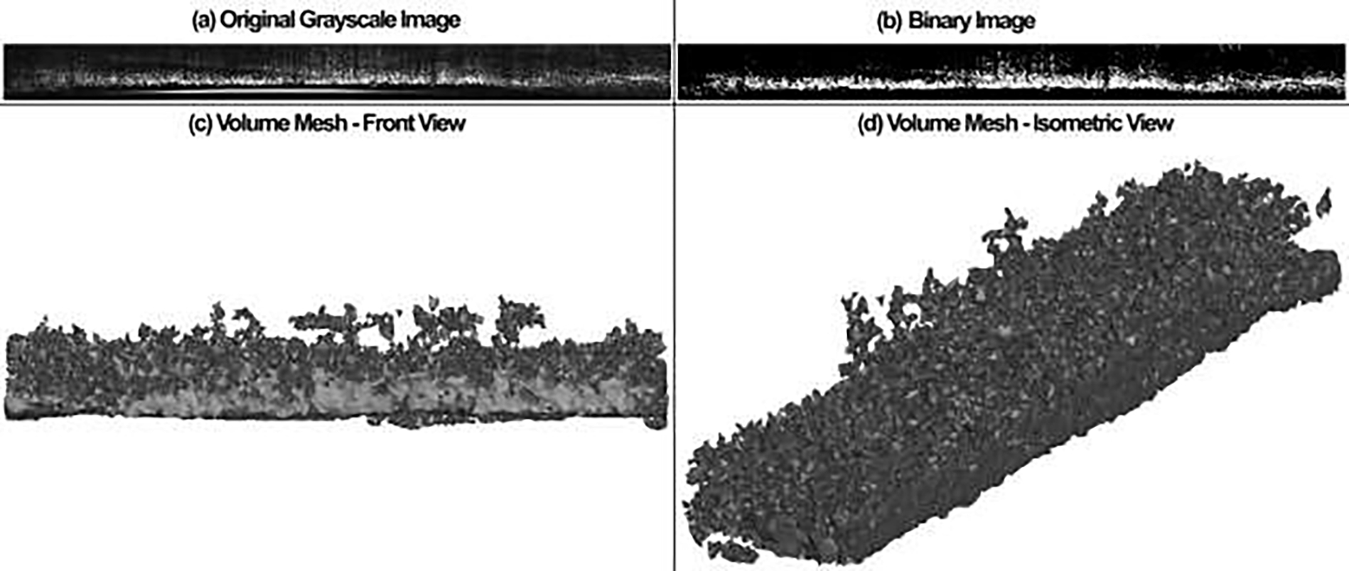
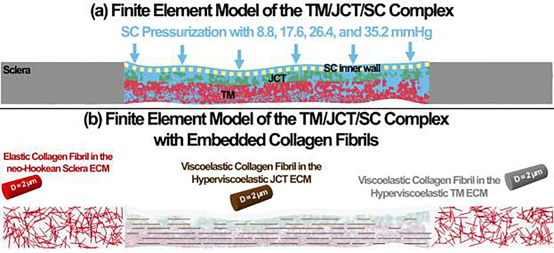
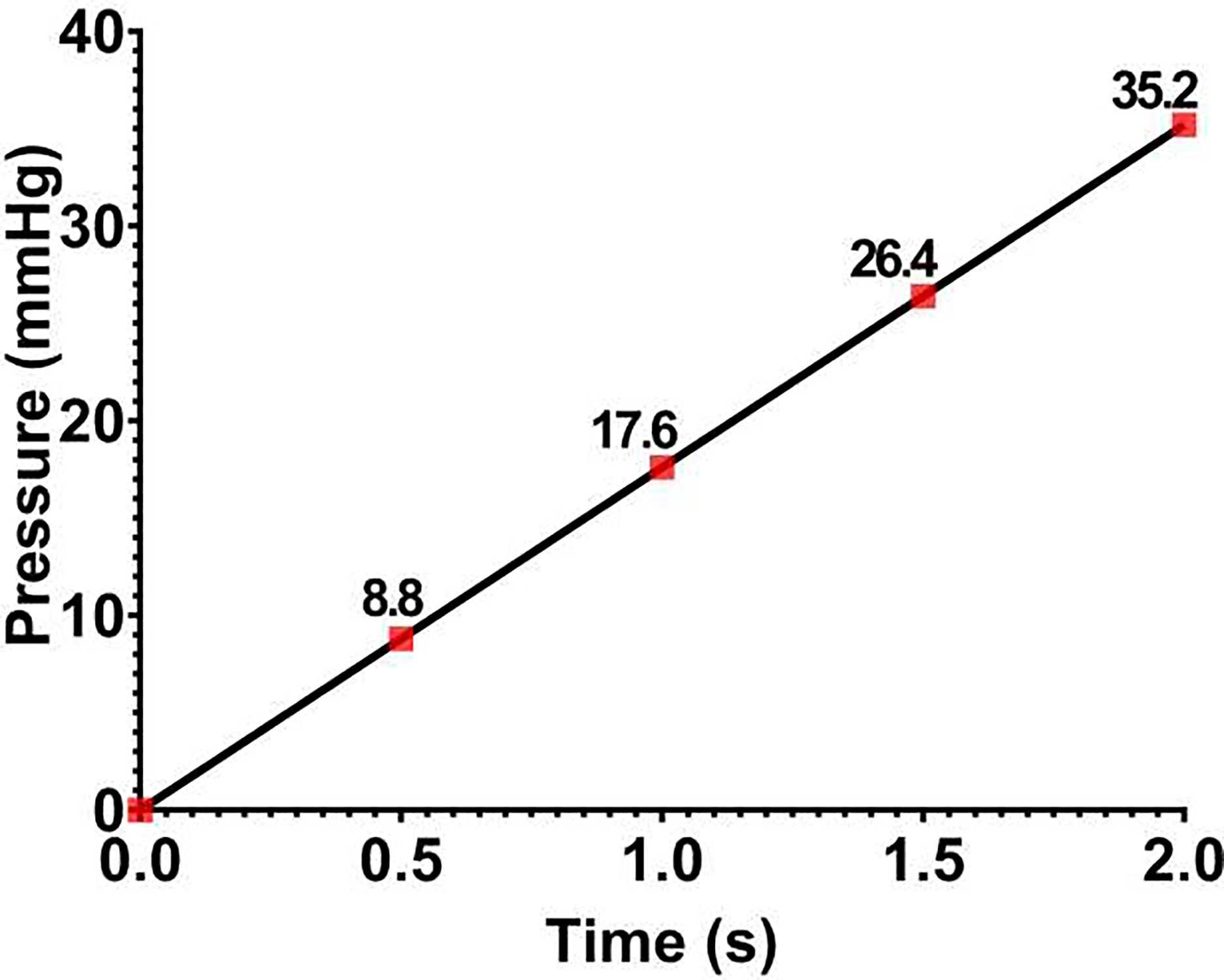
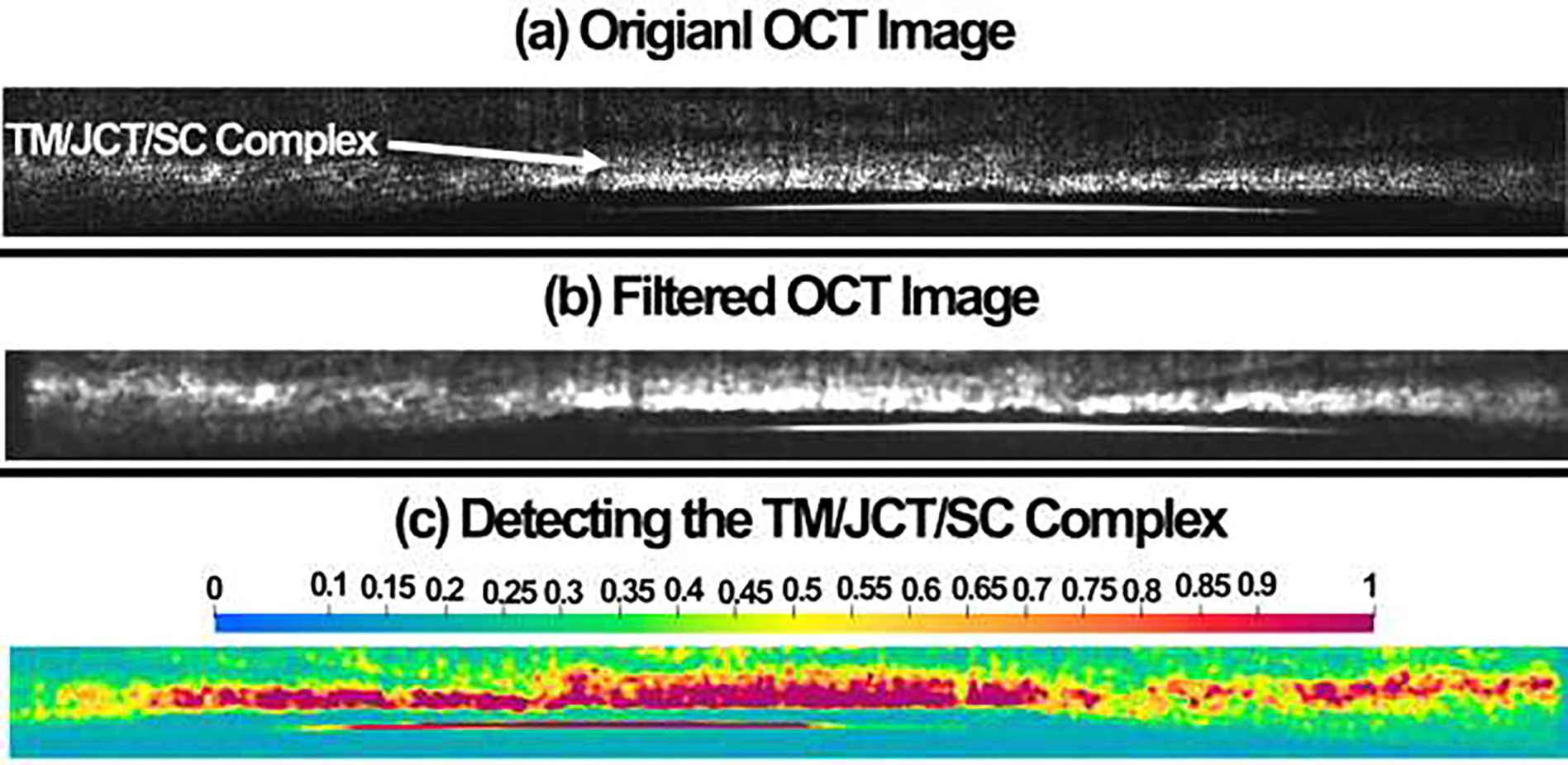
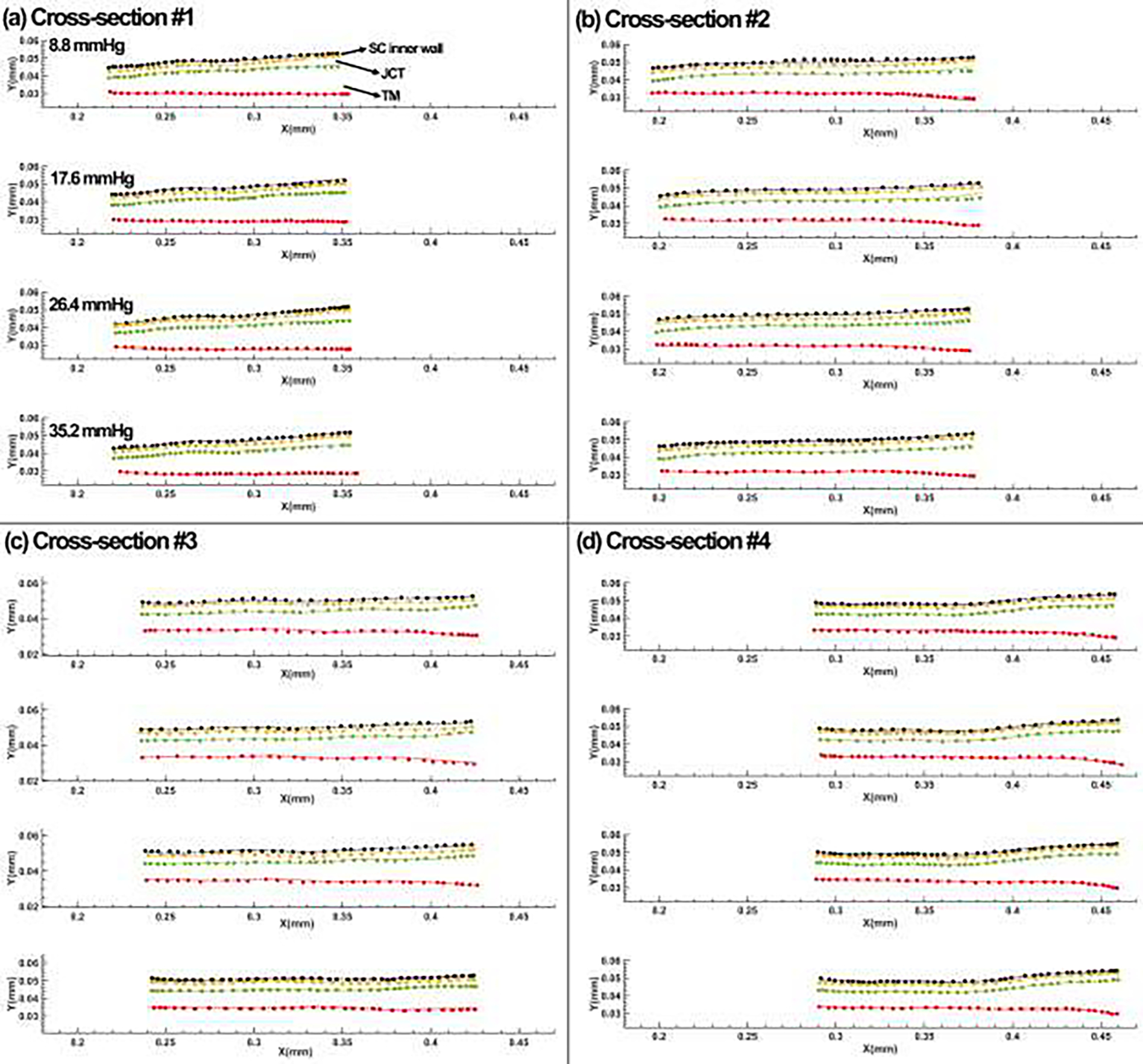
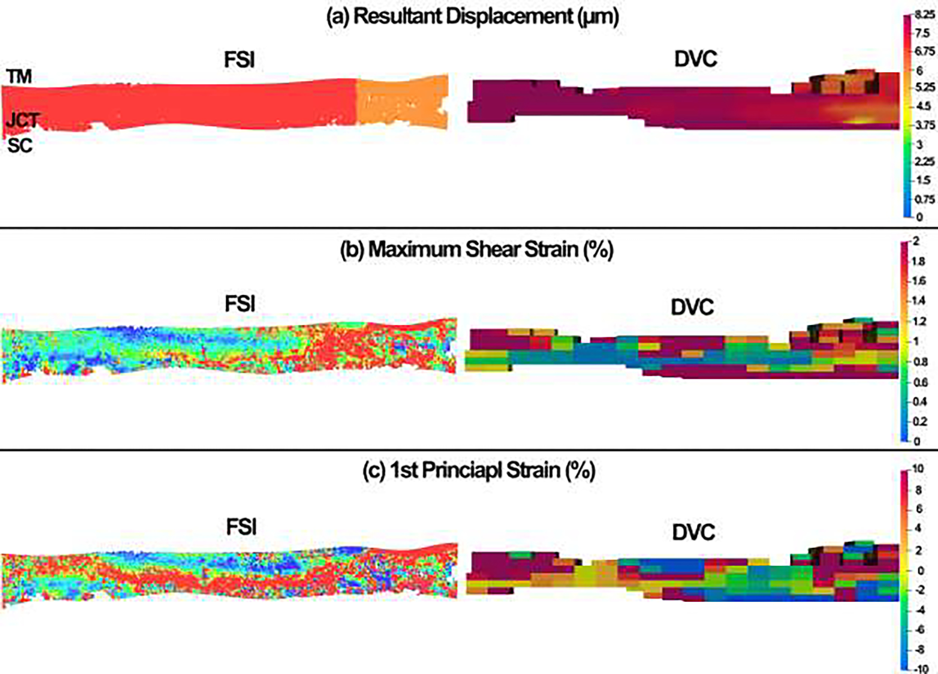
The aqueous humor actively interacts with the trabecular meshwork (TM), juxtacanalicular tissue (JCT), and Schlemm's canal (SC) through a dynamic fluid-structure interaction (FSI) coupling. Despite the fact that intraocular pressure (IOP) undergoes significant fluctuations, our understanding of the hyperviscoelastic biomechanical properties of the aqueous outflow tissues is limited. In this study, a quadrant of the anterior segment from a normal human donor eye was dynamically pressurized in the SC lumen, and imaged using a customized optical coherence tomography (OCT). The TM/JCT/SC complex finite element (FE) with embedded collagen fibrils was reconstructed based on the segmented boundary nodes in the OCT images. The hyperviscoelastic mechanical properties of the outflow tissues' extracellular matrix with embedded viscoelastic collagen fibrils were calculated using an inverse FE-optimization method. Thereafter, the 3D microstructural FE model of the TM, with adjacent JCT and SC inner wall, from the same donor eye was constructed using optical coherence microscopy and subjected to a flow load-boundary from the SC lumen. The resultant deformation/strain in the outflow tissues was calculated using the FSI method, and compared to the digital volume correlation (DVC) data. TM showed larger shear modulus (0.92 MPa) compared to the JCT (0.47 MPa) and SC inner wall (0.85 MPa). Shear modulus (viscoelastic) was larger in the SC inner wall (97.65 MPa) compared to the TM (84.38 MPa) and JCT (56.30 MPa). The conventional aqueous outflow pathway is subjected to a rate-dependent IOP load-boundary with large fluctuations. This necessitates addressing the biomechanics of the outflow tissues using hyperviscoelastic material-model. STATEMENT OF SIGNIFICANCE: While the human conventional aqueous outflow pathway is subjected to a large-deformation and time-dependent IOP load-boundary, we are not aware of any studies that have calculated the hyperviscoelastic mechanical properties of the outflow tissues with embedded viscoelastic collagen fibrils. A quadrant of the anterior segment of a normal humor donor eye was dynamically pressurized from the SC lumen with relatively large fluctuations. The TM/JCT/SC complex were OCT imaged and the mechanical properties of the tissues with embedded collagen fibrils were calculated using the inverse FE-optimization algorithm. The resultant displacement/strain in the FSI outflow model was validated versus the DVC data. The proposed experimental-computational workflow may significantly contribute to understanding of the effects of different drugs on the biomechanics of the conventional aqueous outflow pathway.
Keywords: Finite element method; Hyperviscoelastic; Juxtacanalicular tissue; Optimization algorithm; Schlemm's canal; Trabecular meshwork.
Copyright © 2023 Acta Materialia Inc. Published by Elsevier Ltd. All rights reserved.
Conflict of interest statement
Declaration of Competing Interest The authors declare that they have no known competing financial interests or personal relationships that could have appeared to influence the work reported in this paper.
Figures

References
-
- Johnstone MA, The aqueous outflow system as a mechanical pump: evidence from examination of tissue and aqueous movement in human and non-human primates, J Glaucoma 13(5) (2004) 421–38. - PubMed
-
- Johnstone M, Jamil A, Martin E, Aqueous veins and open angle glaucoma, The Glaucoma Book, Springer; 2010, pp. 65–78.
Publication types
MeSH terms
Substances
Grants and funding
LinkOut - more resources
Full Text Sources
Research Materials
