. 2023 Apr;11(4):e006221.
doi: 10.1136/jitc-2022-006221.

Irreversible electroporation augments β-glucan induced trained innate immunity for the treatment of pancreatic ductal adenocarcinoma

Affiliations

Irreversible electroporation augments β-glucan induced trained innate immunity for the treatment of pancreatic ductal adenocarcinoma

Matthew R Woeste et al. J Immunother Cancer. 2023 Apr.

Abstract

Background: Pancreatic cancer (PC) is a challenging diagnosis that is yet to benefit from the advancements in immuno-oncologic treatments. Irreversible electroporation (IRE), a non-thermal method of tumor ablation, is used in treatment of select patients with locally-advanced unresectable PC and has potentiated the effect of certain immunotherapies. Yeast-derived particulate β-glucan induces trained innate immunity and successfully reduces murine PC tumor burden. This study tests the hypothesis that IRE may augment β-glucan induced trained immunity in the treatment of PC.

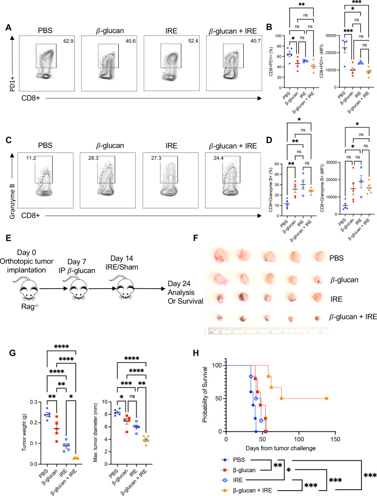
Methods: β-Glucan-trained pancreatic myeloid cells were evaluated ex vivo for trained responses and antitumor function after exposure to ablated and unablated tumor-conditioned media. β-Glucan and IRE combination therapy was tested in an orthotopic murine PC model in wild-type and Rag-/- mice. Tumor immune phenotypes were assessed by flow cytometry. Effect of oral β-glucan in the murine pancreas was evaluated and used in combination with IRE to treat PC. The peripheral blood of patients with PC taking oral β-glucan after IRE was evaluated by mass cytometry.

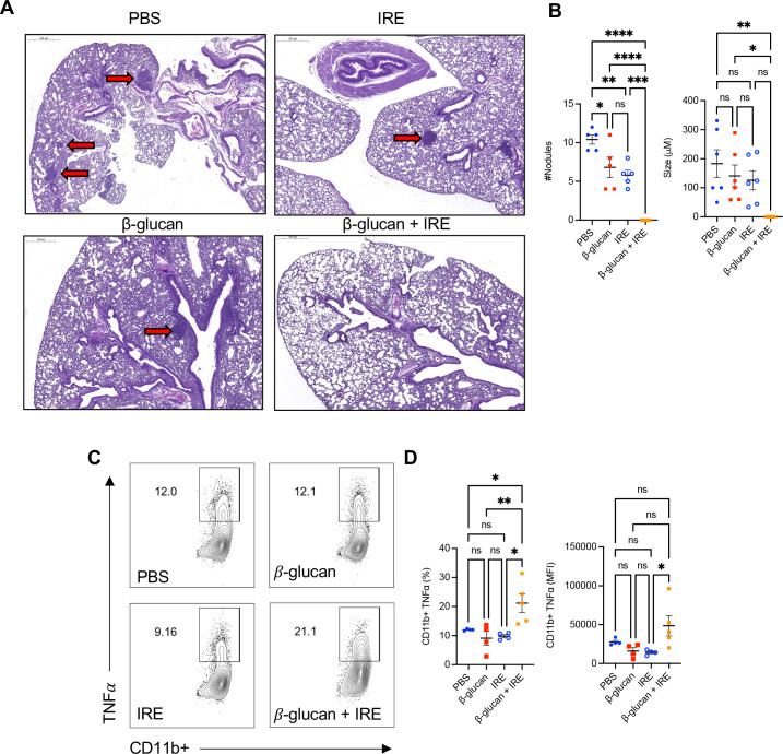
Results: IRE-ablated tumor cells elicited a potent trained response ex vivo and augmented antitumor functionality. In vivo, β-glucan in combination with IRE reduced local and distant tumor burden prolonging survival in a murine orthotopic PC model. This combination augmented immune cell infiltration to the PC tumor microenvironment and potentiated the trained response from tumor-infiltrating myeloid cells. The antitumor effect of this dual therapy occurred independent of the adaptive immune response. Further, orally administered β-glucan was identified as an alternative route to induce trained immunity in the murine pancreas and prolonged PC survival in combination with IRE. β-Glucan in vitro treatment also induced trained immunity in peripheral blood monocytes obtained from patients with treatment-naïve PC. Finally, orally administered β-glucan was found to significantly alter the innate cell landscape within the peripheral blood of five patients with stage III locally-advanced PC who had undergone IRE.

Conclusions: These data highlight a relevant and novel application of trained immunity within the setting of surgical ablation that may stand to benefit patients with PC.

Keywords: Immunity, Innate; Immunotherapy; Phagocytosis; Tumor Microenvironment.


Conflict of interest statement

Competing interests: RCGM is an educational consultant for AngioDynamics.

Figures

Figure 1
Figure 1
IRE releases more MIF from PC tumor cells which elicits a potent trained response in β-glucan-trained pancreatic myeloid cells with enhanced phagocytosis and cytotoxicity. (A) Summarized pancreatic CD11b+TNF-α+ cells from untrained versus β-glucan trained mice on ex vivo restimulation with LPS or increasing doses of rMIF assessed by flow cytometry. n=3 per group. (B) Concentration of MIF (ng/mL) in supernatants after culture of 1×10⁶ KPC cells for 24 hours or ablation of KPC cells as measured by ELISA, n=6 per group. (C) TNF-α and IL-6 (pg/mL) levels measured by ELISA from β-glucan trained CD11b+ pancreatic cells restimulated ex vivo with KPC-conditioned media or IRE-conditioned media for 24 hours. Representative flow cytometry contour plots and quantified percent and MFI of β-glucan trained CD11b+TNF-α+ cells restimulated with media control, KPC-conditioned media or IRE-conditioned media, n=3 per group. (E) β-glucan trained pancreatic CD11b+ cells were stimulated with media alone or KPC supernatant or IRE KPC supernatant and then cocultured with KPCGFP+ tumor cells. Representative flow plots and summarized data are shown. (F) Summarized results from cytotoxicity assay. CD11b+ cells from 7 day β-glucan trained mice were incubated at a ratio of 1:30 KPCLuc+ to CD11b+ cells for 24 hours. Data are representative of two or three independent experiments and presented as mean±SEM. ns, not significant; *p<0.05, **p<0.01, ***p<0.001, ****p<0.0001. IL, interleukin; IRE, irreversible electroporation; LPS, lipopolysaccharide; MIF, migration inhibitory factor; PC, pancreatic cancer; rMIF, recombinant MIF; TNF, tumor necrosis factor.
Figure 2
Figure 2
β-Glucan in combination with IRE reduces PC tumor burden and prolongs survival. (A) Schematic representation of the experimental design. Murine KPC cells were orthotopically implanted into the pancreas at day 0. Mice were treated with one IP β-glucan injection (1 mg/mouse) or PBS control 7 days after tumor challenge. On day 14, sham surgery or IRE ablation was performed. (B) Representative image of orthotopic pancreatic KPC tumors on day 24 post tumor challenge from control and different treatment groups. (C) Tumor weight and maximum tumor diameter (right). PBS n=14, IRE n=15, β-glucan n=14, β-glucan+IRE n=15, respectively. (D) Overall survival. PBS n=5, IRE n=5, β-glucan n=7, β-glucan+IRE n=7, respectively. (E) Quantification of histone modification markers H3K27Ac, H3K4Me3, and H3K27Me3 compared with total H3 in CD11b+ cells measured by ELISA. (F) Arg-1 and IL-6 messenger RNA expression in CD11b+ cells measured by RT-PCR. PBS n=5, IRE n=5, β-glucan n=5, β-glucan+IRE n=5, respectively. Data are representative of two or three independent experiments and presented as mean±SEM. ns, not significant; *p<0.05, **p<0.01, ***p<0.001, ****p<0.0001. Arg-1, arginase 1; IL, interleukin; IP, intraperitoneal; IRE, irreversible electroporation; PBS, phosphate buffered saline; PC, pancreatic cancer; WT, wild-type.
Figure 3
Figure 3
Combined β-glucan and IRE treatment increases total immune cell infiltration in the PC TME and IRE induces a trained innate response in monocytes and macrophages in early tumor progression. Absolute number of live CD45+ (A), CD11b+ (B), CD11b+F4/80+ macrophages (C), CD11b+Ly6G-Ly6C+ monocytes (D), CD11b+Ly6G+Ly6C- neutrophils (E), CD11b+CD11c+MHCII+ dendritic cells (F) per gram of tumor tissue as quantified by flow cytometry. (G) Absolute TNF-α from overall CD11b+ (H) CD11b+F4/80+ macrophages (I) and CD11b+Ly6C+ monocytes per gram of tumor tissue as quantified by flow cytometry. Data are representative of two or three independent experiments and presented as mean±SEM. ns, not significant; *p<0.05, **p<0.01, ***p<0.001, ****p<0.0001. IRE, irreversible electroporation; PBS, phosphate buffered saline; PC, pancreatic cancer; TME, tumor microenvironment; TNF, tumor necrosis factor.
Figure 4
Figure 4
The combination of β-glucan and IRE reduces distant tumor burden late in disease progression. (A) Representative H&E images of lungs harvested from PBS control (n=5), IRE (n=5), β-glucan (n=5), or β-glucan+IRE (n=5) at the time of PBS tumor endpoint. (B) Number of lung nodules per histologic section per group and average measured size (µm) of nodules (right). (C) Representative flow cytometry contour plots of CD11b+TNF-α from PBS control (n=4), IRE (n=4), β-glucan (n=4), or β-glucan+IRE (n=5) cultured in the presence of golgi plug without LPS stimulation. (D) Quantified flow cytometry data in percent CD11b+TNF-α and MFI (right). Data are representative of two independent experiments and presented as mean±SEM. ns, not significant; *p<0.05, **p<0.01, ***p<0.001, ****p<0.0001. IRE, irreversible electroporation; LPS, lipopolysaccharide; MFI, mean fluorescent intensity; PBS, phosphate buffered saline; TNF, tumor necrosis factor.
Figure 5
Figure 5
The antitumor effect of β-glucan and IRE occurs independent of the adaptive immune cells. (A) Representative flow cytometry contour plots of CD8+PD1+ T cells. Cells were first gated on viability and CD45+. (B) Quantified percent and MFI of CD8+PD-1+ T cells. (C) Representative flow cytometry contour plots of CD8+granzyme B+T cells. (D) Summarized percent and MFI of CD8+granzyme B+ expression. PBS n=5, β-glucan n=5, IRE n=4, β-glucan+IRE n=5, respectively. (E) Schematic representation of experimental design using Rag−/− mice. (F) Gross depiction of orthotopic tumors harvested from Rag−/− mice 24 days after tumor challenge. (G) Tumor weight and maximum tumor diameter of tumors depicted above. PBS n=5, IRE n=5 β-glucan n=5 β-glucan+IRE n=5, respectively. (H) Overall survival of Rag−/− KPC-bearing mice treated with different regimens. PBS n=5, IRE n=5 β-glucan n=5 β-glucan+IRE n=6, respectively. Data are presented as mean±SEM. ns, not significant; *p<0.05, **p<0.01, ***p<0.001, ****p<0.0001. IRE, irreversible electroporation; MFI, mean fluorescent intensity; PBS, phosphate buffered saline; PD-1, programmed death protein-1.
Figure 6
Figure 6
Orally administered β-glucan traffics to the murine pancreas, induces trained innate immunity, and prolongs survival in combination with IRE in PC-bearing mice. (A) WT mice were treated with one dose of IP DTAF-β-glucan or oral DTAF-β-glucan for 6 days. On day 7, mice were euthanized and pancreatic myeloid cells were assessed for β-glucan uptake by flow cytometry. Representative flow contour plots are shown. Cells were first gated on viable, CD45+CD11b+ cells. (B) Summarized per cent CD11b+DTAF+ cells and absolute number of CD11b+DTAF+ cells (right) are shown. PBS n=3, IP DTAF-β-glucan n=3, oral DTAF-β-glucan n=4 (C) Representative flow dot plots of viable, CD45+CD11b+ cells from the pancreas of PBS, IP β-glucan, or oral β-glucan treated mice. (D) Summarized per cent CD11b+cells and absolute number of CD11b+ cells (right) are shown. PBS n=4, IP β-glucan n=5, oral β-glucan n=5. (E) Representative flow contour plots of TNF-α producing CD11b+ cells from PBS, IP β-glucan, or oral β-glucan treated mice. (F) Quantified percent CD11b+TNF-α+ cells and MFI (right) are shown. PBS n=4, IP β-glucan n=5, oral β-glucan n=5 (G) Representative flow contour plots of TNF-α producing CD11b+ cells from the pancreas of mice fed six doses of oral β-glucan and exposed to media, KPC, or IRE conditioned media. (H) Summarized per cent and MFI of CD11b+TNF-α+ cells are shown. Media n=4, IP β-glucan n=4, oral β-glucan n=4 (I) Experimental scheme for oral β-glucan in combination with IRE for treatment of the orthotopic KPC model. C57Bl/6 mice were challenged with orthotopic KPC tumors on day 0 and allowed to recover for 1 week before beginning daily neoadjuvant oral β-glucan administration. After 1 week of neoadjuvant oral β-glucan, mice underwent IRE or sham surgery. Oral β-glucan was then continued daily for 4 weeks postoperatively. (J) Overall survival. PBS n=5, IRE n=5, β-glucan n=5, β-glucan+IRE n=5, respectively. Data are representative of two independent experiments and presented as mean±SEM. ns, not significant; *p<0.05, **p<0.01, ***p<0.001, ****p<0.0001. DTAF, 5-((4,6-Dichlorotriazin-2-yl)amino) fluorescein hydrochloride; IP, intraperitoneal; IRE, irreversible electroporation; MFI, mean fluorescent intensity; PBS, phosphate buffered saline; PC, pancreatic cancer; SSC-H, Side Scatter-Height; TNF, tumor necrosis factor; WT, wild-type.
Figure 7
Figure 7
Effect of β-glucan on healthy donor and PC patient peripheral blood mononuclear cells. (A) TNF-α production after in vitro β-glucan trained or untrained healthy donor CD14+ monocytes (n=5) obtained via cell sorting and restimulated with LPS measured by ELISA. (B) Fold change in TNF-α production by in vitro β-glucan-trained healthy donor CD14+ monocytes (n=3) on co-culture with S2013 or IRE-ablated media. (C) TNF-α production after in vitro β-glucan trained or untrained PC treatment naïve CD14+ monocytes (n=5) obtained via cell sorting and restimulated with LPS measured by ELISA. (D) TNF-α production by in vitro β-glucan-trained PC treatment naïve CD14+monocytes (n=3) on co-culture with S2013 or IRE-ablated media. (E) Representative t-SNE plot of the 10 experimental samples identifying 20 distinct cell clusters. (F) Individual t-SNE plots of PBMCs from three representative patients before (baseline) and after IRE and 3 months of oral β-glucan. (G) Quantified percent CD86+ monocytes and Tregs (H) at baseline and after IRE and 3 months of oral β-glucan. (I) Quantified percent of TNF-α production from CD86+ monocytes at baseline and 3 months post IRE and oral β-glucan. PC monocytes were restimulated with LPS (1 ng/mL). Data are presented as mean±SEM. ns, not significant; *p<0.05, **p<0.01, ***p<0.001, ****p<0.0001. IRE, irreversible electroporation; LPS, lipopolysaccharide; NK, natural killer; Mo, monocytes; mo, months; PBMCs, peripheral blood mononuclear cells; PBS, phosphate buffered saline; PC, pancreatic cancer; TNF, tumor necrosis factor; Treg, regulatory T cell; t-SNE, t-distributed Stochastic Neighbor Embedding.

Similar articles

Cited by

References

    1. Siegel RL, Miller KD, Jemal A. Cancer statistics, 2019. CA Cancer J Clin 2019;69:7–34. 10.3322/caac.21551 - DOI - PubMed
    1. Conroy T, Desseigne F, Ychou M, et al. . Folfirinox versus gemcitabine for metastatic pancreatic cancer. N Engl J Med 2011;364:1817–25. 10.1056/NEJMoa1011923 - DOI - PubMed
    1. Pereira SP, Oldfield L, Ney A, et al. . Early detection of pancreatic cancer. Lancet Gastroenterol Hepatol 2020;5:698–710. 10.1016/S2468-1253(19)30416-9 - DOI - PMC - PubMed
    1. Gillen S, Schuster T, Meyer Zum Büschenfelde C, et al. . Preoperative/neoadjuvant therapy in pancreatic cancer: a systematic review and meta-analysis of response and resection percentages. PLoS Med 2010;7:e1000267. 10.1371/journal.pmed.1000267 - DOI - PMC - PubMed
    1. Kamisawa T, Wood LD, Itoi T, et al. . Pancreatic cancer. Lancet 2016;388:73–85. 10.1016/S0140-6736(16)00141-0 - DOI - PubMed

Publication types