Review
. 2023 Apr;11(4):e006401.
doi: 10.1136/jitc-2022-006401.

Barriers to immune cell infiltration in tumors

Affiliations
Review

Barriers to immune cell infiltration in tumors

Marit M Melssen et al. J Immunother Cancer. 2023 Apr.

Abstract

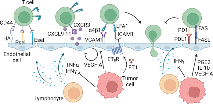
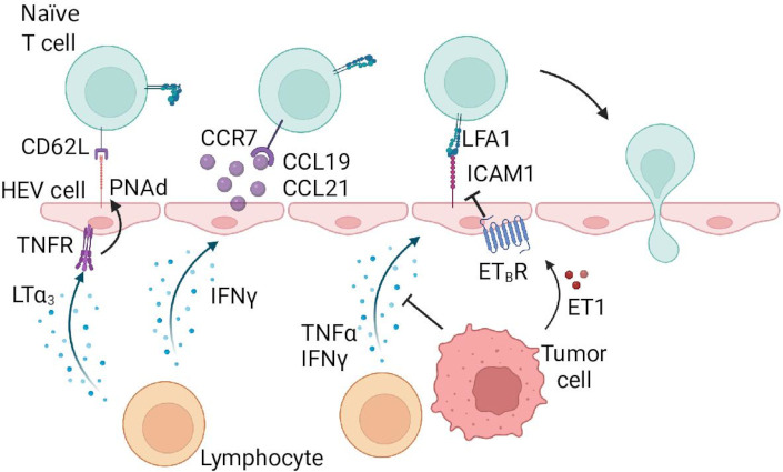
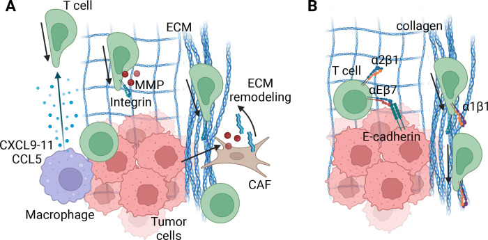
Increased immune cell infiltration into tumors is associated with improved patient survival and predicts response to immune therapies. Thus, identification of factors that determine the extent of immune infiltration is crucial, so that methods to intervene on these targets can be developed. T cells enter tumor tissues through the vasculature, and under control of interactions between homing receptors on the T cells and homing receptor ligands (HRLs) expressed by tumor vascular endothelium and tumor cell nests. HRLs are often deficient in tumors, and there also may be active barriers to infiltration. These remain understudied but may be crucial for enhancing immune-mediated cancer control. Multiple intratumoral and systemic therapeutic approaches show promise to enhance T cell infiltration, including both approved therapies and experimental therapies. This review highlights the intracellular and extracellular determinants of immune cell infiltration into tumors, barriers to infiltration, and approaches for intervention to enhance infiltration and response to immune therapies.

Keywords: CD8-positive T-lymphocytes; cytotoxicity, immunologic; lymphocytes, tumor-infiltrating; tumor microenvironment.


Conflict of interest statement

Competing interests: The following disclosures apply to CLS, but are not related to this work: research support to the University of Virginia from Celldex (funding, drug), GlaxoSmithKline (funding), Merck (funding, drug), 3M (drug), Theraclion (device staff support), Polynoma (PI of clinical trial of melanoma vaccine); funding to the University of Virginia for Scientific Advisory Boards for Immatics and CureVac. Also CLS receives license fee payments through the UVA Licensing and Ventures Group for patents for peptides used in cancer vaccines. No potential conflicts of interest were disclosed by MMM, NDS, and KML.

Figures

Figure 1
Figure 1
Effector T cell homing in tumors. The first step in effector T cell extravasation into peripheral tissues involves tethering and rolling on the endothelial cells, mediated by homing receptors CD44, P-selectin ligand (PSL), and E-selectin ligand (ESL) on the T cell and homing receptor ligands, hyaluronic acid (HA), P-selectin (Psel), and E-selectin (Esel) on the vasculature. Inflammatory molecules interferon (IFN)γ and tumor necrosis factor (TNF)α expressed in the tumor microenvironment (TME) can upregulate these homing receptor ligands, thereby facilitating the recruitment of T cells. Next, inflammatory cytokines CXCL9-11 expressed by endothelial cells, and other cells in the TME, bind to CXCR3, activating integrins α4β1 and lymphocyte function-associated antigen 1 (LFA1). These integrins subsequently bind to vascular cell adhesion molecule 1 (VCAM-1) and intracellular adhesion molecule 1 (ICAM-1) on the vasculature, allowing firm adhesion and arrest of the T cell, followed by extravasation through the endothelial cell layer into the tissue. In this process, tumor cells can inhibit expression of inflammatory cytokines and chemokines in the TME, thereby indirectly suppressing the expression of homing ligands. Additionally, tumor cells express factors that can directly signal within the endothelial cells to downregulate homing receptor ligands, or upregulate T cell suppression molecules directly on the vasculature. Made with Biorender.com.
Figure 2
Figure 2
Naïve T cell homing in tumors. Similar to effector T cells, naïve T cells require multiple signals to extravasate through the vasculature. Contrastingly, naïve T cells require specialized vasculature called high-endothelial venules (HEV), which are capable of expressing the proper receptors and chemokines. Tethering and rolling is initiated by peripheral node addressin (PNAd) binding to CD62L on the T cell. Chemokines CCL19 and CCL21 activate lymphocyte function-associated antigen-1 (LFA1) integrin, and facilitate firm adhesion and arrest by binding of activated LFA1 to intracellular adhesion molecule 1 (ICAM-1). PNAd, CCL19, and CCL21 get upregulated by factors expressed in the tissue microenvironment, LTα3 induces PNAd expression through TNFR signaling, and interferon ((IFN)γ induces CCL19 and CCL21 expression. Tumor cells can inhibit expression of these cytokines, as well as directly downregulate ICAM-1 expression through endothelin-1-endothelin receptor B (ETBR) signaling. Made with Biorender.com.
Figure 3
Figure 3
T cell migration in the tumor microenvironment (TME). (A). Speed and direction of T cells in tumors relies on various factors, including, but likely not limited to, chemokine gradients, extracellular matrix (ECM) organization and integrin expression and migratory capacity of the T cell. Tumor cells or myeloid cells in close proximity to tumor cells can express CXCL9-11 and/or CCL5, which directs effector T cells to the tumor cells directly. When the ECM is organized in a relaxed, diffuse meshwork of ECM molecules, similar to normal epithelial tissues, T cells may migrate along them in an integrin-dependent manner. Local expression of matrix metalloproteinases (MMPs) will furthermore guide their migration. However, cancer-associated fibroblasts (CAFs) often remodel the ECM into a dense and disorganized matrix, rendering T cells unable to leave and get ‘stuck’ in stromal areas of the tumor. Due to high variability from tumor to tumor, the exact specifics of the organization and structure likely determine the ultimate effect the ECM has on T cell motility and function, and requires further investigation. (B) Retention of T cells into a tissue microenvironment relies on ECM and cell-binding integrins. These include, but are likely not limited to, α1β1 and α2β1 binding to collagen and αEβ7 binding to E-cadherin on epithelial and/or tumor cells. Additionally, adhesion of α1β1 to collagen increases T cell motility, providing both a retention and migration mechanism. In tumors with localized, dense collagen this may lead to rapid motility, thereby effectively distracting the T cell from forming long-lasting engagement with target cells. Made with Biorender.com.

Similar articles

Cited by

References

    1. Erdag G, Schaefer JT, Smolkin ME, et al. . Immunotype and immunohistologic characteristics of tumor-infiltrating immune cells are associated with clinical outcome in metastatic melanoma. Cancer Res 2012;72:1070–80. 10.1158/0008-5472.CAN-11-3218 - DOI - PMC - PubMed
    1. Bogunovic D, O’Neill DW, Belitskaya-Levy I, et al. . Immune profile and mitotic index of metastatic melanoma lesions enhance clinical staging in predicting patient survival. Proc Natl Acad Sci U S A 2009;106:20429–34. 10.1073/pnas.0905139106 - DOI - PMC - PubMed
    1. Mihm MCJ, Clemente CG, Cascinelli N. Tumor infiltrating lymphocytes in lymph node melanoma metastases: a histopathologic prognostic indicator and an expression of local immune response. Lab Invest 1996;74:43–7. - PubMed
    1. Wu X, Li J, Connolly EM, et al. . Combined anti-VEGF and anti-CTLA-4 therapy elicits humoral immunity to galectin-1 which is associated with favorable clinical outcomes. Cancer Immunol Res 2017;5:446–54. 10.1158/2326-6066.CIR-16-0385 - DOI - PMC - PubMed
    1. Hamid O, Schmidt H, Nissan A, et al. . A prospective phase II trial exploring the association between tumor microenvironment biomarkers and clinical activity of ipilimumab in advanced melanoma. J Transl Med 2011;9:204. 10.1186/1479-5876-9-204 - DOI - PMC - PubMed

Publication types