. 2023 Apr 18;13(1):6341.
doi: 10.1038/s41598-023-33507-2.

CBGA ameliorates inflammation and fibrosis in nephropathy

Affiliations

CBGA ameliorates inflammation and fibrosis in nephropathy

Sayuri Suzuki et al. Sci Rep. .

Abstract

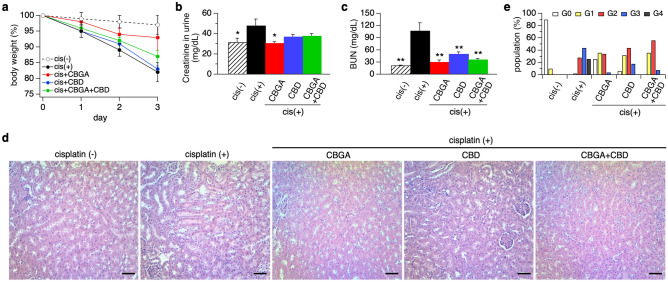
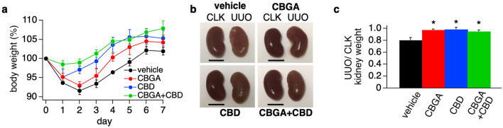
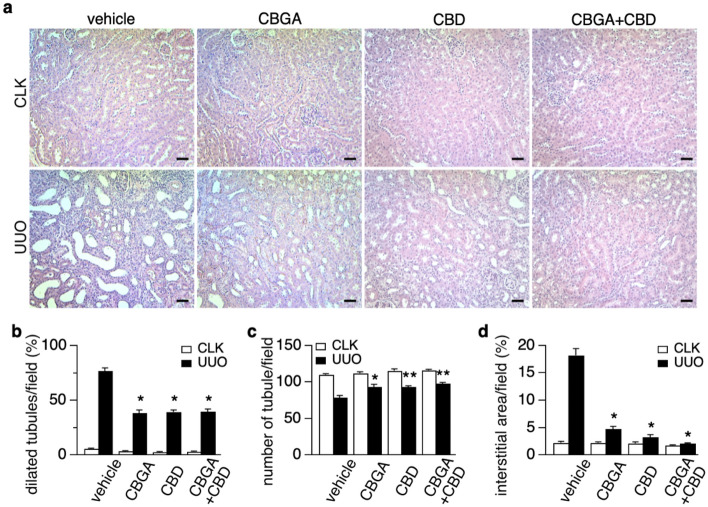
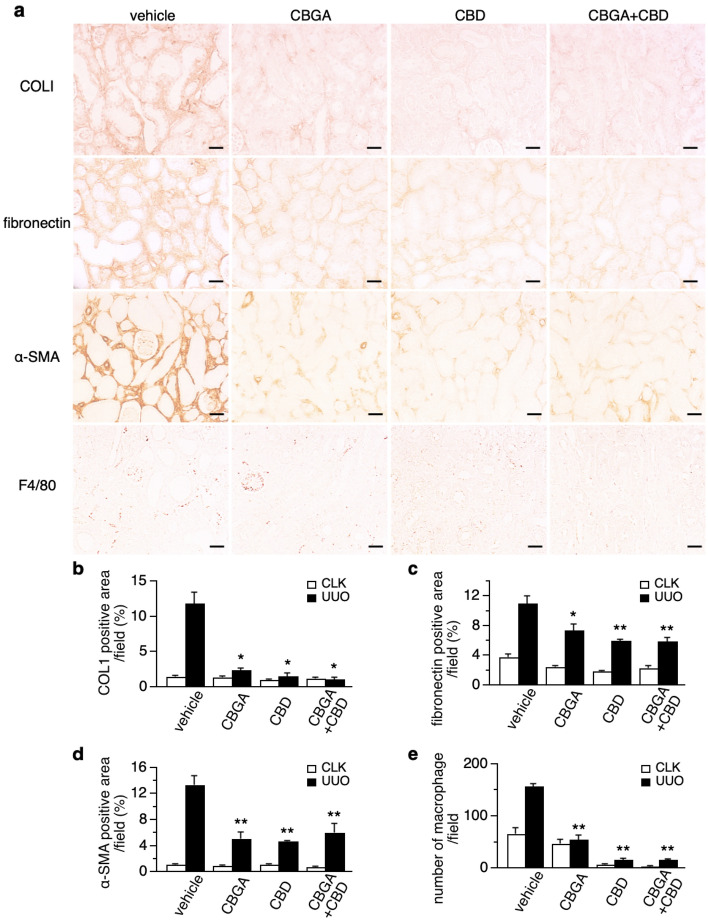
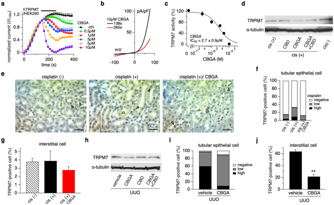
Cannabidiol (CBD) is thought to have multiple biological effects, including the ability to attenuate inflammatory processes. Cannabigerols (CBGA and its decarboxylated CBG molecule) have pharmacological profiles similar to CBD. The endocannabinoid system has recently emerged to contribute to kidney disease, however, the therapeutic properties of cannabinoids in kidney disease remain largely unknown. In this study, we determined whether CBD and CBGA can attenuate kidney damage in an acute kidney disease model induced by the chemotherapeutic cisplatin. In addition, we evaluated the anti-fibrosis effects of these cannabinoids in a chronic kidney disease model induced by unilateral ureteral obstruction (UUO). We find that CBGA, but not CBD, protects the kidney from cisplatin-induced nephrotoxicity. CBGA also strongly suppressed mRNA of inflammatory cytokines in cisplatin-induced nephropathy, whereas CBD treatment was only partially effective. Furthermore, both CBGA and CBD treatment significantly reduced apoptosis through inhibition of caspase-3 activity. In UUO kidneys, both CBGA and CBD strongly reduced renal fibrosis. Finally, we find that CBGA, but not CBD, has a potent inhibitory effect on the channel-kinase TRPM7. We conclude that CBGA and CBD possess reno-protective properties, with CBGA having a higher efficacy, likely due to its dual anti-inflammatory and anti-fibrotic effects paired with TRPM7 inhibition.


Conflict of interest statement

The authors declare no competing interests.

Figures

Figure 1
Figure 1
CBGA and CBD prevent kidney functional loss and damage in the cisplatin-induced acute nephropathy mouse model. (a) Body weight of mice was measured daily before administration of CBGA (10 mg/kg, red circles), CBD (10 mg/kg, blue circles), CBGA + CBD (each 10 mg/kg, green circles) from day 0 to day 3. The cisplatin (+) group was injected daily with vehicle in cisplatin-administered mice as a control (cis (+), black circles). Cisplatin was injected 2 h after the first cannabinoid administration at day 0. The animal group without cisplatin administration received vehicle instead of cisplatin as a negative control against cisplatin-induced nephropathy (cis (−), n = 4, white circles). Body weight of each day was normalized by the weight before cannabinoid injection at day 0 and the average is indicated (n = 5). (b) Creatinine levels were determined at day 3 in urine. (c) BUN levels were determined at day 3. (d) Representative pictures of HE staining taken from kidney sections in each treatment group (magnification × 200). Scale bars represent 50 µm. (e) The grading of tubular injury was quantified from HE staining and plotted as follows: G0 (grade 0, none, 0%, white bars); G1 (grade1, weak, ≤ 20%, yellow bars); G2 (grade 2, mild, > 20 to ≤ 50%, red bars); G3 (grade 3, moderate, > 50 to ≤ 80%, blue bars); G4 (grade 4, strong, > 80%, black bars). *p < 0.05, **p < 0.01 vs. cisplatin (+) group.
Figure 2
Figure 2
CBGA reduced the mRNA expression of inflammatory cytokines and proteins in cisplatin-induced acute nephropathy. The mRNA levels of TNFα (a), IL-6 (b), CXCL10 (c), IL-2 (d), ICAM-1 (e), MCP-1 (f), CRP (g) and ET-1 (h) were measured in kidneys of the cisplatin (+) group (black bars), the CBGA treatment group (red bars), the CBD treatment group (blue bars), and the CBGA + CBD treatment group (green bars) at day 3 (see numeric data in Supplementary Table S1). Shaded bars represent the non-cisplatin treatment group as a control group. *p < 0.05, **p < 0.01 vs. cisplatin (+) group.
Figure 3
Figure 3
CBGA and CBD reduced apoptosis in cisplatin-induced acute nephropathy. (a) Representative pictures of TUNEL-positive apoptotic cells in kidneys from non-cisplatin administered mice or cisplatin-induced nephropathy mice treated with CBGA, CBD or CBGA + CBD (magnification × 200). Scale bars represent 50 µm. (The pictures of high magnification are shown in Supplementary Fig. S1). (b) Quantitative analysis of TUNEL-positive renal tubular epithelial cells assessed in kidneys from cisplatin (+) group (black bar), CBGA treatment group (red bar), CBD treatment group (blue bar), CBGA + CBD treatment group (green bar) at day 3. Shaded bar represents the non-cisplatin treatment group as control. (c) Caspase-3 activity in kidneys was determined using Caspase-3 assay kit (see “Materials and methods”). (d) The levels of full-length PARP1 and cleaved PARP1 protein expression were assessed in kidney tissue from cisplatin-induced acute nephropathy using western blotting (full image is shown in Supplementary Fig. S2). The quantitative percentages are indicated under each picture. **p < 0.01 vs. cisplatin (+) treatment group.
Figure 4
Figure 4
CBGA and CBD prevent kidney atrophy in the UUO mouse model. (a) Body weight of mice was measured daily before administration of CBGA (10 mg/kg, red circles), CBD (10 mg/kg, blue circles) and CBGA + CBD (each 10 mg/kg, green circles) from post-UUO surgery day 0 to day 6 (n = 5) and before sacrifice at day 7. The non-treatment group was injected daily with vehicle as a control (n = 4, black circles). Body weight of each day was normalized by the weight before UUO surgery, and the average is indicated. (b) Representative images of CLK (left in each picture) and UUO kidneys (right in each picture) isolated from UUO mice at day 7 after UUO surgery. Scale bars represent 5 mm. (c) The ratio of UUO kidney weight at day 7. Each UUO kidney weight was normalized by the weight of the corresponding CLK kidney, and the ratio is indicated. Black bar represents the vehicle treatment as a control group, red bar is CBGA treatment group, blue bar is CBD treatment group, and green bar represents CBGA + CBD treatment group. *p < 0.05 vs. vehicle treatment.
Figure 5
Figure 5
CBGA and CBD protect renal morphology in the UUO kidneys. (a) Representative images of HE stainings taken from CLK (upper panels) and UUO (lower panels) kidney sections (magnification × 200) from mice treated with vehicle, CBGA, CBD, CBGA + CBD. Scale bars represent 50 µm. (b, c) The number of dilated tubules (b) and total tubules (c) were counted and evaluated statistically in the CLK (white bars) and UUO kidneys (black bars) at day 7 after UUO. (d) The percentage of the interstitial area in the CLK (white bars) and UUO kidneys (black bars) were quantified using the sections stained for collagen type I (see Fig. 6a). *p < 0.05, **p < 0.01 vs. UUO kidneys treated with vehicle group.
Figure 6
Figure 6
Renal fibrosis is attenuated by CBGA and CBD treatment. (a) Representative images of immunostainings for collagen type I, fibronectin, α-SMA and F4/80 from UUO kidneys treated with vehicle, CBGA, CBD and CBGA + CBD (magnification × 200). Scale bars represent 50 µm. (see also Supplementary Fig. S3 for CLK kidneys) (b–d) The average percentage of the collagen type I-positive area (b), fibronectin-positive area (c), α-SMA-positive area (d) in CLK (white bars) and UUO kidneys (black bars) with vehicle treatment, CBGA, CBD and CBGA + CBD treatment. The intensity of staining in the interstitium was computed using ImageJ software in 10 representative non-overlapping slides. (e) F4/80 antibody was used to detect macrophage on immunostaining, F4/80-positive cells in interstitium were counted as number of macrophages. *p < 0.05, **p < 0.01 vs. UUO kidneys treated with vehicle group.
Figure 7
Figure 7
CBGA inhibits TRPM7 currents in HEK293 cells and reduces TRPM7 expression in kidney damage. (a) Inhibitory effects of CBGA on TRPM7 currents was investigated by whole-cell patch-clamp recordings from HEK293 cells overexpressing TRPM7. Average TRPM7-mediated outward currents at + 80 mV extracted from ramp currents delivered at 0.5 Hz and plotted as a function of time. Concentrations of 0.3 µM (green circles, n = 5), 1 µM (red circles, n = 7), 3 µM (blue circles, n = 7), 5 µM (orange circles, n = 7), and 10 µM (purple circles, n = 6), were applied from 140 to 260 s (black bar). Standard Ringer without CBGA contained acetonitrile as vehicle and was used as control (black circles, n = 8). (b) Average I/V curve of TRPM7 currents extracted before (138 s, black line) and after (260 s, red line) 10 µM CBGA application. (c) The dose–response curve of CBGA on TRPM7 channels. Data points for the curve were obtained from the normalized currents in panel (a) at 260 s. (d) TRPM7 protein expression was examined in kidney tissues from cisplatin-administered mice treated with CBD, CBGA and CBGA + CBD using western blotting (Full images are shown in Supplementary Fig. S5a). (e) Representative pictures of immunostaining to TRPM7 (magnification × 400) in kidneys from non-cisplatin treated mice (left panel), cisplatin-treated mice (middle panel) and cisplatin/CBGA-treated mice (right panel). Scale bars represent 50 µm. Green arrow heads indicate TRPM7-low expression and red arrow heads are high expression levels of TRPM7, yellow arrow heads indicate TRPM7-negative (TRPM7 staining not detectable) in renal tubular epithelial cells. (f) The percentage of TRPM7-positive cells in tubular epithelial cells in kidneys from cisplatin-treated mice. TRPM7-high expression (black bars), TRPM7-low expression (gray bars) and non-detectable TRPM7 staining (white bars) in tubular epithelial cells assessed in a total of 10 non-overlapping fields. (g) The percentage of TRPM7-positive interstitial cells assessed in the same fields as in panel (f). (h) TRPM7 protein expression was examined in cortical UUO kidney tissue from mice treated with vehicle, CBGA, CBD and CBGA + CBD using western blot (Full images are shown in Supplementary Fig. S6a). (i) The percentage of TRPM7 in tubular epithelial cells from UUO kidneys treated with CBGA or not. Same experimental protocol as in (f). Representative pictures are shown in Supplementary Fig. S6c. (j) The percentage of TRPM7-positive interstitial cells assessed in the same fields as in panel (i). **p < 0.01 vs. vehicle treatment group.

References

    1. Arany I, Safirstein RL. Cisplatin nephrotoxicity. Semin. Nephrol. 2003;23:460–464. doi: 10.1016/S0270-9295(03)00089-5. - DOI - PubMed
    1. Perše M, Večerić-Haler Ž. Cisplatin-induced rodent model of kidney injury: Characteristics and challenges. Biomed. Res. Int. 2018;2018:1–29. doi: 10.1155/2018/1462802. - DOI - PMC - PubMed
    1. Sharp CN, Siskind LJ. Developing better mouse models to study cisplatin-induced kidney injury. Am. J. Physiol. Renal Physiol. 2017;313:F835–F841. doi: 10.1152/ajprenal.00285.2017. - DOI - PMC - PubMed
    1. Manohar S, Leung N. Cisplatin nephrotoxicity: A review of the literature. J. Nephrol. 2018;31:15–25. doi: 10.1007/s40620-017-0392-z. - DOI - PubMed
    1. Suzuki S, Ohashi N, Kitagawa M. Roles of the Skp2/p27 axis in the progression of chronic nephropathy. Cell. Mol. Life Sci. 2013;70:3277–3287. doi: 10.1007/s00018-012-1232-x. - DOI - PMC - PubMed

Publication types