. 2023 Apr 18;13(1):6277.
doi: 10.1038/s41598-023-33508-1.

Tumor resident memory CD8 T cells and concomitant tumor immunity develop independently of CD4 help

Affiliations

Tumor resident memory CD8 T cells and concomitant tumor immunity develop independently of CD4 help

Terry R Medler et al. Sci Rep. .

Abstract

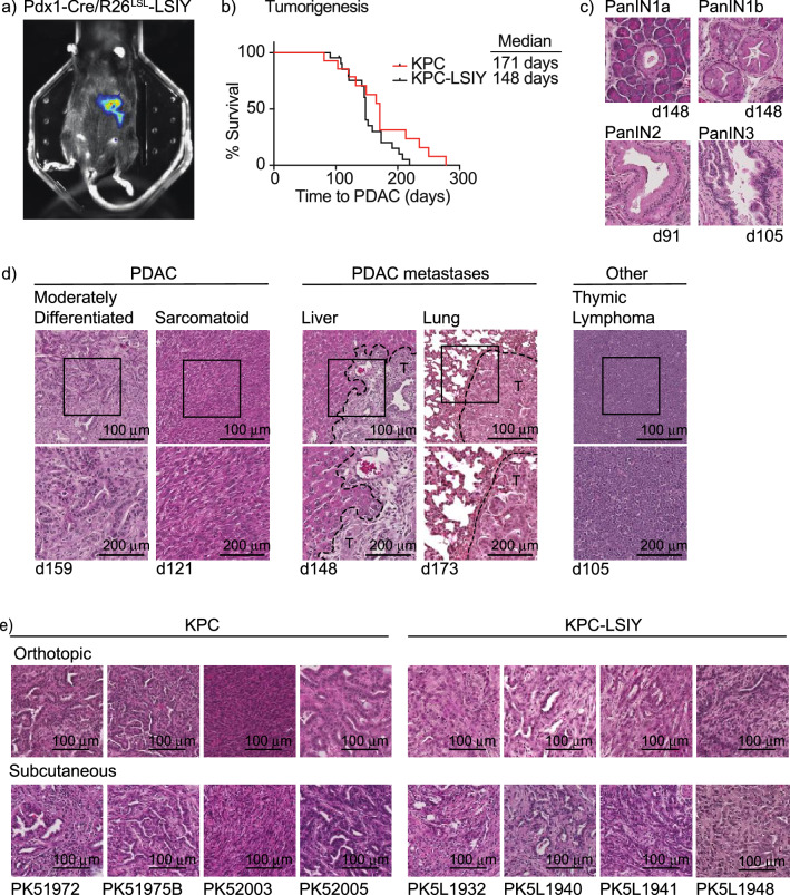
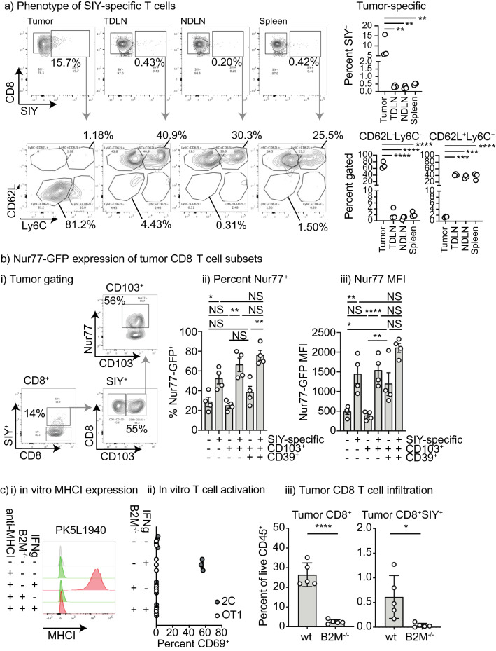
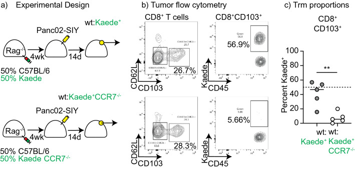
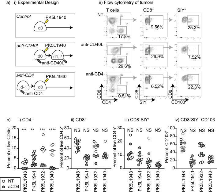
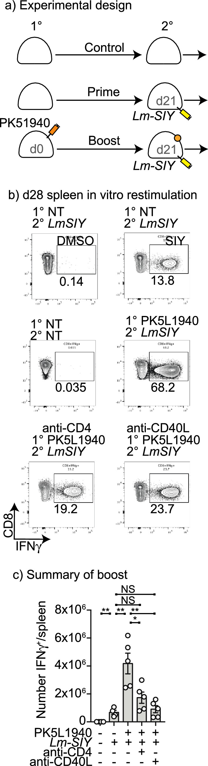
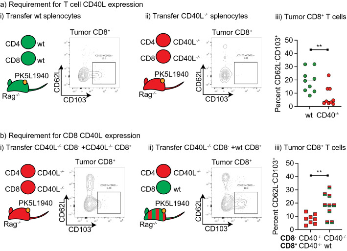
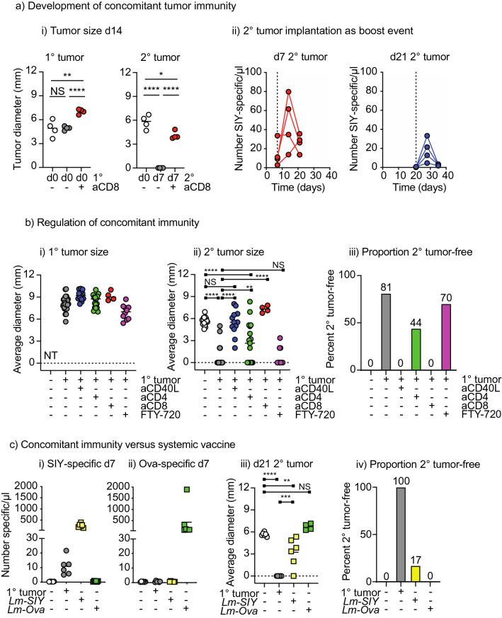
Tissue resident memory (Trm) CD8 T cells infiltrating tumors represent an enriched population of tumor antigen-specific T cells, and their presence is associated with improved outcomes in patients. Using genetically engineered mouse pancreatic tumor models we demonstrate that tumor implantation generates a Trm niche that is dependent on direct antigen presentation by cancer cells. However, we observe that initial CCR7-mediated localization of CD8 T cells to tumor draining lymph nodes is required to subsequently generate CD103+ CD8 T cells in tumors. We observe that the formation of CD103+ CD8 T cells in tumors is dependent on CD40L but independent of CD4 T cells, and using mixed chimeras we show that CD8 T cells can provide their own CD40L to permit CD103+ CD8 T cell differentiation. Finally, we show that CD40L is required to provide systemic protection against secondary tumors. These data suggest that CD103+ CD8 T cell formation in tumors can occur independent of the two-factor authentication provided by CD4 T cells and highlight CD103+ CD8 T cells as a distinct differentiation decision from CD4-dependent central memory.


Conflict of interest statement

MJG receives research funding from Bristol Myers Squibb and VIR Biotechnology. KY receives research funding from Bristol Myers Squibb and consulting fees/stock from Synthis Therapeutics. MRC receives consulting fees from Roche. All other authors have no conflicts to declare. Research funding is not directly related to the topic of this manuscript. Funders had no role in the representation of the data or the preparation of the manuscript.

Figures

Figure 1
Figure 1
Characterization of tumors in KPC-LSIY mice. (A) In vivo luciferase expression in Pdx1-cre + /LSIY + by MuriGlo-assisted CT scan. (B) Survival curves of KPC mice compared to KPC-LSIY mice. (C) Representative histology of premalignant progression in KPC-LSIY mice. (D) Representative histology of moderately differentiated and sarcomatoid PDAC phenotypes, liver metastasis, lung metastasis, as well as thymic lymphoma and in KPC-LSIY mice. (E) Representative histology of orthotopic and subcutaneous tumors using KPC (PK5) and KPC-LSIY (PK5L)-derived tumor cell lines.
Figure 2
Figure 2
Direct interaction of tumor antigen specific T cells with antigen in tumors. (a) Analysis of CD8 T cells in tumor and peripheral lymphoid organs. PK5L1940 tumors were implanted sc. into C57BL/6 mice and tumors, tumor-draining lymph node (TDLN), non-draining lymph node (NDLN), and spleen were analyzed for SIY-specific CD8 T cells at d14. Subplots show CD62L and Ly6C expression on SIY-specific CD8 T cell populations in the lymphoid organs versus the tumor. (b) PK5L1940 tumors were implanted sc. into Nur77-GFP mice and tumor infiltrating T cells were analyzed by flow cytometry. (i) representative flow plots. The SIY-specific T cell population of infiltrating CD8 T cells was identified by pentamer staining, and CD103 + and CD103 + CD39 + subpopulations were identified and Nur77-GFP expression was determined in each cell type. Graphs show (ii) Percent Nur77-GFP + and (iii) MFI of Nur77-GFP in each group. (c) PK5L1940 cancer cells were deleted of B2M expression using CRISPR/Cas9. (i) MHCI expression in cancer cells was detected by flow cytometry compared to isotype control antibody (gray), with cells untreated (green) or pre-treated with IFNg (red). (ii) PK5L1940 or PK5L1940-B2M−/− were co-cultured with SIY-specific 2C cells or Ova-specific OT1 cells and assessed for expression of the activation marker CD69. (iii) PK5L1940 or PK5L1940-B2M−/− were implanted sc. into C57BL/6 mice and harvested at d14 and analyzed for infiltrating CD8 T cells and SIY-specific CD8 T cells by flow cytometry. Key: NS = not significant; * = p < 0.05; ** = p < 0.01; *** = p < 0.001; **** = p < 0.0001. Results are representative of two or more experiments.
Figure 3
Figure 3
Role of direct presentation in Trm formation. (a) Rag−/− mice were reconstituted with a 1:1 mix of splenocytes from wt and Kaede mice, or a 50:50 mice of splenocytes from wt and Kaede CCR7−/− mice. Following reconstitution, mice were implanted sc. with tumors and tumor-infiltrating CD8+CD103+ T cells were analyzed for Kaede expression after 14 days. (b) Representative gating on CD103+ T cells, and Kaede expression by these cells. (c) Summary of percent of CD103+ cells that are Kaede+ in mice given Kaede or Kaede CCR7−/− cells. Key: ** = p < 0.01. Results are representative of two or more experiments.
Figure 4
Figure 4
CD4 depletion minimally impacts CD8 T cell responses to tumor implantation. (a) PK5L1940 pancreatic adenocarcinoma cells were injected sc. into C57BL/6 mice, C57BL/6 mice treated with anti-CD40L on d0, 1, and 2, or C57BL/6 mice treated with anti-CD4 on d-1. ii) Representative flow cytometry of tumor infiltrating T cells 14d following implantation. Plots show gated CD90.2+ T cells, and subgates for CD8+ T cells, and SIY-pentamer+ CD8 T cells. (b) Summary of T cell infiltration of PK5L1948, PK5L1941, PK5L1932, and PK5L1940 tumors derived from Pdx-Cre+/− Kras(G12D)+/− Trp53(R172H)+/− Luciferase-SIY + mice implanted sc. into C57BL/6 mice or C57BL/6 mice treated with anti-CD4 on d-1. Graphs show tumor the proportion of CD4, CD8, CD8 SIY-specific T cells, and CD103 expression on CD8 SIY-specific T cells. Key: NS = not significant; * = p < 0.05; ** = p < 0.01; *** = p < 0.001; **** = p < 0.0001. Results are representative of two or more experiments.
Figure 5
Figure 5
Decreased tumor-specific memory following anti-CD40L or anti-CD4 treatment at tumor implantation. (a) PK5L1940 pancreatic adenocarcinoma cells were injected sc. into C57BL/6 mice or C57BL/6 mice treated with anti-CD40L on d0, 1, and 2, or C57BL/6 mice treated with anti-CD4 on d-1. Control mice received no treatment. (b) Mice were treated with Lm-SIY on d21 and analyzed for SIY-specific T cells in the spleen on d28 by intracellular cytokine staining for IFNg following SIY peptide stimulation. (c) Quantitative analysis of SIY-specific cells in the spleen at d28.
Figure 6
Figure 6
CD8 T cell can provide CD40L for Trm formation. (a) Rag−/− mice were adoptively transferred with splenocytes from wt or CD40L−/− mice, then following reconstitution, implanted sc. with PK51940 tumors. d14 tumors were harvested and analyzed for tumor infiltrating CD8 T cell phenotypes. Flow plots show representative CD103 expression in tumor CD8 T cells from mice reconstituted with (i) wt or (ii) CD40L−/− splenocytes. (iii) Summary of CD103 expression in tumor T cells. (b) Rag−/− mice were reconstituted with CD8-depleted CD40L−/− splenocytes, along with purified CD40L−/− CD8+ splenocytes, or purified wt CD8+ splenocytes. Following reconstitution, mice were implanted sc. with PK51940 tumors. d14 tumors were harvested and analyzed for tumor infiltrating CD8 T cell phenotypes. Flow plots show representative CD103 expression in tumor CD8 T cells from mice reconstituted with (i) CD40L−/− CD8 T cells, or (ii) wt CD8 T cells. (iii) Summary of CD103 expression in tumor T cells. . Key: ** = p < 0.01. Results are the combination of two experiments.
Figure 7
Figure 7
Concomitant tumor immunity is dependent on CD40L. (a)(i) Mice were implanted with a primary sc. PK5L1940 tumor on d0 and a secondary sc. tumor on the opposite flank at d0 or d7 (with and without CD8 depletion). Graphs show the size of the primary (1°) and secondary tumor (2°) at d14. (ii) Mice bearing a primary sc. PK5L1940 tumor were injected with a secondary tumor sc. on d7 or d21. The number of SIY-specific CD8 T cells were determined 0, 7 and 14d following secondary tumor implantation. (b) Mice were implanted with a primary sc. PK5L1940 tumor or no treatment at d0, and a secondary sc. tumor on d7. Groups of mice also received anti-CD40L on d0, 1, and 2, anti-CD4 on d-1, or anti-CD8 on d6. Additional groups received FTY-720 IP on days 6–11. Graphs show the size of the (i) primary (1°) and (ii) secondary tumor (2°) on d21. (iii) Proportion of mice secondary tumor free on d21. (c) Mice were implanted with a primary sc. PK5L1940 tumor or no treatment at d0 or were vaccinated with Lm-SIY or Lm-Ova IV on d0. Mice received a secondary sc. tumor on d7. Peripheral blood was analyzed for (i) SIY-specific T cells or (ii) ova-specific T cells on d7. (iii) Size of secondary tumors on d21. (iv) proportion of mice tumor-free on d21. Key: NS = not significant; * = p < 0.05; ** = p < 0.01; *** = p < 0.001; **** = p < 0.0001. Results are the combination of 4–5 experiments.

Similar articles

Cited by

References

    1. Ridge JP, Di Rosa F, Matzinger P. A conditioned dendritic cell can be a temporal bridge between a CD4+ T-helper and a T-killer cell. Nature. 1998;393:474–478. doi: 10.1038/30989. - DOI - PubMed
    1. Bennett SR, et al. Help for cytotoxic-T-cell responses is mediated by CD40 signalling. Nature. 1998;393:478–480. doi: 10.1038/30996. - DOI - PubMed
    1. Schoenberger SP, Toes RE, van der Voort EI, Offringa R, Melief CJ. T-cell help for cytotoxic T lymphocytes is mediated by CD40-CD40L interactions. Nature. 1998;393:480–483. doi: 10.1038/31002. - DOI - PubMed
    1. Behr FM, et al. Tissue-resident memory CD8(+) T cells shape local and systemic secondary T cell responses. Nat Immunol. 2020;21:1070–1081. doi: 10.1038/s41590-020-0723-4. - DOI - PubMed
    1. Moran AE, Polesso F, Weinberg AD. Immunotherapy expands and maintains the function of high-affinity tumor-infiltrating CD8 T cells in situ. J. Immunol. 2016;197:2509–2521. doi: 10.4049/jimmunol.1502659. - DOI - PMC - PubMed

Publication types