Review
. 2023 Jul;24(7):421-441.
doi: 10.1038/s41576-023-00587-9. Epub 2023 Apr 18.

Primary cilia as dynamic and diverse signalling hubs in development and disease

Affiliations
Review

Primary cilia as dynamic and diverse signalling hubs in development and disease

Pleasantine Mill et al. Nat Rev Genet. 2023 Jul.

Abstract

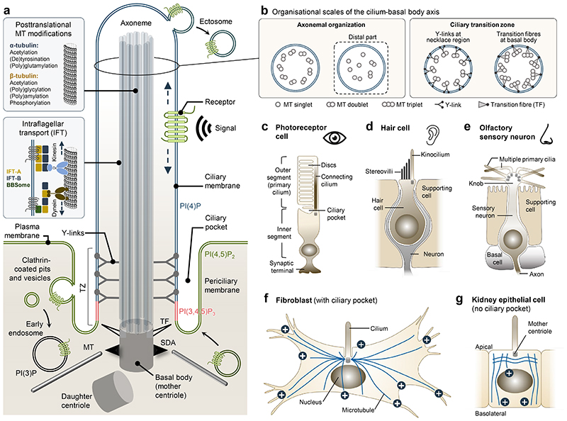
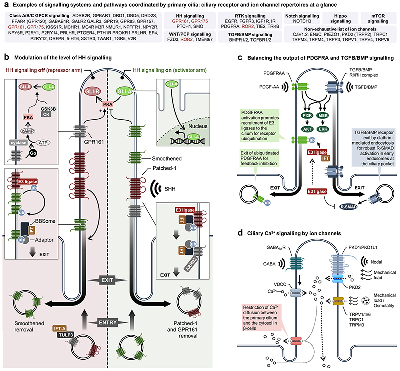
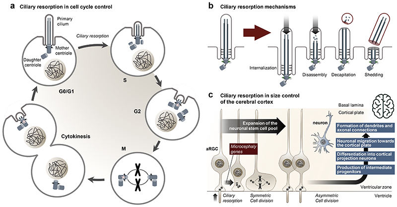
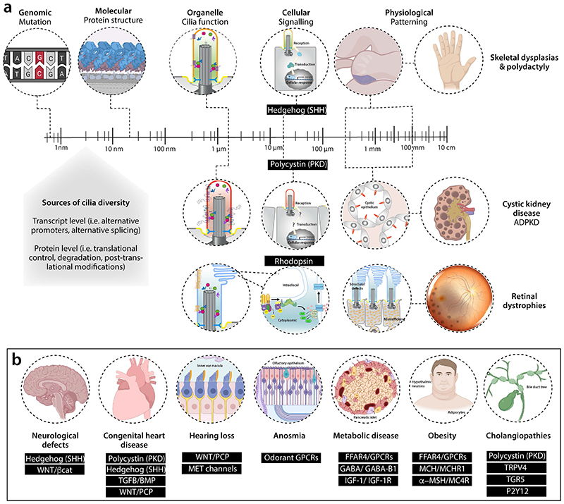
Primary cilia, antenna-like sensory organelles protruding from the surface of most vertebrate cell types, are essential for regulating signalling pathways during development and adult homeostasis. Mutations in genes affecting cilia cause an overlapping spectrum of >30 human diseases and syndromes, the ciliopathies. Given the immense structural and functional diversity of the mammalian cilia repertoire, there is a growing disconnect between patient genotype and associated phenotypes, with variable severity and expressivity characteristic of the ciliopathies as a group. Recent technological developments are rapidly advancing our understanding of the complex mechanisms that control biogenesis and function of primary cilia across a range of cell types and are starting to tackle this diversity. Here, we examine the structural and functional diversity of primary cilia, their dynamic regulation in different cellular and developmental contexts and their disruption in disease.


Conflict of interest statement

Competing interests

The authors declare no competing interests.

Figures

Figure 1
Figure 1. Architecture of primary cilia and main ciliary sub compartments.
a, Schematic of a generic primary cilium showing the main ciliary subcompartments. b, Cross-sectional views of the cilium-basal body axis (from tip to base) showing the changes in microtubule (MT) arrangement along the axis. The panel 1b right image presented in the box for axonemal organization illustrates the compromised 9+0 MT organization of unstructured bundles of MT singlets and doublets that can be observed at the distal region of the cilium in certain cell types. c, d, e, Diagrams of the indicated vertebrate cell types illustrating the diverse types of non-motile, sensory cilia found in these cells. f, g, Depictions of a fibroblast and polarized kidney epithelial cell showing the presence and absence, respectively, of a ciliary pocket, and the different cytoplasmic MT organisation (blue lines; MT plus ends are marked with “+”) in the two cell types. Abbreviations: IFT, intraflagellar transport; SDA, subdistal appendage; TF, transition fibre; TZ, transition zone.
Figure 2
Figure 2. Overview of signalling pathways coordinated by primary cilia.
a, Overview of receptors and ion channels in ciliary signalling pathways. Receptors in red are listed twice, as they can be categorized in more than one signalling system. b, Overview of Hedgehog (HH) signalling. In the absence of sonic hedgehog (SHH) (in the repressor arm of HH signalling), the receptor patched-1 (PTCH1) is enriched in the ciliary membrane, preventing ciliary enrichment of smoothened (SMO) through WWP1 (E3-ligase)-mediated ubiquitination and ciliary exit by retrograde IFT. The class A GPCR, GPR161, is targeted to the cilium by tubby-like protein 3 (TULP3) and IFT-A to activate adenylate cyclases via G-proteins (Gα), leading to increased ciliary levels of cAMP. cAMP activates protein kinase A (PKA), which in complex with glycogen synthase kinase 3 β (GSK3β) and casein kinases (CK) promotes the limited proteolytic cleavage of full-length and activator versions of GLI2/3 transcription factors (GLI-A) into their repressor form (GLI-R). In the presence of SHH (in the activator arm of HH signalling), PTCH1 and GPR161 exit the cilium, allowing enrichment of ciliary SMO, which promotes formation of GLI-A. Exit of PTCH1 and GPR161 similarly relies on their ubiquitination and removal by BBSome-assisted retrograde IFT; GPR161 ubiquitination being controlled at the level of beta-arrestin 2 (ARRB2). Both GLI-A and GLI-R translocate from the cilium into the nucleus to induce and repress transcriptional activation of HH target genes, respectively. c, Overview of ciliary control of platelet-derived growth factor α (PDGFRα) and transforming growth factor β (TGF-β)/bone morphogenetic protein (BMP) signalling. Following activation of PDGFRα and downstream signalling via PI3K-AKT and MEK1/2-ERK1/2 pathways, E3 ligases of the CBL family ubiquitinate the receptor for internalization and feedback inhibition. TGFB/BMP signalling operates in the cilium via both canonical (R-SMAD) and non-canonical (e.g. PI3K-AKT and MEK1/2-ERK1/2 pathways). Robust canonical signalling relies on ciliary exit of activated TGFB receptors to activate R-SMADs, which are inhibited by E3-ligase SMURF1 at the ciliary pocket. d) Examples of stimulation modes (chemosensation, mechanosensation and osmolality) for ciliary Ca2+ signalling regulated by GPCRs and ion channels. Please see main text for further details. Abbreviations Ub: ubiquitination.
Figure 3
Figure 3. Ciliary dynamics.
a, Assembly and disassembly of primary cilia are tightly coordinated with the cell cycle. A single primary cilium assembles from the mother centriole in G0/G1 phase, and the cilium is resorbed during the G1/S and M phases to liberate duplicated centrioles for mitotic spindle pole formation. At the end of cytokinesis, the daughter cell inheriting the oldest mother centriole (marked with an asterisk), will begin forming a new primary cilium prior to the other daughter cells. b, Schematic illustration of different modes by which a cilium can be resorbed. c, During development of the cerebral cortex, apical radial glia cells (aRGC) primary cilia, which project into the ventricular lumen, are resorbed to allow expansion of the neural stem cell pool by symmetric cell divisions. By asymmetric cell divisions, aRGCs subsequently form intermediate progenitors that differentiate into cortical projecting neurons that migrate towards the cortical plate to form the dendrites and axonal connections. Consequently, dysfunction in the timely resorption of aRGC primary cilia is linked to proliferation–differentiation decision defects and reduced size of the cerebral cortex such as in microcephaly.
Figure 4
Figure 4. Challenge of ciliopathies — biology across scales.
a, Schematic of how variant identification in a patient with ciliopathy is just the start of the challenge. Genetic changes are the same across all cell types. Understanding how these variants disturb different types of cilia structurally and functionally in terms of signalling readouts is our knowledge gap across cell types and developmental times, one that we need to address to understand patient phenotypes. Possible sources of tissue-specific phenotypes are highlighted. We focus on the best characterized cilia-dependent signalling defects resulting from ciliopathy mutations in different tissue types, including skeleton, kidney epithelia and photoreceptors resulting in ciliopathic disease. For the kidney, we have focused on autosomal dominant polycystic kidney disease through PKD1/2 signalling but other renal diseases include autosomal recessive polycystic kidney disease and nephronophthisis, the latter involving additional signaling defects (see Table 1). b, Examples of the signalling pathways disrupted in different tissue types in the ciliopathies. Abbreviations: ADPKD, autosomal dominant polycystic kidney disease; FFAR4, Free Fatty Acid Receptor 4; GABA, gamma-aminobutyric acid; GPCRs, G protein coupled receptors; IGF, insulin-like growth factor; MCH, melanin-concentrating hormone; MET, mechano-electrical transduction; MSH, melanocyte-stimulating hormone; P2Y12, purinergic receptor P2Y; PKD, Polycystin; SHH, Sonic Hedgehog; TGFB/BMP, Transforming Growth Factor Beta/ Bone Morphogenetic Protein; TGR5, Takeda G protein-coupled receptor 5; TRPV4, transient receptor potential vanilloid-type 4; WNT/ βcat, canonical Wingless/Integrated Beta catenin pathway; WNT/PCP, non-canonical Wingless/Integrated Planar Cell Polarity pathway.

References

    1. Niederlova V, Modrak M, Tsyklauri O, Huranova M, Stepanek O. Meta-analysis of genotype-phenotype associations in Bardet-Biedl syndrome uncovers differences among causative genes. Human Mutation. 2019;40:2068–2087. doi: 10.1002/humu.23862. - DOI - PubMed
    1. Reiter JF, Leroux MR. Genes and molecular pathways underpinning ciliopathies. Nat Rev Mol Cell Biol. 2017;18:533–547. doi: 10.1038/nrm.2017.60. - DOI - PMC - PubMed
    1. Wheway G, Mitchison HM, Genomics England Research, C Opportunities and Challenges for Molecular Understanding of Ciliopathies-The 100,000 Genomes Project. Front Genet. 2019;10:127. doi: 10.3389/fgene.2019.00127. - DOI - PMC - PubMed
    1. Chandra B, Tung ML, Hsu Y, Scheetz T, Sheffield VC. Retinal ciliopathies through the lens of Bardet-Biedl Syndrome: Past, present and future. Prog Retin Eye Res. 2022;89:101035. doi: 10.1016/j.preteyeres.2021.101035. - DOI - PubMed
    1. Van De Weghe JC, Gomez A, Doherty D. The Joubert-Meckel-Nephronophthisis Spectrum of Ciliopathies. Annu Rev Genomics Hum Genet. 2022;23:301–329. doi: 10.1146/annurev-genom-121321-093528. - DOI - PMC - PubMed