. 2023 Apr 18;6(1):403.
doi: 10.1038/s42003-023-04640-5.

Pigmentation and TYRP1 expression are mediated by zinc through the early secretory pathway-resident ZNT proteins

Affiliations

Pigmentation and TYRP1 expression are mediated by zinc through the early secretory pathway-resident ZNT proteins

Takumi Wagatsuma et al. Commun Biol. .

Abstract

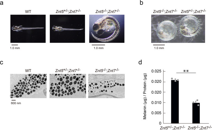
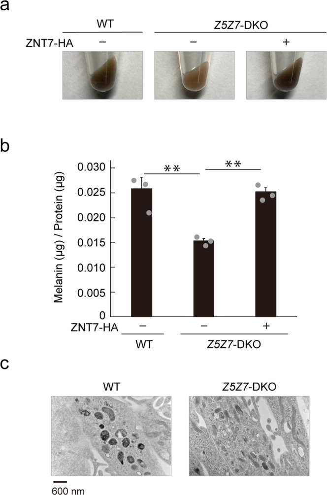
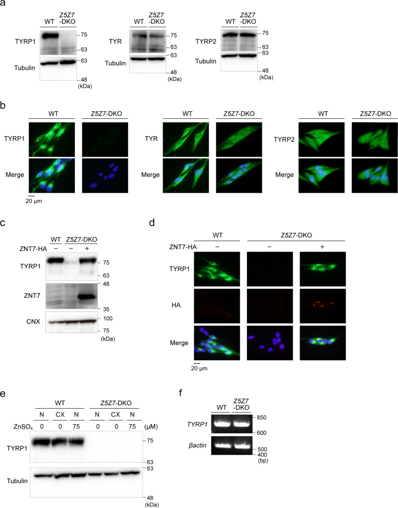
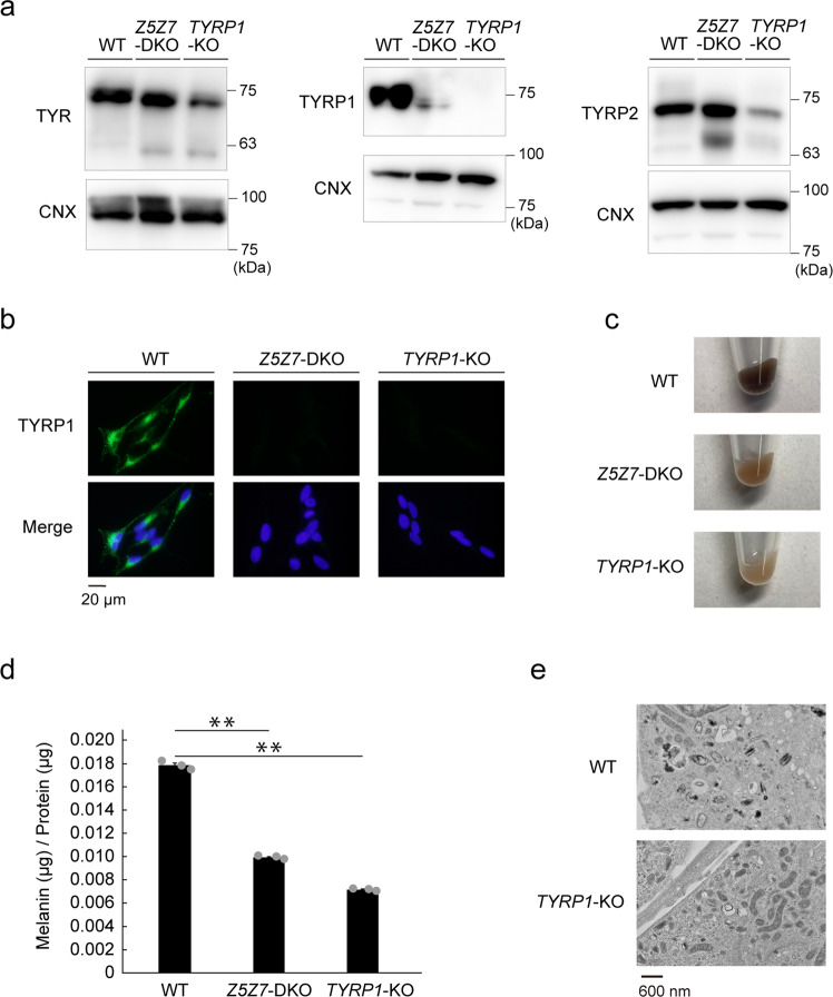
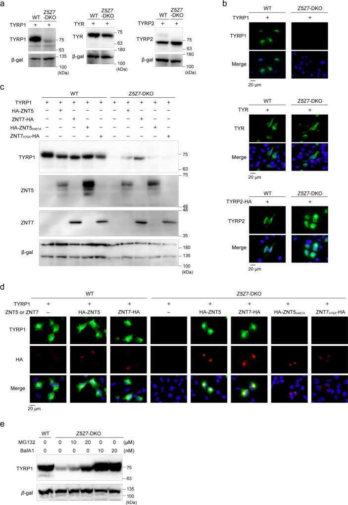
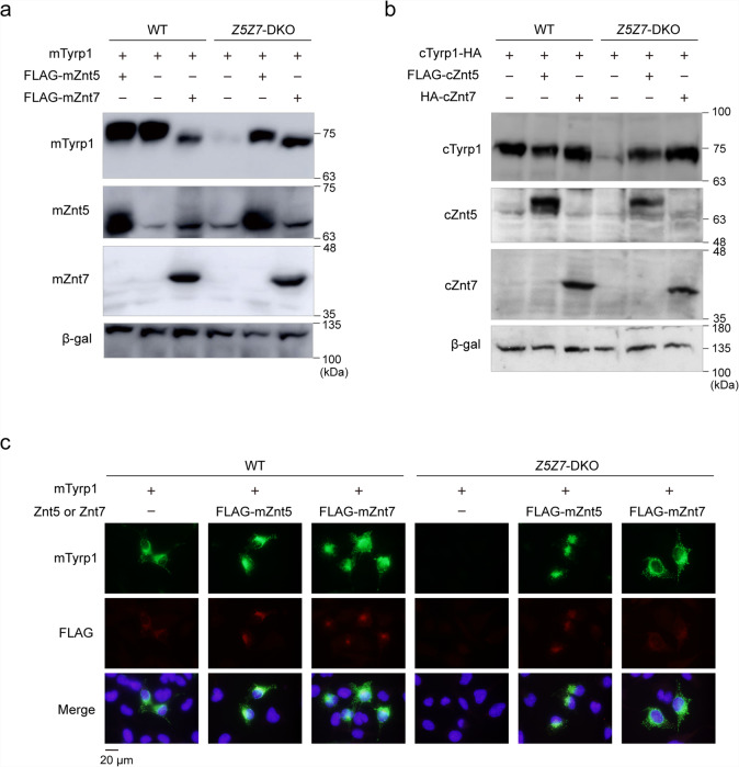
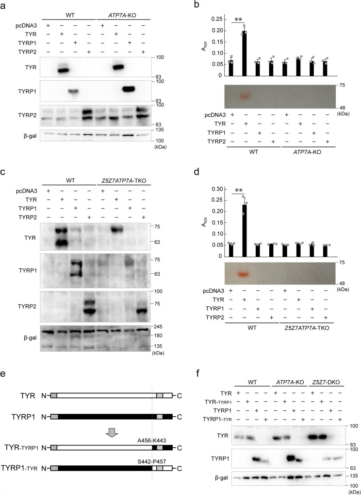
Tyrosinase (TYR) and tyrosinase-related proteins 1 and 2 (TYRP1 and TYRP2) are essential for pigmentation. They are generally classified as type-3 copper proteins, with binuclear copper active sites. Although there is experimental evidence for a copper cofactor in TYR, delivered via the copper transporter, ATP7A, the presence of copper in TYRP1 and TYRP2 has not been demonstrated. Here, we report that the expression and function of TYRP1 requires zinc, mediated by ZNT5-ZNT6 heterodimers (ZNT5-6) or ZNT7-ZNT7 homodimers (ZNT7). Loss of ZNT5-6 and ZNT7 function results in hypopigmentation in medaka fish and human melanoma cells, and is accompanied by immature melanosomes and reduced melanin content, as observed in TYRP1 dysfunction. The requirement of ZNT5-6 and ZNT7 for TYRP1 expression is conserved in human, mouse, and chicken orthologs. Our results provide novel insights into the pigmentation process and address questions regarding metalation in tyrosinase protein family.


Conflict of interest statement

The authors declare no competing interests.

Figures

Fig. 1
Fig. 1. Znt5-6 and Znt7 are required for melanogenesis in medaka fish.
a Representative dorsal views of whole larvae at 8–9 dpf for wild-type (WT), Znt5+/-;Znt7-/-, and Znt5-/-;Znt7-/- medaka. The Znt5-/-;Znt7-/- medaka did not hatch, and thus is shown after manual removal of the chorion. b Representative lateral views of embryos at 8–9 dpf for Znt5-/-;Znt7-/- (left) and Znt5+/-;Znt7-/- (right) medaka before hatching. c Transmission electron microscopy (TEM) shows irregular melanosomes with less electron dense pigment in Znt5-/-;Znt7-/- medaka compared with those in WT and Znt5+/-;Znt7-/- medaka. d Melanin content was decreased in Znt5-/-;Znt7-/- mutant medaka compared with that in Znt5+/-;Znt7-/- littermate (n = 3). Statistical significance was analyzed by Student’s t-test. **p < 0.01. In ac, each analysis was performed on more than three individual medaka, and in d, the experiments were performed at least three times, and representative results are shown.
Fig. 2
Fig. 2. Melanin synthesis is decreased in Mewo-Z5Z7-DKO cells.
a Pellets of wild-type (WT) Mewo and Mewo-Z5Z7-DKO cells, and of Mewo-Z5Z7-DKO cells stably re-expressing ZNT7-HA. b Melanin content was decreased in Mewo-Z5Z7-DKO cells compared with that in WT Mewo cells; the decrease was recovered upon expression of ZNT7 (n = 3). Statistical significance was determined using the one-way analysis of variance (ANOVA) followed by Tukey’s post hoc test. **p < 0.01. c Transmission electron microscopy (TEM) showing irregular melanosomes with less electron dense pigment in Mewo-Z5Z7-DKO cells compared with those in WT Mewo cells. Each experiment, except for that in c, was performed at least three times, and representative results from independent experiments are shown. Panels in c show representative results for at least three cells.
Fig. 3
Fig. 3. TYPR1 expression is substantially decreased in Mewo-Z5Z7-DKO cells.
a, b Expression of TYRP1, but not TYR or TYRP2, was substantially decreased in Mewo-Z5Z7-DKO cells. Immunoblotting (a) and immunofluorescence staining (b). c, d Expression of TYRP1 in Mewo-Z5Z7-DKO cells was reversed by stable re-expression of ZNT7. Immunoblotting (c) and immunofluorescence staining (d). e Effects of zinc status on the expression of TYRP1. Wild-type (WT) Mewo and Mewo-Z5Z7-DKO cells were cultured in normal medium (N), normal medium supplemented with ZnSO4 (75 μM), or in zinc-deficient medium (CX) generated using Chelex-treated FCS for 24 h. f TYRP1 mRNA levels were not decreased in Mewo-Z5Z7-DKO cells. In a, c, and e, Tubulin or CNX was detected as a loading control. Each experiment was performed at least three times, and representative results from independent experiments are shown.
Fig. 4
Fig. 4. Mewo-Z5Z7-DKO cells show similar defects as in Mewo-TYRP1-KO cells.
a Analysis of TYRP1, TYR, and TYR2 expression by immunoblotting in wild-type (WT) Mewo, Mewo-Z5Z7-DKO and Mewo-TYRP1-KO cells. Note the loss of TYRP1, but not TYR and TYRP2, in both Mewo-Z5Z7-DKO and Mewo-TYRP1-KO cells. b Immunofluorescence analysis confirmed a loss of TYRP1 in both Mewo-Z5Z7-DKO and Mewo-TYRP1-KO cells. c Pellets of Mewo-Z5Z7-DKO and Mewo-TYRP1-KO cells exhibit hypopigmentation compared to WT Mewo cells. d Melanin content was decreased in Mewo-TYRP1-KO cells, similar to that in Mewo-Z5Z7-DKO cells (n = 3). Statistical significance was determined using the one-way analysis of variance (ANOVA) followed by Tukey’s post hoc test. **p < 0.01. e TEM showed irregular melanosomes with less electron dense contents in Mewo-TYRP1-KO, compared with WT Mewo cells. Each experiment except for (e) was performed at least three times, and representative results from independent experiments are shown. Panels in e were performed using at least three cells, and representative results are shown.
Fig. 5
Fig. 5. TYRP1 expression is dependent on functional ZNT5 or ZNT7 transporters.
a Immunoblot analysis of TYRP1, TYR, and TYRP2 in wild-type (WT) SK-MEL-2 cells and SK-Z5Z7-DKO cells transfected with plasmids encoding TYRP1, TYR and TYRP2. Note that TYRP1, but not TYR and TYRP2, was substantially decreased in SK-Z5Z7-DKO cells. b Confirmation of substantial reduction in TYRP1 expression using immunofluorescence staining. c Immunoblot and d immunofluorescence microscopy showing restoration of TYRP1 expression in SK-Z5Z7-DKO cells following re-expression of wild type ZNT5 or ZNT7, but not of zinc transport-incompetent mutants of ZNT5 and ZNT7 (ZNT5H451A and ZNT7H70A). e Bafilomycin A1 (BafA1) treatment stabilizes the expression of TYRP1, transiently transfected in SK-Z5Z7-DKO cells. SK-Z5Z7-DKO cells were treated with the indicated concentrations of MG132 and BafA1 for 6 h. In c, d, TYR, TYRP1, and TYRP2 plasmids and ZNT plasmids were transfected at a ratio of 1:10. In a, c, and e, β-galactosidase (β-gal) was used as a transfection control. Each experiment was performed at least three times, and representative results from independent experiments are shown.
Fig. 6
Fig. 6. Restoration of mouse or chicken Tyrp1 expression in SK-Z5Z7-DKO cells following re-expression of mouse or chicken Znt5 or Znt7.
a, b Expression of recombinant mouse (a) and chicken (b) Tyrp1 was substantially decreased in SK-Z5Z7-DKO cells compared to wild-type (WT) SK-MEL-2 cells, but stabilized by expression of recombinant mouse or chicken Znt5 or Znt7. In b, cTyrp1, tagged with HA at the C-terminus, was used. c Immunofluorescence staining of mouse Tyrp1 expressed in WT SK-MEL-2 or SK-Z5Z7-DKO cells. Tyrp1 and Znt plasmids were transfected at a ratio of 1:10. Each experiment was performed at least three times, and representative results from independent experiments are shown.
Fig. 7
Fig. 7. TYRP1 expression is not dependent on the ATP7A copper transporter.
a Immunoblot analysis shows that the expression of TYRP1, TYR, and TYRP2 in SK-ATP7A-KO cells was comparable with levels in WT SK-MEL-2 cells. b TYR activity was substantially decreased in SK-ATP7A-KO cells. L-DOPA oxidase activity (upper graph, n = 3) and tyrosinase zymography (lower panel) were performed. c, d Loss of TYRP1 expression in SK-Z5Z7-DKO cells (c) and loss of TYR activity in SK-ATP7A-KO cells (d) were not altered in SK-Z5Z7ATP7A-TKO cells. e Schematic representation of the chimeric constructs analyzed in f. f Domain exchange analysis between TYR and TYRP1. TYRP1-TYR chimera mutant showed the same property as that of TYRP1 expressed in SK-ATP7A-KO and SK-Z5Z7-DKO cells. In a, c, and f, β-galactosidase (β-gal) was used as a transfection control. In b and d, statistical significance was determined using the one-way analysis of variance (ANOVA) followed by Tukey’s post hoc test. **p < 0.01. Each experiment was performed at least three times, and representative results from independent experiments are shown.

Similar articles

Cited by

References

    1. Yamaguchi Y, Hearing VJ. Melanocytes and their diseases. Cold Spring Harb. Perspect Med. 2014;4:a017046. doi: 10.1101/cshperspect.a017046. - DOI - PMC - PubMed
    1. Solano F. On the metal cofactor in the tyrosinase family. Int. J. Mol. Sci. 2018;19:633. doi: 10.3390/ijms19020633. - DOI - PMC - PubMed
    1. Tomita Y, Takeda A, Okinaga S, Tagami H, Shibahara S. Human oculocutaneous albinism caused by single base insertion in the tyrosinase gene. Biochem. Biophys. Res. Commun. 1989;164:990–996. doi: 10.1016/0006-291X(89)91767-1. - DOI - PubMed
    1. Zhang KH, et al. Oculocutaneous albinism type 3 (OCA3): analysis of two novel mutations in TYRP1 gene in two Chinese patients. Cell Biochem. Biophys. 2011;61:523–529. doi: 10.1007/s12013-011-9234-0. - DOI - PubMed
    1. Pennamen P, et al. Dopachrome tautomerase variants in patients with oculocutaneous albinism. Genet. Med. 2021;23:479–487. doi: 10.1038/s41436-020-00997-8. - DOI - PubMed

Publication types