. 2023 Apr 18;23(1):356.
doi: 10.1186/s12885-023-10845-y.

Analysis of patients with colorectal cancer shows a specific increase in serum anti-ING1 autoantibody levels

Affiliations

Analysis of patients with colorectal cancer shows a specific increase in serum anti-ING1 autoantibody levels

Takahiro Arasawa et al. BMC Cancer. .

Abstract

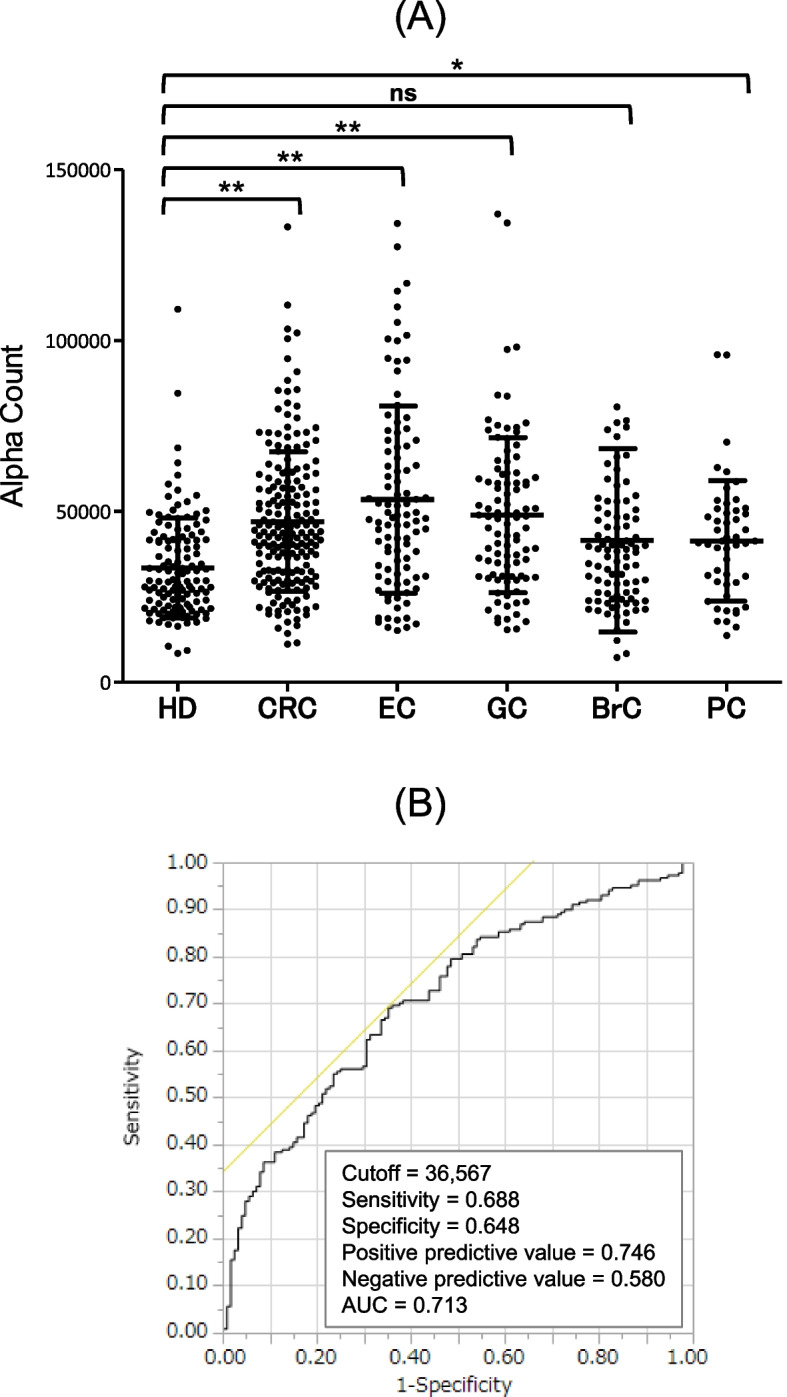
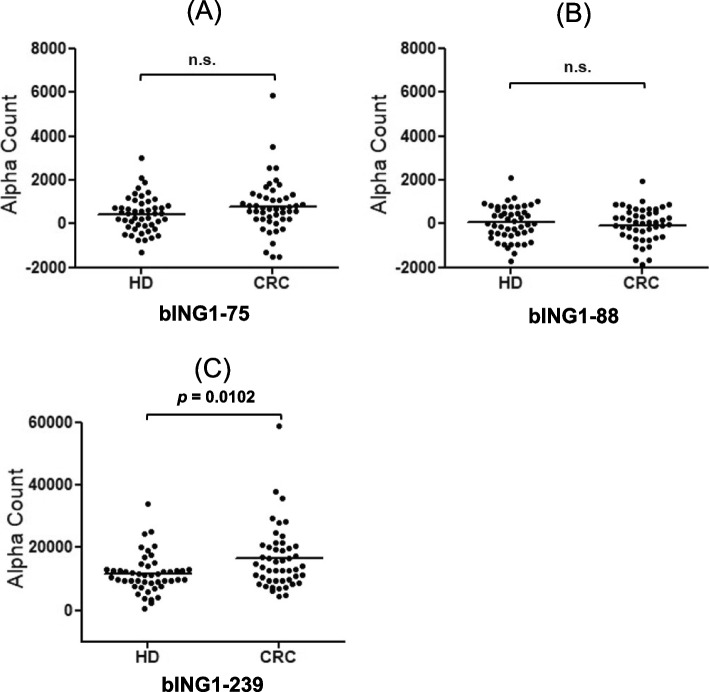
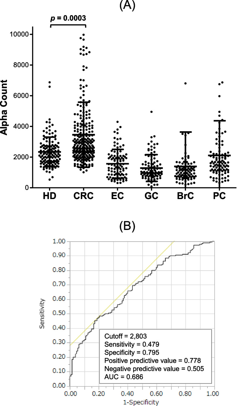
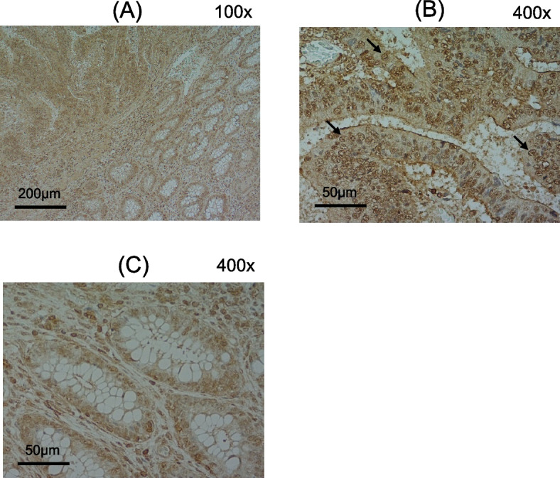
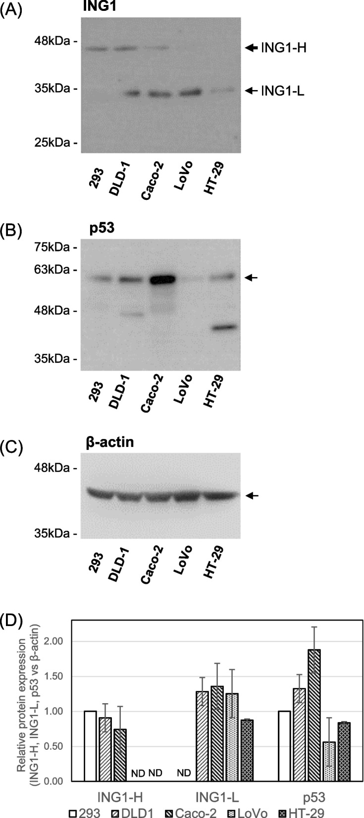
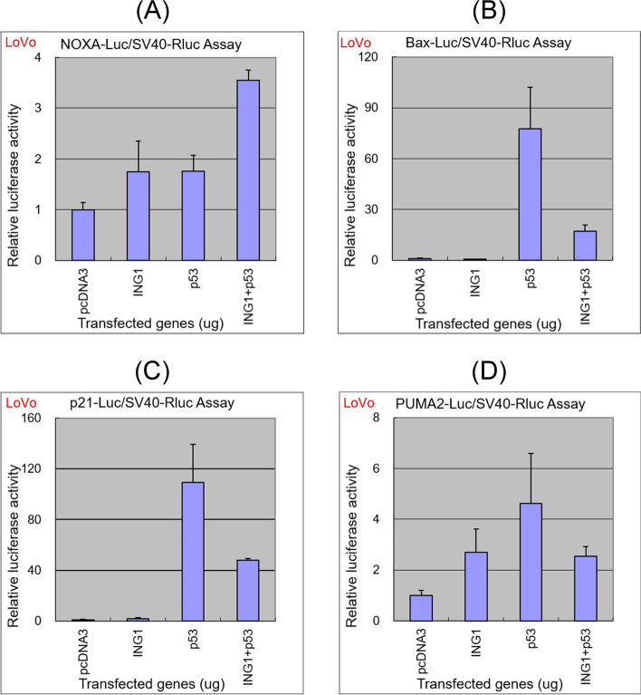
Colorectal cancer (CRC) is the third most prevalent cancer in the world, yet the sensitivity and specificity of biomarkers for CRC diagnosis are insufficient. In the present study, we performed a protein microarray screening method to identify antibody markers for CRC. Inhibitor of growth family 1 (ING1) was identified as a candidate tumor antigen for CRC using protein microarrays (ProtoArray). Subsequent amplified luminescence proximity homogeneous assay-linked immunosorbent assay using recombinant ING1 protein showed that the serum levels of anti-ING1 antibodies were increased not only in patients with CRC but also in those with esophageal cancer (EC), gastric cancer (GC), breast cancer (BrC), and pancreatic cancer (PC) compared with those of healthy donors (HDs). Antibodies against the ING1 amino acids between 239 and 253 were present at significantly higher levels in patients with CRC than in those with EC, GC, BrC, or PC. Anti-ING1 antibody levels were significantly higher in the patients with CRC at any stages than in the HDs. Immunohistochemical staining revealed higher expression of ING1 protein in CRC cells than in the adjacent normal tissues. In luciferase reporter assays using a CRC cell line, ING1 augmented p53-mediated NOXA promoter activity but attenuated p53-stimulated Bax, p21, and PUMA promoter activities. Consequently, serum anti-ING1 antibodies can be used for sensitive and specific diagnoses of CRC.

Keywords: Antibody; Colorectal cancer; Inhibitor of growth protein 1; Protein array analysis; Tumor biomarker.


Conflict of interest statement

The authors declare no competing interests.

Figures

Fig. 1
Fig. 1
Presence of anti-ING1 antibodies in sera of patients with CRC. The reactivity of anti-ING1 antibodies against ING1 protein were examined by western blot analysis. Glutathione-S-transferase (GST) (lane 1) and GST-ING1 proteins (lane 2) were electrophoresed on sodium dodecyl sulfate–polyacrylamide gels. Anti-GST antibody (Rockland, Gilbertsville, PA), serum from healthy donors (HDs), or sera from patients with colorectal cancer (CRC) (CRC #1 and #2) were used as primary antibodies. One of the 2 independent repeat experiments is shown. Molecular sizes are shown at the left. Asterisks represent the degradation products of GST-ING1 protein. The full blot images are shown in Additional file 2: Figure S1
Fig. 2
Fig. 2
Comparison of anti-ING1 antibody levels between HDs and patients with solid cancers. A Serum antibody levels against ING1 protein were quantified by amplified luminescence proximity homogeneous assay-linked immunosorbent assay (AlphaLISA), and compared between HDs and patients with CRC, esophageal cancer (EC), gastric cancer (GC), breast cancer (BrC), and pancreatic cancer (PC). The Alpha photon counts represent the antibody levels, and are shown in a scatter dot plot. Horizontal lines represent the averages, and the error bars represent standard deviations (SDs). Differences were examined with Dunn’s multiple comparison test following a Kruscal-Wallis test (p < 0.001). *, p < 0.05; **, p < 0.001; ns, not significant. Data are summarized in Additional file 1: Table S3. B CRC detection using s-ING1-Abs was assessed using receiver operating characteristic (ROC) analyses. Numbers in the figures indicate cutoff values for Alpha counts, sensitivity, specificity, and area under the curve (AUC)
Fig. 3
Fig. 3
Serum antibody levels against ING1 peptides. Serum antibody levels were examined using AlphaLISA with the biotinylated peptides, bING1-75 (A), bING1-88 (B), and bING1-239 (C) as antigens. Preliminary experiments were performed after random selection of 48 serum samples from HDs and 48 from patients with CRC. The p value was calculated with the Mann–Whitney U test. n.s., not significant
Fig. 4
Fig. 4
Comparison of anti-ING1-peptide antibody levels between HDs and patients with solid cancers. A Serum antibody levels against the bING1-239 peptide were compared between HDs and patients with solid cancers. The levels examined by AlphaLISA are shown in a scatter dot plot. Horizontal lines represent the averages, and the error bars represent SDs as described in the legends of Fig. 2. The p value was calculated with Dunn’s multiple comparison tests following a Kruscal-Wallis test (p < 0.001). Data are summarized in Additional file 1: Table S3. B CRC detection using anti-bING1 peptide antibodies was assessed using ROC analysis. Numbers in the figures indicate cutoff values, sensitivity, specificity, and AUC value
Fig. 5
Fig. 5
Comparison of s-ING1-Ab levels among stages of CRC. Serum antibody levels against the bING1-239 peptide were compared among pathological properties including A pathological stages, B lymphatic invasion (ly), and C venous invasion (v) in 133 patients with CRC. Patients treated with chemotherapy and/or palliative colostomy only or with neo adjuvant therapy were excluded. The sample number of each stage and invasion status are shown in Additional file 1: Table S4. Comparisons were made using Dunn’s multiple comparison test following a Kruscal-Wallis test (p < 0.001). (A), and the Mann–Whitney U test (B, C)
Fig. 6
Fig. 6
Kaplan–Meier overall survival curves for patients with or without serum bING1-239 antibodies. The cutoff value was determined by ROC analysis. The p value was calculated using log-rank and generalized Wilcoxon tests
Fig. 7
Fig. 7
Immunohistochemical staining of normal and cancer tissues from patients with colorectal cancer. Immunohistochemical staining was performed on 6 resection specimens randomly selected from 192 patients with CRC. One representative result is shown. A cancer (upper left) and normal (lower right) tissue; B cancer tissue; C normal tissue. Some of typical localizations in nuclei are indicated by arrows in (B)
Fig. 8
Fig. 8
The expression of ING1 and p53 proteins in CRC cell lines. The expression levels of ING1 and p53 protein as well as a loading control, β-actin, in human embryonic kidney, 293 cells, and CRC cell lines (DLD-1, Caco-2, LoVo, and HT-29) (10 μg protein of total cell extracts) were examined by western blotting using anti-human ING1 (C19, sc-7566, Santa Cruz Biotechnology) (A), anti-human p53 (DO-1, sc-126, Santa Cruz Biotechnology) (B), and anti-β-actin (C11, sc-1615, Santa Cruz Biotechnology) antibodies (C). The positions of p47-ING1 (ING1-H), p33-ING1 (ING1-L), p53, and β-actin proteins are indicated by arrows. D Quantification of the results for ING1-H, ING1-L, and p53 versus β-actin. ING1-H/β-actin and p53/β-actin were normalized to 1.0. Values are expressed as the average ± SD of 3 independent experiments. ND, not detectable
Fig. 9
Fig. 9
Luciferase assays. LoVo CRC cells were seeded and transfected with the luciferase reporter plasmids NOXA-Luc (A), Bax-Luc (B), p21-Luc (C), and PUMA2-Luc (D) with or without the expression plasmids of ING1 and/or p53. Cells were harvested 48 h after transfection and luciferase activities in the cell lysates were measured. Relative luciferase activities in cells transfected with ING1 and/or p53 versus those in cells transfected with empty vector pcDNA3 are shown. Each column and bar represent the average and SD, respectively, of 3 independent experiments

Similar articles

Cited by

References

    1. Ferlay J, Colombet M, Soerjomataram I, Mathers C, Parkin DM, Piñeros M, Znaor A, Bray F. Estimating the global cancer incidence and mortality in 2018: GLOBOCAN sources and methods. Int J Cancer. 2018;144:1941–1953. doi: 10.1002/ijc.31937. - DOI - PubMed
    1. Mandel JS, Bond JH, Church TR, Snover DC, Bradley GM, Schuman LM, Ederer F. Reducing Mortality from colorectal cancer by screening for fecal occult blood. Minnesota Colon Cancer Control Study. N Engl J Med. 1993;328:1365–71. doi: 10.1056/NEJM199305133281901. - DOI - PubMed
    1. Zamcheck N, Pusztaszeri G. CEA, AFP and other potential tumor markers. CA Cancer J Clin. 1975;25:204–214. doi: 10.3322/canjclin.25.4.204. - DOI - PubMed
    1. Kuusela P, Jalanko H, Roberts P, Sipponen P, Mecklin JP, Pitkänen R, Mäkelä O. Comparison of CA 19–9 and carcinoembryonic antigen (CEA) levels in the serum of patients with colorectal diseases. Br J Cancer. 1984;49:135–139. doi: 10.1038/bjc.1984.25. - DOI - PMC - PubMed
    1. Shimada H, Takeda A, Arima M, Okazumi S, Matsubara H, Nabeya Y, Funami Y, Hayashi H, Gunji Y, Suzuki T, et al. Serum p53 antibody is a useful tumor marker in superficial esophageal squamous cell carcinoma. Cancer. 2000;89:1677–1683. doi: 10.1002/1097-0142(20001015)89:8<1677::AID-CNCR5>3.0.CO;2-9. - DOI - PubMed

MeSH terms

Substances