Case Reports
. 2023 Apr 18;18(1):47.
doi: 10.1186/s13000-023-01336-6.

Synchronous thyroid cancer and malignant struma ovarii: concordant mutations and microRNA profile, discordant loss of heterozygosity loci

Affiliations
Case Reports

Synchronous thyroid cancer and malignant struma ovarii: concordant mutations and microRNA profile, discordant loss of heterozygosity loci

Gabriella T Seo et al. Diagn Pathol. .

Abstract

Background: Struma ovarii is an unusual ovarian teratoma containing predominantly thyroid tissue. Less than 10% of cases undergo malignant transformation in the thyroid tissue and are considered malignant struma ovarii (MSO). MSO have been reported with concurrent thyroid lesions, but molecular data is lacking.

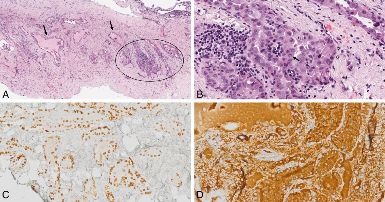
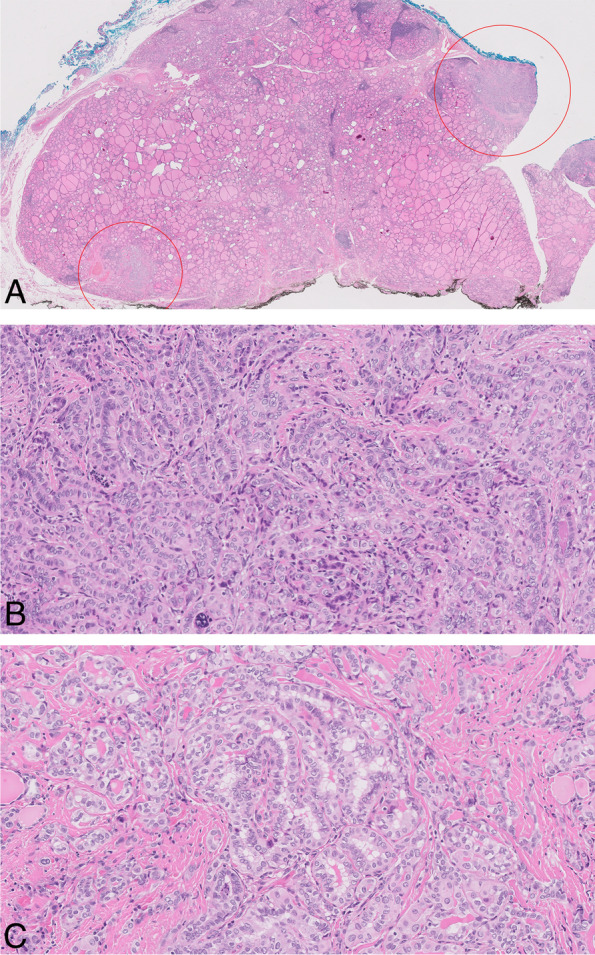
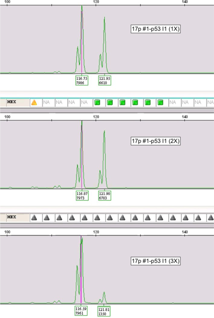
Case presentation: A 42-year-old female developed MSO and synchronous multifocal subcentimeter papillary thyroid carcinoma (PTC). The patient underwent a salpingo-oophrectomy, thyroidectomy, and low-dose radioactive iodine ablation. Both the thyroid subcentimeter PTC and MSO were positive for BRAF V600E mutation, and microRNA expression profiles were similar across all tumor deposits. However, only the malignant component demonstrated extensive loss of heterozygosity (LOH) involving multiple tumor suppressor gene (TSG) chromosomal loci.

Conclusions: We present the first reported case of MSO with synchronous multifocal subcentimeter PTC in the thyroid containing concordant BRAF V600E mutations and resulting with discordant LOH findings. This data suggests that loss of expression in tumor suppressor gene(s) may be an important contributor to phenotypic expression of malignancy.

Keywords: Loss of heterozygosity; Malignant struma ovarii; Papillary thyroid cancer; microRNA.


Conflict of interest statement

G.S.: No conflicts to disclose. J.M.: No conflicts to disclose. D.K.: No conflicts to disclose. J.F.: No conflicts to disclose. G.M.: No conflicts to disclose. M.M.: No conflicts to disclose. E.D.: No conflicts to disclose. A.M.: No conflicts to disclose. D.A.: No conflicts to disclose. M.D.: No conflicts to disclose. C.S.: No conflicts to disclose. A.B.: No conflicts to disclose. M.L.U: Dr. Mark Urken is the Medical Advisor of the THANC Foundation. M.B.W.: No conflicts to disclose. S.F.: No conflicts to disclose.

Figures

Fig. 1
Fig. 1
Malignant Struma Ovarii. A Low power view (0.4x) of H&E section from the left ovary shows thyroid follicles of various sizes and shapes embedded in an inflamed fibrotic stroma. Note the malignant aggregate of thyroid tissue (circled) and adjacent poorly formed thyroid follicles (arrow). B High power image (20x) of the malignant component with nuclear pleomorphism, nuclear overlapping, irregular nuclear contour, and nuclear pseudoinclusions (arrow). These cells are positive for TTF-1 (C) and thyroglobulin (D). Magnification in panel C and D is demonstrated by a scale bar
Fig. 2
Fig. 2
Primary Thyroid Carcinoma. A Low power view (0.5x) of H&E section from left thyroid lobe shows two foci of papillary thyroid carcinoma (PTC) present in a background of Hashimoto’s thyroiditis. Focus #1 is indicated by a rectangle and focus #2 is indicated by a circle. B High power view (20x) of PTC focus #1. C High power view (20x) of PTC focus #2. The neoplastic cells demonstrate characteristic PTC nuclear features (nuclear enlargement, elongation, overlapping, chromatin clearing, irregular nuclear contour, nuclear grooves and nuclear pseudoinclusions). Focus #1 also shows tall cell features with tall cell component estimated 10–20%
Fig. 3
Fig. 3
Example of LOH in MSO. TP53 intron 1 pentanucleotide microsatellite. Top- non-papillary thyroid cancer struma ovarii target. Middle- Thyroid gland microcarcinoma. Bottom- Papillary thyroid carcinoma in struma ovarii showing major loss of the larger polymorphic allele

References

    1. Siegel MR, Wolsky RJ, Alvarez EA, Mengesha BM. Struma ovarii with atypical features and synchronous primary thyroid cancer: a case report and review of the literature. Arch Gynecol Obstet. 2019;300(6):1693–1707. doi: 10.1007/s00404-019-05329-z. - DOI - PubMed
    1. Krishnamurthy A, Ramshankar V, Vaidyalingam V, Majhi U. Synchronous papillary carcinoma thyroid with malignant struma ovarii: A management dilemma. Indian J Nucl Med. 2013;28(4):243–245. doi: 10.4103/0972-3919.121975. - DOI - PMC - PubMed
    1. Gomes-Lima CJ, Nikiforov YE, Lee W, Burman KD. Synchronous independent papillary thyroid carcinomas in struma ovarii and the thyroid gland with different RAS mutations. J Endocr Soc. 2018;2(8):944–948. doi: 10.1210/js.2018-00132. - DOI - PMC - PubMed
    1. Goffredo P, Sawka AM, Pura J, Adam MA, Roman SA, Sosa JA. Malignant struma ovarii: a population-level analysis of a large series of 68 patients. Thyroid. 2015;25(2):211–215. doi: 10.1089/thy.2014.0328. - DOI - PubMed
    1. Lupo MA, Walts AE, Sistrunk JW, Giordano TJ, Sadow PM, Massoll N, Campbell R, Jackson SA, Toney N, Narick CM, et al. Multiplatform molecular test performance in indeterminate thyroid nodules. Diagn Cytopathol. 2020;48(12):1254–1264. doi: 10.1002/dc.24564. - DOI - PMC - PubMed

Publication types