. 2023 Aug 3;15(4):mjad025.
doi: 10.1093/jmcb/mjad025.

CSF2 upregulates CXCL3 expression in adipocytes to promote metastasis of breast cancer via the FAK signaling pathway

Affiliations

CSF2 upregulates CXCL3 expression in adipocytes to promote metastasis of breast cancer via the FAK signaling pathway

Xi He et al. J Mol Cell Biol. .

Abstract

Recent studies have demonstrated that cancer-associated adipocytes (CAAs) in the tumor microenvironment are involved in the malignant progression of breast cancer. However, the underlying mechanism of CAA formation and its effects on the development of breast cancer are still unknown. Here, we show that CSF2 is highly expressed in both CAAs and breast cancer cells. CSF2 promotes inflammatory phenotypic changes of adipocytes through the Stat3 signaling pathway, leading to the secretion of multiple cytokines and proteases, particularly C-X-C motif chemokine ligand 3 (CXCL3). Adipocyte-derived CXCL3 binds to its specific receptor CXCR2 on breast cancer cells and activates the FAK pathway, enhancing the mesenchymal phenotype, migration, and invasion of breast cancer cells. In addition, a combination treatment targeting CSF2 and CXCR2 shows a synergistic inhibitory effect on adipocyte-induced lung metastasis of mouse 4T1 cells in vivo. These findings elucidate a novel mechanism of breast cancer metastasis and provide a potential therapeutic strategy for breast cancer metastasis.

Keywords: CSF2; CXCL3; FAK; breast cancer metastasis; cancer-associated adipocytes.


Figures

Figure 1
Figure 1
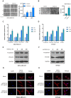
CSF2 is highly expressed in adipocytes and tumor cells after co-culture. (A and B) The expression (A) and secretion (B) of CSF2 in preadipocytes co-cultured with MDA-MB-231 cells for 0, 12, 24, and 48 h were measured by q-PCR and ELISA, respectively. (C and D) The expression (C) and secretion (D) of CSF2 in adipocytes co-cultured with MDA-MB-231 cells for 0, 12, 24, and 48 h. (E and F) Adipocytes were co-cultured with different breast cancer cells or normal mammary epithelial cells (MCF10A), and the expression (E) and secretion (F) of CSF2 were measured. (G and H) The expression (G) and secretion (H) of CSF2 in MDA-MB-231 and BT549 cells co-cultured with adipocytes. The relevant MDA-MB-231 or BT549 cells cultured alone served as the control. (I) The mRNA expression levels of CSF2 in normal mammary and breast cancer tissues were measured by q-PCR. ***P < 0.001, **P < 0.01, *P < 0.05, and NS (P > 0.05); n = 3.
Figure 2
Figure 2
CSF2 activates Stat3 to upregulate CXCL3 expression in adipocytes co-cultured with breast cancer cells. (A) The mRNA expression levels of CSF2Rα and CSF2Rβ in adipocytes co-cultured with MDA-MB-231 cells were measured by q-PCR. (B and C) Cytokine expression (B) and secretion (C) in adipocytes treated with rhCSF2 (20 ng/ml) were detected by q-PCR and ELISA, respectively. (D) The mRNA expression levels of IL-6, CSF2, and CXCLs in normal and paracancerous adipose tissues. (E) Adipocytes were stimulated with rhCSF2 for 12 h and then treated with stattic (10 μM). The mRNA expression levels of CXCLs, IL-6, and IL-1β in adipocytes were detected. (F) Adipocytes were treated with rhCSF2 and stattic for 15 min, and Stat3 phosphorylation was analyzed by western blotting. (G and H) Adipocytes were co-cultured with MDA-MB-231 cells in the presence or absence of CSF2-neutralizing antibody (2 μg/ml). The expression of CXCLs (G) and Stat3 phosphorylation (H) in adipocytes were analyzed by q-PCR and western blotting, respectively. ###P < 0.001, ##P < 0.01, and #P < 0.05 compared to the control group; ***P < 0.001, **P < 0.01, and *P < 0.05 compared to experimental group; n = 3.
Figure 3
Figure 3
rhCXCL3 promotes the mesenchymal phenotype, migration, and invasion of breast cancer cells. (A and B) MDA-MB-231 cells were treated with rhCXCL3 (20 ng/ml). Cell migration and invasion were measured by the wound-healing assay (A) and the Transwell invasion assay (B), respectively. (C and D) MDA-MB-231 (C) and BT549 (D) cells were treated with rhCXCL3 for 12 and 24 h, and the mRNA expression levels of EMT markers were detected by q-PCR. (E and F) MDA-MB-231 (E) and BT549 (F) cells were treated with rhCXCL3 for 24 and 48 h, and the protein expression levels of N-cadherin and Vimentin were detected by western blotting. (G and H) MDA-MB-231 (G) and BT549 (H) cells were treated with 20 and 40 ng/ml rhCXCL3 for 24 h, and the cytoskeleton was stained with TRITC-phalloidin. Scale bar, 20 μm. ***P < 0.001, **P < 0.01, and *P < 0.05; n = 3.
Figure 4
Figure 4
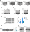
CXCL3/CXCR2 signaling activates FAK to enhance the mesenchymal phenotype of breast cancer cells. (A) MDA-MB-231 and BT549 cells were treated with rhCXCL3 at doses of 20, 40, 80, and 160 ng/ml for 30 min. FAK phosphorylation was analyzed by western blotting. (B) Breast cancer cells were treated with 20 ng/ml rhCXCL3 for 5, 15, 30, and 60 min, and FAK phosphorylation was analyzed. (C) The protein expression levels of CXCR1 and CXCR2 in MCF10A, MBA-MB-231, BT549, MCF-7, and T47D cells. (D) MDA-MB-231 and BT549 cells were treated with rhCXCL3 in the presence or absence of SB225002 (0.1 μM) or reparixin (0.1 μM), and FAK phosphorylation was analyzed. (E and F) MDA-MB-231 cells were treated with rhCXCL3 and/or PF573228 (5 μM). Cell migration (E) and invasion (F) were measured. (G) MDA-MB-231 and BT549 cells were treated with rhCXCL3 and/or PF573228 for 30 min. FAK phosphorylation was analyzed. ###P < 0.001, ##P < 0.01, and #P < 0.05 compared to the control group; ***P < 0.001, **P < 0.01, and *P < 0.05 compared to the experimental group; n = 3.
Figure 5
Figure 5
Downregulation of FAK inhibits CXCL3-induced migration and invasion of breast cancer cells. (A) FAK protein levels in MDA-MB-231 cells transfected with siRNAs were measured by western blotting. (B and C) MDA-MB-231 cells transfected with siRNAs were treated with rhCXCL3. Cell migration (B) and invasion (C) were measured. (D and E) MDA-MB-231 (D) and BT549 (E) cells were treated with rhCXCL3 and/or PF573228, and the mRNA expression levels of EMT markers were detected by q-PCR. (F) MDA-MB-231 and BT549 cells were treated with rhCXCL3 and/or PF573228, and the protein expression levels of N-cadherin and Vimentin were detected by western blotting. ###P < 0.001, ##P < 0.01, and #P < 0.05 compared to the control group; ***P < 0.001, **P < 0.01, and *P < 0.05 compared to the experimental group; n = 3.
Figure 6
Figure 6
Both CXCL3-neutralizing antibody and PF573228 inhibit CAA-CM-promoted migration, invasion, and FAK phosphorylation of breast cancer cells. (A and B) MDA-MB-231 cells treated with Adi-CM, CAA-CM, or control medium were incubated with CXCL3-neutralizing antibody (2 μg/ml) or its IgG control. Cell migration (A) and invasion (B) were measured. (C) MDA-MB-231 and BT549 cells were treated with Adi-CM, CAA-CM, or control medium for 30 min. FAK phosphorylation was measured by western blotting. (D) MDA-MB-231 and BT549 cells treated with Adi-CM or CAA-CM were cultured with or without CXCL3-neutralizing antibody. FAK phosphorylation was analyzed. (E and F) MDA-MB-231 cells treated with Adi-CM or CAA-CM were incubated with or without PF573228, and cell migration (E) and invasion (F) were measured. (G and H) MDA-MB-231 (G) and BT549 (H) cells were treated with Adi-CM or CAA-CM for 30 min, with or without PF573228. FAK phosphorylation was analyzed. ###P < 0.001, ##P < 0.01, and #P < 0.05 compared to the control group; ***P < 0.001, **P < 0.01, and *P < 0.05 compared to the experimental group; n = 3.
Figure 7
Figure 7
CSF2 upregulates CXCL3 expression in adipocytes to promote the migration and invasion of breast cancer cells via FAK signaling. (A and B) MDA-MB-231 (A) and BT549 (B) cells were cultured with CAA-CM in the presence or absence of CSF2-neutralizing antibody for 30 min. FAK phosphorylation was analyzed by western blotting. (C and D) MDA-MB-231 cells were incubated with CSF2-CM followed by treatment with SB225002 or CXCL3-neutralizing antibody. The migration (C) and invasion (D) were measured. (E and F) MDA-MB-231 (E) and BT549 (F) cells were treated with CSF2-CM followed by incubation with SB225002 or CXCL3-neutralizing antibody for 30 min. FAK phosphorylation was analyzed. (G) H&E staining and IHC images showing the expression of CSF2, CXCL3, and p-FAK in normal mammary and breast cancer tissues. Scale bar, 200 μm. (H and I) IHC images were analyzed with Image-ProPlus, and the relative integral optic density (IOD) of each group was calculated to indicate the protein expression level (n = 10). ###P < 0.001, ##P < 0.01, and #P < 0.05 compared to the control group; ***P < 0.001, **P < 0.01, and *P < 0.05 compared to the experimental group; n = 3.
Figure 8
Figure 8
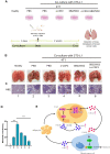
CSF2-neutralizing antibody and CXCR2 inhibitor synergistically inhibit adipocyte-induced lung metastasis of breast cancer cells in mice. (A) Schematic diagram presenting the development of lung metastasis of breast cancer cells in a mouse model, by Figdraw (www.figdraw.com accessed on April 6, 2023). (B and C) Representative photos (B) and H&E staining images (C) of the lungs from healthy and 4T1 cell-bearing mice. Black arrows indicate typical metastatic lung nodules. (D) Quantification of metastatic nodules in mouse lungs of each group. (E) Schematic diagram showing the role of the CSF2/CXCL3/FAK signaling axis in promoting breast cancer metastasis, by Figdraw (www.figdraw.com accessed on July 29, 2022). ***P < 0.001, **P < 0.01, and *P < 0.05; n = 6.

Similar articles

Cited by

References

    1. Al-Alwan L.A., Chang Y., Mogas A.et al. (2013). Differential roles of CXCL2 and CXCL3 and their receptors in regulating normal and asthmatic airway smooth muscle cell migration. J. Immunol. 191, 2731–2741. - PMC - PubMed
    1. Al-Khalaf H.H., Al-Harbi B., Al-Sayed A.et al. (2019). Interleukin-8 activates breast cancer-associated adipocytes and promotes their angiogenesis- and tumorigenesis-promoting effects. Mol. Cell. Biol. 39, e00332-18. - PMC - PubMed
    1. Anderson N.M., Simon M.C. (2020). The tumor microenvironment. Curr. Biol. 30, R921–R925. - PMC - PubMed
    1. Boyer S., Lee H.J., Steele N.et al. (2022). Multiomic characterization of pancreatic cancer-associated macrophage polarization reveals deregulated metabolic programs driven by the GM-CSF–PI3K pathway. eLife 11, e73796. - PMC - PubMed
    1. Chuang H.H., Zhen Y.Y., Tsai Y.C.et al. (2022). FAK in cancer: from mechanisms to therapeutic strategies. Int. J. Mol. Sci. 23, 1726. - PMC - PubMed

Publication types