Review
. 2021 Jan 19;3(2):169-188.
doi: 10.1007/s42995-020-00082-8. eCollection 2021 May.

One cell at a time: droplet-based microbial cultivation, screening and sequencing

Affiliations
Review

One cell at a time: droplet-based microbial cultivation, screening and sequencing

Beiyu Hu et al. Mar Life Sci Technol. .

Abstract

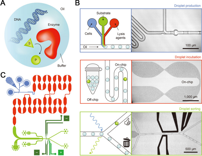
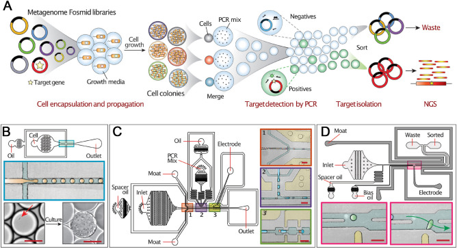
Microbes thrive and, in turn, influence the earth's environment, but most are poorly understood because of our limited capacity to reveal their natural diversity and function. Developing novel tools and effective strategies are critical to ease this dilemma and will help to understand their roles in ecology and human health. Recently, droplet microfluidics is emerging as a promising technology for microbial studies with value in microbial cultivating, screening, and sequencing. This review aims to provide an overview of droplet microfluidics techniques for microbial research. First, some critical points or steps in the microfluidic system are introduced, such as droplet stabilization, manipulation, and detection. We then highlight the recent progress of droplet-based methods for microbiological applications, from high-throughput single-cell cultivation, screening to the targeted or whole-genome sequencing of single cells.

Keywords: Antibiotic resistance; Droplet microfluidics; Microbial cultivation; Single-cell screening; Single-cell sequencing; Uncultured microorganisms.


Conflict of interest statement

Conflict of interestThe authors declare there is no conflict of interest between them.

Figures

Fig. 1
Fig. 1
Schematic illustration of the applications of microfluidics in microbiological research at six aspects in this review
Fig. 2
Fig. 2
Schematic illustrates surfactant molecules to stabilize aqueous droplets. The amphiphilic molecules were added to the oil phase to maintain droplets as a stable micro-reactor
Fig. 3
Fig. 3
Droplet-based devices for the cultivation of uncultured microbes. a Schematic illustrates the principle for parallel microbial cultivation and Fluorescence In Situ Hybridization (FISH) identification (Reprinted with permission from Liu et al. . Lab Chip 9: 2153–2162. Copyright (2009) Royal Society of Chemistry). b Schematic of mass activated droplet sorting (MADS) system and the zoom-in microscopic images of the devices showing droplets operation (Reprinted with permission from Holland-Moritz et al. . Angew Chem Int Edit 59: 4470–4477. Copyright (2020) John Wiley & Sons). c Schematic of Microfluidic streak plate (MSP) platform (Reprinted with permission from Chen et al. . J Hazard Mater 366: 512–519. Copyright (2019) Elsevier). d Schematic of semi-automated droplet picker (Fig. 3D, and 3F reprinted with permission from Hu et al. . Lab Chip 20: 363–372. Copyright (2020) Royal Society of Chemistry) e Left, heatmap of amplicon sequencing for comparison of the original community, pooled cells from agar plates, and pooled cells from MSP. Right, parallel comparison of isolated species and number of isolates for agar plate and MSP cultivations (Reprinted with permission from Jiang et al. . Appl Environ Microb 82: 2210–2218. Copyright (2016) American Society for Microbiology). f MSP isolated microorganisms from deep-sea sediments samples
Fig. 4
Fig. 4
Microfluidic droplet-based co-cultivation and its application. a The workflow of droplet co-cultivation. b The cultivation results of microbial droplets co-cultivation with W- and Y- Strains (Adapted from Park et al. . PLOS ONE 6: e17019. followed the terms of the Creative Commons Attribution License). c Schematic of using a co-cultivation strategy for bear oral microbiome screening. d The microfluidic droplets with the encapsulation of Staphylococcus aureus (S. aureus) and oral microbes after cultivation (scale bar 50 μm) (Fig. 4C and 4D reprinted from Terekhov et al. . Proc Natl Acad Sci USA 115:9551–9556. followed Creative Commons Attribution-NonCommercial-NoDerivatives License 4.0 (CC BY-NC-ND))
Fig. 5
Fig. 5
Microfluidic droplet-based platform for rapid antibiotic susceptibility tests (ASTs). a Schematic of a droplet array-based gradient AST system. Methicillin-susceptible Staphylococcus aureus (MSSA) was tested with serial gradients of antibiotics include LVF (levofloxicin), OXA (oxicillin), VCM (vancomycin), and AMP (ampicillin) (Reprinted with permission from Boedicker et al. . Lab Chip 8: 1265–1272. Copyright (2008) Royal Society of Chemistry). b An integrated microfluidic chip illustrates the principle and workflow for droplet-based high-resolution dose–response profiling (Reprinted with permission from Kaushik et al. . Biosens Bioelectron 97: 260–266. Copyright (2017) Elsevier). c Stationary nanoliter droplets array fixed with microwell illustrates the workflow for AST (Reprinted with permission from Shemesh et al. . Proc Natl Acad Sci USA 111: 11293–11298. Copyright (2014) National Academy of Sciences, USA.). d Schematic illustrates a chip-free platform of multichannel dynamic interfacial printing (MC-DIP) for AST (Reprinted with permission from Liao et al. . ACS Appl Mater Interfaces 9: 43545−43552. Copyright (2017) American Chemical Society)
Fig. 6
Fig. 6
Schematic representation of the integrated microfluidic chip for the directed evolution of aldolases. a Schematic showing the use of aqueous droplets as bioreactors for enzyme directed evolution. b Schematic of the integrated chip, the droplets generation module is shown in blue, the droplets incubation module is shown in red, and the droplet sorting module is shown in green. “-” indicates the negative electrode and “ + ” is the positive electrode. c Zoom-in schematics and images of the functional modules of the fully automated FADS system (Reprinted with permission from Obexer et al. . Nat Chem 9: 50–56. Copyright (2017) Springer Nature)
Fig. 7
Fig. 7
Schematic principle of Emulsion, Paired Isolation, and Concatenation PCR (epicPCR) for the association of functional genes with phylogenetic status at the single-cell level. A Microbial cells in acrylamide suspension are mixed into emulsion oil. The emulsion droplets are polymerized into polyacrylamide beads containing single cells. The emulsion is broken, and the cells in the polyacrylamide beads are treated enzymatically to expose the genomic DNA by destroying cell walls, membranes, and protein components. B Polyacrylamide-trapped, permeabilized microbial cells are encapsulated into an emulsion with fusion PCR reagents. C Fusion PCR first amplifies a target gene with an overhang of 16S rRNA gene homology. With a limiting concentration of overhang primer, the target gene amplicon will anneal and extend into the 16S rRNA gene, forming a fusion product that continues to amplify from a reverse 16S rRNA gene primer. D The fused amplicons only form in the emulsion compartments where a given microbial cell has the target functional gene. e After breaking the emulsion, the fused amplicons are prepared for next-generation sequencing. The resulting DNA sequences are concatemers of the functional gene and the 16S rRNA gene of the same cell (Reprinted from Spencer et al. . ISME J 10: 427–436. followed Creative Commons Attribution 4.0 International License)
Fig. 8
Fig. 8
Microfluidic Automated Plasmid Library Enrichment (MAPLE) workflow and associated microfluidic devices. a Overview of MAPLE workflow. b The droplet maker device used for encapsulating single cells. Top, device schematic; middle, enlarged view of the cross junction where droplets are generated; bottom, an image of a single bacterium (red arrow) in the droplet before culture (left) and resulting colony after incubation (right). Scale bar = 20 µm. c Droplet merging device. Left, device schematic. Right, inserts showing magnified views of the three numbered regions. Insert 1, reinjection of close-packed droplets containing cell colonies spaced out by oil flow. Insert 2, pairing of colony droplets (orange) with PCR reagent droplets (blue) at a ∼1:1 ratio. Insert 3, the entrance of droplet pairs into merging zone for electro-coalescence. Scale bar = 100 µm. d Droplet sorter device used for sorting fluorescently positive droplets. Top, device schematic. Bottom, inserts showing the junction where droplets are sorted. If a droplet passing the laser (light spot) has a fluorescence signal exceeding the threshold, then the electrode (yellow bar) activates, applying a dielectrophoretic force to pull it into the ‘sorted’ channel. Scale bar = 100 µm (Reprinted with permission from Xu et al. . Nucleic Acids Res 48: e48. Copyright (2020) Oxford University Press)
Fig. 9
Fig. 9
Single-cell whole-genome amplification and sequencing using fluorescence-activated cell sorting (FACS) coupled with interfacial nanoinjection (INJ) technique
Fig. 10
Fig. 10
Microfluidic and biochemical workflow to generate a single-cell genomic sequencing (SiC-seq) library. a Generating barcode droplets by encapsulating random DNA oligos at limiting dilution and amplification by in-droplet PCR (SYBR-stained for visualization). b Cells are encapsulated at limiting dilution with molten agarose to generate agarose microgels, each contains a single cell. c The single-cell genomes are purified through a series of bulk enzymatic and detergent lysis steps. d Microgels are re-encapsulated in droplets containing tagmentation reagents. e The droplets containing tagmented genomes are merged sequentially with PCR reagents and barcode droplets at a 1:1 ratio, followed by PCR to splice barcodes to genomic fragments (Reprinted with permission from Lan et al. . Nat Biotechnol 35: 640–646. Copyright (2017) Springer Nature)

Similar articles

Cited by

References

    1. Abate AR, Weitz DA. Faster multiple emulsification with drop splitting. Lab Chip. 2011;11:1911–1915. doi: 10.1039/c0lc00706d. - DOI - PubMed
    1. Abate AR, Hung T, Mary P, Agresti JJ, Weitz DA. High-throughput injection with microfluidics using picoinjectors. Proc Natl Acad Sci USA. 2010;107:19163–19166. doi: 10.1073/pnas.1006888107. - DOI - PMC - PubMed
    1. Agresti JJ, Antipov E, Abate AR, Ahn K, Rowat AC, Baret JC, Marquez M, Klibanov AM, Griffiths AD, Weitz DA. Ultrahigh-throughput screening in drop-based microfluidics for directed evolution. Proc Natl Acad Sci U S A. 2010;107:4004–4009. doi: 10.1073/pnas.0910781107. - DOI - PMC - PubMed
    1. Autour A, Ryckelynck M. Ultrahigh-throughput improvement and discovery of enzymes using droplet-based microfluidic screening. Micromachines. 2017;8:128. doi: 10.3390/mi8040128. - DOI
    1. Bai Y, He X, Liu D, Patil SN, Bratton D, Huebner A, Hollfelder F, Abell C, Huck WTS. A double droplet trap system for studying mass transport across a droplet-droplet interface. Lab Chip. 2010;10:1281–1285. doi: 10.1039/b925133b. - DOI - PubMed

LinkOut - more resources