. 2023 Apr;12(4):e12323.
doi: 10.1002/jev2.12323.

Convection and extracellular matrix binding control interstitial transport of extracellular vesicles

Affiliations

Convection and extracellular matrix binding control interstitial transport of extracellular vesicles

Peter A Sariano et al. J Extracell Vesicles. 2023 Apr.

Abstract

Extracellular vesicles (EVs) influence a host of normal and pathophysiological processes in vivo. Compared to soluble mediators, EVs can traffic a wide range of proteins on their surface including extracellular matrix (ECM) binding proteins, and their large size (∼30-150 nm) limits diffusion. We isolated EVs from the MCF10 series-a model human cell line of breast cancer progression-and demonstrated increasing presence of laminin-binding integrins α3β1 and α6β1 on the EVs as the malignant potential of the MCF10 cells increased. Transport of the EVs within a microfluidic device under controlled physiological interstitial flow (0.15-0.75 μm/s) demonstrated that convection was the dominant mechanism of transport. Binding of the EVs to the ECM enhanced the spatial concentration and gradient, which was mitigated by blocking integrins α3β1 and α6β1. Our studies demonstrate that convection and ECM binding are the dominant mechanisms controlling EV interstitial transport and should be leveraged in nanotherapeutic design.

Keywords: diffusion; exosome; gradient; integrin binding; spatial concentration.


Conflict of interest statement

There are no conflicts of interest to declare.

Figures
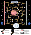
FIGURE 1
FIGURE 1
Extracellular vesicle transport and binding in the interstitium. A heterogeneous population of extracellular vesicles (EV) are secreted by all cell types (small black arrows), including tumor cells, and can directly enter the blood or lymphatic circulations or be transported by convection through the extracellular matrix by interstitial flow. Interstitial flow is generated as water leaks from the blood capillaries (blue arrows) into the interstitium. A small portion of the leak is reabsorbed near the venular end of the blood capillary (green arrows), and the remaining by the lymphatic capillaries (green arrows) generating a net interstitial flow from the blood capillaries to the lymphatic capillaries. As EVs are transported through the interstitium, they have the potential to bind (and release) to the ECM, thus impacting the population of EVs that are absorbed by the blood and lymphatic capillaries (black dashed arrows), and also establishing the spatial distribution in the interstitium of bound and free EVs. (constructed, in part, in BioRender).
FIGURE 2
FIGURE 2
Isolation and characterization of EVs. (a) Nanoparticle tracking analysis (NTA) revealed expected EV size distribution and adequate concentration for in vitro studies as well as (b) increasing secretion with malignancy when normalized by cell number. (c) Transmission electron microscopy (TEM) demonstrated sufficient EV concentration and a characteristic cup‐shape morphology. (d) Loading the same concentration of EVs revealed increasing expression of integrin α3, α6, and β1 with parent cell malignancy by ExoView analysis. Percentage integrin positive of all tetraspanin positive EVs is shown in parentheses. *p = 0.0319; ***p < 0.0001; One‐way ANOVA, Tukey post‐hoc.
FIGURE 3
FIGURE 3
EV matrix binding is impacted by parent cell malignancy. (a) FRAP recovery fitted curves were consistently slower and lower for malignant MCF10CA1 EVs across all treatment conditions, followed by MCF10DCIS and MCF10A EVs. Recoveries generally appeared to display asymptotic behaviour by t = 300 sec. Error bars = 95% CI; n = 9 replicates per curve; n* = 8 replicates for MCF10CA1 curve. (b) Fitted Kd parameter was consistent with behaviour in FRAP curves. MCF10CA1 EVs exhibited lowest Kd values, indicating the highest levels of EV binding to the matrix. (c) Molecular diffusivity was broadly independent of cell line or treatment condition. (d) The bound fraction (Ceq) was assessed for each integrin binding state. Highest EV binding fractions were observed with MCF10CA1 EVs in all conditions compared to EVs from MCF10A and MCF10DCIS. One‐way Anova, Tukey post‐hoc. *p < 0.05, **p < 0.01, ***p < 0.001; One‐way ANOVA, Tukey post‐hoc.
FIGURE 4
FIGURE 4
Finite element models reveal interstitial accumulation of high affinity binding EVs. (a) A range of physiologically relevant interstitial flow velocities (v, 0.5 – 1 μm/s) were established by varying hydrostatic pressure boundary conditions at port inlets and outlets. (b) Bound EV concentration profiles after T = 30 min with a 0.5 μm/s flow velocity reveal accumulation of bound EVs in compartments 2a and 2b (Fig. S1) as binding affinity increases. Concentration line profiles (white arrows) demonstrated (c) flatter spatial EV concentration profiles with increasing flow velocity as well as higher levels of bound EVs with high affinity binding parameters. (d) Comparing diffusivity of a small molecule dextran with EVs demonstrated differences in spatial profiles, but no significant difference when D < 10‐12 m2/s which signifies EV transport is convection‐limited.
FIGURE 5
FIGURE 5
Interstitial EV accumulation with increased malignancy and high affinity integrin binding. (a‐d) Representative images demonstrated a range of EV binding profiles. Most consistent high binding was observed with MCF10CA1 EVs treated with Mn2+ (b, right column, white arrows) for high affinity binding. Differences in background fluorescent intensity were evident but were normalized for quantitative analysis.
FIGURE 6
FIGURE 6
EV convective transport is affected by parent cell malignancy and integrin activation state. (a) Line profiles were used to assess EV concentration after T = 30 min of flow. (b‐h) Comparisons of concentration profiles by integrin activation state showed highest differences for high affinity MCF10CA1 and MCF10DCIS EV binding. Blocking conditions reduced the formation of spatial gradients. Physiologic binding conditions showed minimal differences between EV conditions. Error bars = 95% CI. n = 2‐4 devices per curve (average devices per condition = 3); n = 21‐80 ports per curve (average ports per curve = 52).

References

    1. Aisenbrey, E. A. , & Murphy, W. L. (2020). Synthetic alternatives to Matrigel. Nature Reviews Materials, 5, 539–551. - PMC - PubMed
    1. Almaqwashi, A. A. , Paramanathan, T. , Lincoln, P. , Rouzina, I. , Westerlund, F. , & Williams, M. C. (2014). Strong DNA deformation required for extremely slow DNA threading intercalation by a binuclear ruthenium complex. Nucleic Acids Research, 42, 11634–11641. - PMC - PubMed
    1. Aumailley, M. (2013). The laminin family. Cell Adhesion & Migration, 7, 48–55. - PMC - PubMed
    1. Becker, A. , Thakur, B. K. , Weiss, J. M. , Kim, H. S. , Peinado, H. , & Lyden, D. (2016). Extracellular vesicles in cancer: Cell‐to‐cell mediators of metastasis. Cancer Cell, 30, 836–848. Preprint at 10.1016/j.ccell.2016.10.009 - DOI - PMC - PubMed
    1. Behbod, F. , Kittrell, F. S. , Lamarca, H. , Edwards, D. , Kerbawy, S. , Heestand, J. C. , Young, E. , Mukhopadhyay, P. , Yeh, H.‐W. , Allred, D. C. , Hu, M. , Polyak, K. , Rosen, J. M. , & Medina, D. (2009). An intraductal human‐in‐mouse transplantation model mimics the subtypes of ductal carcinoma in situ. Breast Cancer Res 11, R66. 10.1186/bcr2358 - DOI - PMC - PubMed

Publication types