

Integrative roles of human amygdala subdivisions: Insight from direct intracerebral stimulations via stereotactic EEG
- PMID: 37073861
- PMCID: PMC10203795
- DOI: 10.1002/hbm.26300
Integrative roles of human amygdala subdivisions: Insight from direct intracerebral stimulations via stereotactic EEG
Abstract
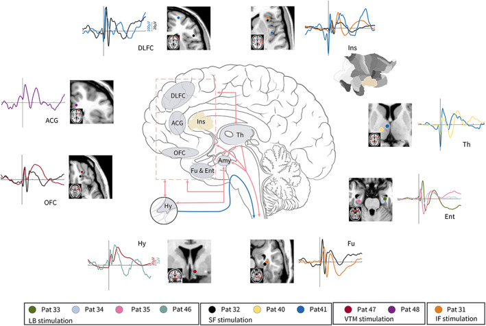
Substantial studies of human amygdala function have revealed its importance in processing emotional experience, autonomic regulation, and sensory information; however, the neural substrates and circuitry subserving functions have not been directly mapped at the level of the subnuclei in humans. We provide a useful overview of amygdala functional characterization by using direct electrical stimulation to various amygdala regions in 48 patients with drug-resistant epilepsy undergoing stereoelectroencephalography recordings. This stimulation extends beyond the anticipated emotional, neurovegetative, olfactory, and somatosensory responses to include visual, auditory, and vestibular sensations, which may be explained by the functional connectivity with cortical and subcortical regions due to evoked amygdala-cortical potentials. Among the physiological symptom categories for each subnucleus, the most frequently evoked neurovegetative symptoms were distributed in almost every subnucleus. Laterobasal subnuclei are mainly associated with emotional responses, somatosensory responses, and vestibular sensations. Superficial subnuclei are mainly associated with emotional responses and olfactory and visual hallucinations. Our findings contribute to a better understanding of the functional architecture of the human amygdala at the subnuclei level and as a mechanistic basis for the clinical practice of amygdala stimulation in treating patients with neuropsychiatric disorders.
Keywords: SEEG; electrical stimulation; function; human amygdala; subnuclei.
© 2023 The Authors. Human Brain Mapping published by Wiley Periodicals LLC.
Conflict of interest statement
The authors declare no conflict of interest.
Figures


Similar articles
-
Odorants elicit evoked potentials in the human amygdala.Cereb Cortex. 2001 Jul;11(7):619-27. doi: 10.1093/cercor/11.7.619. Cereb Cortex. 2001. PMID: 11415964
-
Multimodal responses induced by cortical stimulation of the parietal lobe: a stereo-electroencephalography study.Brain. 2015 Sep;138(Pt 9):2596-607. doi: 10.1093/brain/awv187. Epub 2015 Jun 29. Brain. 2015. PMID: 26129769
-
Simultaneous stereo-EEG and high-density scalp EEG recordings to study the effects of intracerebral stimulation parameters.Brain Stimul. 2022 May-Jun;15(3):664-675. doi: 10.1016/j.brs.2022.04.007. Epub 2022 Apr 12. Brain Stimul. 2022. PMID: 35421585
-
Functional mapping of the human insula: Data from electrical stimulations.Rev Neurol (Paris). 2019 Mar;175(3):150-156. doi: 10.1016/j.neurol.2018.12.003. Epub 2019 Mar 1. Rev Neurol (Paris). 2019. PMID: 30827578 Review.
-
Central control of cardiac activity as assessed by intra-cerebral recordings and stimulations.Neurophysiol Clin. 2023 Apr;53(2):102849. doi: 10.1016/j.neucli.2023.102849. Epub 2023 Mar 1. Neurophysiol Clin. 2023. PMID: 36867969 Review.
Cited by
-
Amygdalar involvement in respiratory dysfunction.Front Physiol. 2024 Aug 28;15:1424889. doi: 10.3389/fphys.2024.1424889. eCollection 2024. Front Physiol. 2024. PMID: 39263625 Free PMC article. Review.
-
Brainwide mesoscale functional networks revealed by focal infrared neural stimulation of the amygdala.Natl Sci Rev. 2024 Dec 24;12(4):nwae473. doi: 10.1093/nsr/nwae473. eCollection 2025 Apr. Natl Sci Rev. 2024. PMID: 40170996 Free PMC article.
-
Transcriptome analysis identifies an ASD-Like phenotype in oligodendrocytes and microglia from C58/J amygdala that is dependent on sex and sociability.Behav Brain Funct. 2024 Jun 19;20(1):14. doi: 10.1186/s12993-024-00240-3. Behav Brain Funct. 2024. PMID: 38898502 Free PMC article.
-
Advances in epileptic network findings of hypothalamic hamartomas.J Cent Nerv Syst Dis. 2024 Mar 5;16:11795735241237627. doi: 10.1177/11795735241237627. eCollection 2024. J Cent Nerv Syst Dis. 2024. PMID: 38449707 Free PMC article. Review.
-
Electrical stimulation of the peripheral and central vestibular system.Curr Opin Neurol. 2024 Feb 1;37(1):40-51. doi: 10.1097/WCO.0000000000001228. Epub 2023 Oct 25. Curr Opin Neurol. 2024. PMID: 37889571 Free PMC article. Review.
References
-
- Armio, R. L. , Laurikainen, H. , Ilonen, T. , Walta, M. , Salokangas, R. K. R. , Koutsouleris, N. , Hietala, J. , & Tuominen, L. (2020). Amygdala subnucleus volumes in psychosis high‐risk state and first‐episode psychosis. Schizophrenia Research, 215, 284–292. 10.1016/j.schres.2019.10.014 - DOI - PubMed
-
- Avecillas‐Chasin, J. M. , Justo, M. , Levinson, S. , Koek, R. , Krahl, S. E. , Chen, J. W. Y. , Lee, S. J. , Langevin, J.‐P. , & Bari, A. (2020). Structural correlates of emotional response to electrical stimulation of the amygdala in subjects with PTSD. Brain Stimulation, 13(2), 424–426. 10.1016/j.brs.2019.12.004 - DOI - PMC - PubMed
Publication types
MeSH terms
LinkOut - more resources
Full Text Sources
