. 2023 May 1;129(5):1237-1248.
doi: 10.1152/jn.00016.2023. Epub 2023 Apr 19.

Persistent inflammation selectively activates opioid-sensitive phasic-firing neurons within the vlPAG

Affiliations

Persistent inflammation selectively activates opioid-sensitive phasic-firing neurons within the vlPAG

Kylie B McPherson et al. J Neurophysiol. .

Abstract

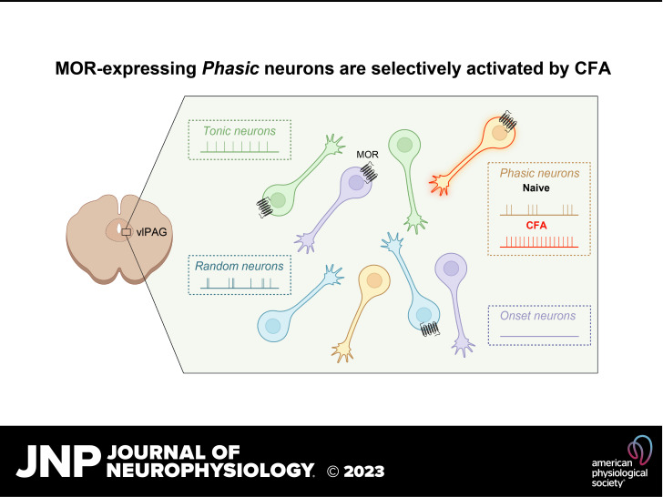
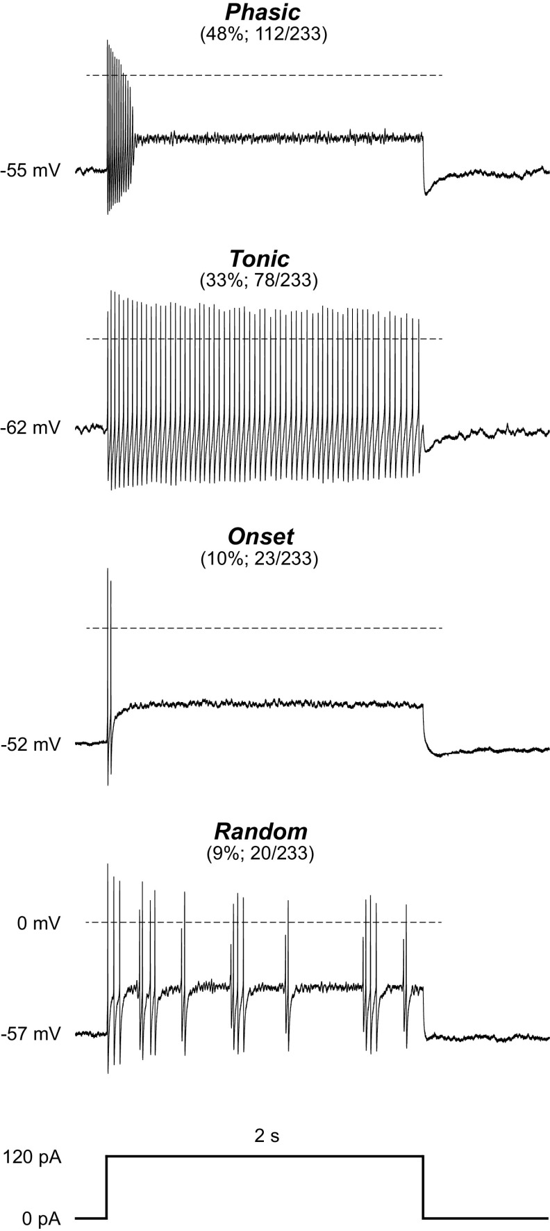
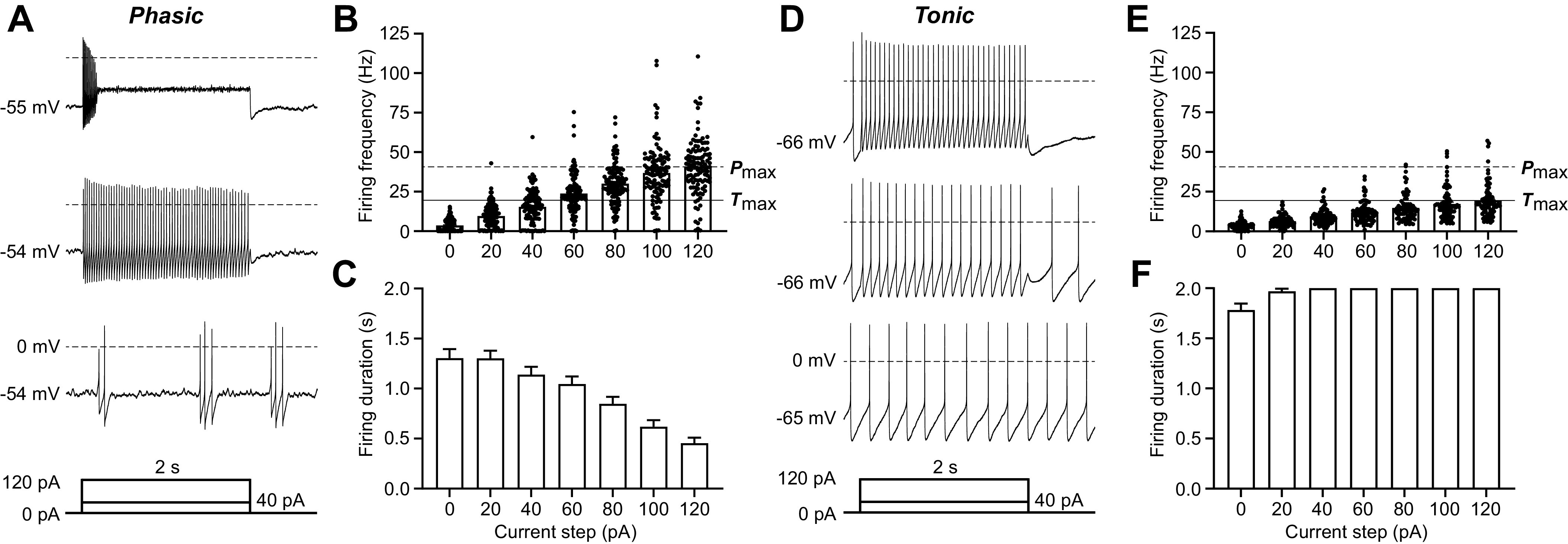
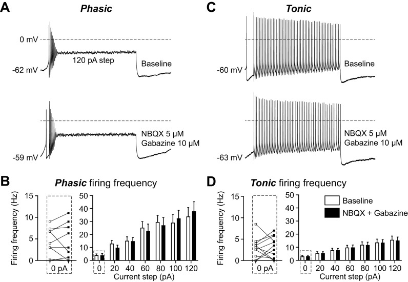
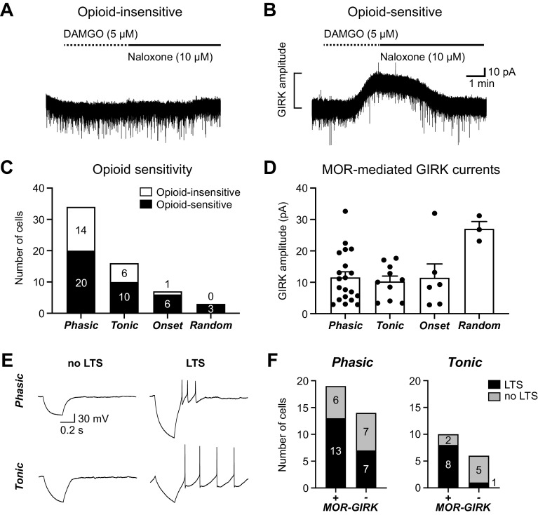
The ventrolateral periaqueductal gray (vlPAG) is a key brain area within the descending pain modulatory pathway and an important target for opioid-induced analgesia. The vlPAG contains heterogeneous neurons with respect to neurotransmitter content, receptor and channel expression, and in vivo response to noxious stimuli. This study characterizes intrinsic membrane properties of vlPAG neurons to identify neuron types that respond to inflammation and determine whether the pain-responsive neurons are inhibited by opioids. Surveying 382 neurons identified four neuron types with distinct intrinsic firing patterns: Phasic (48%), Tonic (33%), Onset (10%), and Random (9%). Mu-opioid receptor (MOR) expression was determined by the ability of a selective MOR agonist (DAMGO) to activate G protein-coupled inwardly rectifying potassium channel (GIRK) currents. Opioid-sensitive neurons were observed within each neuron type. Opioid sensitivity did not correlate with other intrinsic firing features, including low-threshold spiking that has been previously proposed to identify opioid-sensitive GABAergic neurons in the vlPAG of mice. Complete Freund's adjuvant (CFA)-induced acute inflammation (2 h) had no effect on vlPAG neuron firing patterns. However, persistent inflammation (5-7 days) selectively activated Phasic neurons through a significant reduction in their firing threshold. Opioid-sensitive neurons were strongly activated compared with the opioid-insensitive Phasic neurons. Overall, this study provides a framework to further identify neurons activated by persistent inflammation so that they may be targeted for future pain therapies.NEW & NOTEWORTHY Intrinsic firing properties define four distinct vlPAG neuron populations, and a subset of each population expresses MORs coupled to GIRK channels. Persistent, but not acute, inflammation selectively activates opioid-sensitive Phasic vlPAG neurons. Although the vlPAG is known to contribute to the descending inhibition of pain, the activation of a single physiologically defined neuron type in the presence of persistent inflammation represents a mechanism by which the vlPAG participates in descending facilitation of pain.

Keywords: descending pain modulation; electrophysiology; firing properties; inflammatory pain; vlPAG.


Conflict of interest statement

No conflicts of interest, financial or otherwise, are declared by the authors.

Figures

None
Graphical abstract
Figure 1.
Figure 1.
The ventrolateral periaqueductal gray (vlPAG) contains 4 neuron types defined by distinct firing patterns. Representative traces from vlPAG neurons from naive rats showing 4 distinct neuron types based on their response to a 2-s-long 120-pA depolarizing current step. The proportion of neurons exhibiting the firing pattern out of the total neurons recorded in naive rats and the resulting percentage are shown in parentheses. Resting membrane potential is noted at the beginning of each trace, and the dotted line represents 0 mV. One hundred twelve Phasic neurons were observed from 29 female (56 neurons) and 21 male (50 neurons) naive rats; 78 Tonic neurons were observed from 22 female (38 neurons) and 19 male (37 neurons) naive rats; 23 Onset neurons were observed from 9 female (15 neurons) and 6 male (7 neurons) naive rats; 20 Random neurons were observed from 8 female (12 neurons) and 5 male (7 neurons) naive rats. A subset of cells from Sprague-Dawley (SD) rats was grouped with the Long-Evans (LE) cells after showing no differences in firing patterns observed and prevalence of neuron types (nSD = 33, nLE = 200; chi-square = 6.34, df = 3, P = 0.096).
Figure 2.
Figure 2.
Defining features of the 2 most common ventrolateral periaqueductal gray (vlPAG) neuron types: Phasic and Tonic neurons. A: representative trace of a recording from a Phasic vlPAG neuron from a naive rat in response to 0-pA (bottom trace), 40-pA (middle trace), and 120-pA (top trace) current injections. Current injections were 2 s long after a 50-ms delay, followed by 250-ms return to baseline (as shown in the current protocol schematic below the traces). B: firing frequency of all Phasic neurons for current steps ranging from 0 pA to 120 pA in 20-pA increments (ntotal = 112; nfemale = 56, nmale = 50). The dashed line labeled Pmax indicates the average firing frequency at the maximally depolarizing current step (120 pA) for Phasic neurons. The solid line labeled Tmax indicates the average firing frequency at the maximally depolarizing current step (120 pA) for Tonic neurons. C: total firing duration of all Phasic neurons throughout each of the 2-s depolarizing current steps (ntotal = 112; nfemale = 56, nmale = 50). D: representative trace of a recording from a Tonic vlPAG neuron from a naive rat in response to 0-pA (bottom trace), 40-pA (middle trace), and 120-pA (top trace) current injections. E: firing frequency of all Tonic neurons for current steps ranging from 0 pA to 120 pA in 20-pA increments (ntotal = 78; nfemale = 38, nmale = 37). F: compiled data showing the total firing duration of all Tonic neurons throughout each of the 2-s depolarizing current steps (ntotal = 78; nfemale = 38, nmale = 37).
Figure 3.
Figure 3.
The firing patterns of Phasic and Tonic neurons are maintained in the absence of synaptic inputs. A: representative traces of a recording from a Phasic ventrolateral periaqueductal gray (vlPAG) neuron from a naive rat in response to 120-pA current injections before (top) and after (bottom) synaptic blockers for glutamate [2,3-dioxo-6-nitro-7-sulfamoyl-benzo[f]quinoxaline (NBQX) 5 µM] and GABA (gabazine 10 µM). B: compiled data showing the firing frequency of all Phasic neurons for current steps ranging from 0 pA to 120 pA in 20-pA increments before (white bars) and after (black bars) synaptic blockers (2-way ANOVA, effect of synaptic blockers, F1,18 = 0.002, P = 0.96; n = 10). Individual experiments from the 0-pA step, showing the effect of synaptic blockers on spontaneous firing frequency (white squares are baseline and black squares are after blockers), are shown on left (Wilcoxon test, P = 0.8125). C: representative traces of a recording from a Tonic vlPAG neuron from a naive rat in response to 120-pA current injections before (top) and after (bottom) synaptic blockers. D: compiled data showing the firing frequency of all Tonic neurons for all current steps before and after synaptic blockers (2-way ANOVA, effect of synaptic blockers, F1,20 = 0.0007, P = 0.98; n = 11). Individual experiments from the 0-pA step, showing the effect of synaptic blockers on spontaneous firing frequency, are shown on left (Wilcoxon test, P = 0.85).
Figure 4.
Figure 4.
Opioid-mediated G protein-coupled inwardly rectifying potassium channel (GIRK) currents are observed in all 4 neuron types. A: a representative trace from an opioid-insensitive ventrolateral periaqueductal gray (vlPAG) neuron in which the selective mu-opioid receptor (MOR) agonist DAMGO (5 µM) did not elicit an outward GIRK current. B: a representative trace from an opioid-sensitive vlPAG neuron in which DAMGO (5 µM) elicited an outward GIRK current that was reversed by the MOR antagonist naloxone (10 µM). C: number of neurons exhibiting DAMGO-mediated GIRK current responses (opioid sensitive) compared with those that did not (opioid insensitive) in each neuron type (n = 60). Opioid insensitivity was observed in 21 neurons from 16 naive rats (nfemale = 7, nmale = 9), and opioid sensitivity was observed in 39 neurons from 31 naive rats (nfemale = 18, nmale = 13). D: DAMGO-mediated GIRK current amplitudes (pA) for opioid-sensitive neurons in each neuron type (1-way ANOVA, main effect of neuron type, F3,35 = 3.9, P = 0.02; Tukey’s post hoc test Phasic vs. Random P = 0.01, Tonic vs. Random P = 0.01, and Onset vs. Random P = 0.04; n = 39). E: sample whole cell traces in response to 500-ms-long −50-pA hyperpolarizing current steps, demonstrating neurons with and without low-threshold spikes (LTS) in both Phasic and Tonic neuron populations. F: the proportion of LTS and non-LTS neurons in opioid-sensitive (MOR-GIRK+) and opioid-insensitive (MOR-GIRK−) groups of Phasic (left; Fisher’s exact test, P = 0.47) and Tonic (right; Fisher’s exact test, P = 0.035) neurons.
Figure 5.
Figure 5.
Persistent inflammation selectively activates opioid-sensitive Phasic neurons. A: region of interest [ventrolateral periaqueductal gray (vlPAG)] outlined and representative image location demonstrated with a dashed box on a diagram of bregma −8.28 mm (data are quantified from the full rostral-caudal axis of vlPAG); cerebral aqueduct is labeled “Aq.” Representative images from Fos immunohistochemistry of tissue from naive rats or rats euthanized 2 h or 6 days after Complete Freund’s adjuvant (CFA) injection to the hind paw. Black arrowheads indicate Fos+ nuclei that were above our quantification threshold. White arrowheads are Fos– neurons below our detection threshold. Scale bar, 50 µm for all images. B: average Fos+ nuclei/mm2 for naive rats and those 2 h or 6 days after CFA injections (3 males and 3 females in the naive group, 2 males and 2 females in each of the CFA-treated groups). C: spontaneous firing frequency (no current injection) of Tonic neurons is unaltered by acute or persistent inflammation whether the CFA groups are compared to all naive recordings (Kruskal–Wallis test, P = 0.29; nnaive = 78, nfemale = 38, nmale = 37; nCFA 2 h = 27, nfemale = 20, nmale = 7; nCFA 5-7days = 35, nfemale = 16, nmale = 19) or a subset of naive recordings from the same time frame as the CFA recordings (data not shown; Kruskal–Wallis test, P = 0.28; nnaive = 25, nfemale = 13, nmale = 12; nCFA 2 h = 27, nfemale = 20, nmale = 7; nCFA 5-7days = 35, nfemale = 16, nmale = 19). D: persistent inflammation significantly increases the spontaneous firing frequency (no current injection) of Phasic neurons compared with all naive recordings (Kruskal–Wallis test, P = 0.0031; multiple comparisons, naive vs. CFA 5–7 days, P = 0.0056 and CFA 2 h vs. CFA 5–7 days, P = 0.0189; nnaive = 112, nfemale = 56, nmale = 50; nCFA 2 h = 20, nfemale = 15, nmale = 5; nCFA 5-7days = 41, nfemale = 19, nmale = 23) or a subset of naive recordings from the same time frame as the CFA recordings (data not shown; Kruskal–Wallis test, P = 0.0011; multiple comparisons, naive vs. CFA 5–7 days, P = 0.0023 and CFA 2 h vs. CFA 5–7 days, P = 0.020; nnaive = 34, nfemale = 18, nmale = 15; nCFA 2 h = 20, nfemale = 15, nmale = 5; nCFA 5-7days = 41, nfemale = 19, nmale = 22). E: the same proportion of opioid-sensitive Phasic neurons is observed in the naive and persistent inflammation conditions (Fisher’s exact test, P > 0.99). F: DAMGO-mediated G protein-coupled inwardly rectifying potassium channel (GIRK) current amplitudes (pA) of opioid-sensitive Phasic neurons are unchanged by persistent inflammation (unpaired t test, t27 = 1.379, P = 0.89, nnaive = 20, nCFA 5-7days = 8). G: comparing the spontaneous firing frequency of opioid-sensitive and -insensitive Phasic neurons before and after persistent inflammation reveals a significant effect of CFA treatment (2-way ANOVA, P = 0.025) and an interaction between treatment and opioid sensitivity (2-way ANOVA, P = 0.028).

Similar articles

Cited by

References

    1. Bodnar RJ. Supraspinal circuitry mediating opioid antinociception: antagonist and synergy studies in multiple sites. J Biomed Sci 7: 181–194, 2000. doi:10.1007/BF02255465. - DOI - PubMed
    1. Heinricher MM, Ingram SL. Brainstem and nociceptive modulation. In: The Senses: A Comprehensive Reference (2nd ed.), edited by Fritzsch B. Oxford, UK: Elsevier, 2020, vol. 5, p. 249–271.
    1. Silva C, McNaughton N. Are periaqueductal gray and dorsal raphe the foundation of appetitive and aversive control? A comprehensive review. Prog Neurobiol 177: 33–72, 2019. doi:10.1016/j.pneurobio.2019.02.001. - DOI - PubMed
    1. Bandler R, Keay KA, Floyd N, Price J. Central circuits mediating patterned autonomic activity during active vs. passive emotional coping. Brain Res Bull 53: 95–104, 2000. doi:10.1016/s0361-9230(00)00313-0. - DOI - PubMed
    1. Reynolds DV. Surgery in the rat during electrical analgesia induced by focal brain stimulation. Science 164: 444–445, 1969. doi:10.1126/science.164.3878.444. - DOI - PubMed

Publication types

Substances