

Dosimetric variability across a library of computational tumor phantoms
- PMID: 37074039
- PMCID: PMC10152122
- DOI: 10.2967/jnumed.122.264916
Dosimetric variability across a library of computational tumor phantoms
Abstract
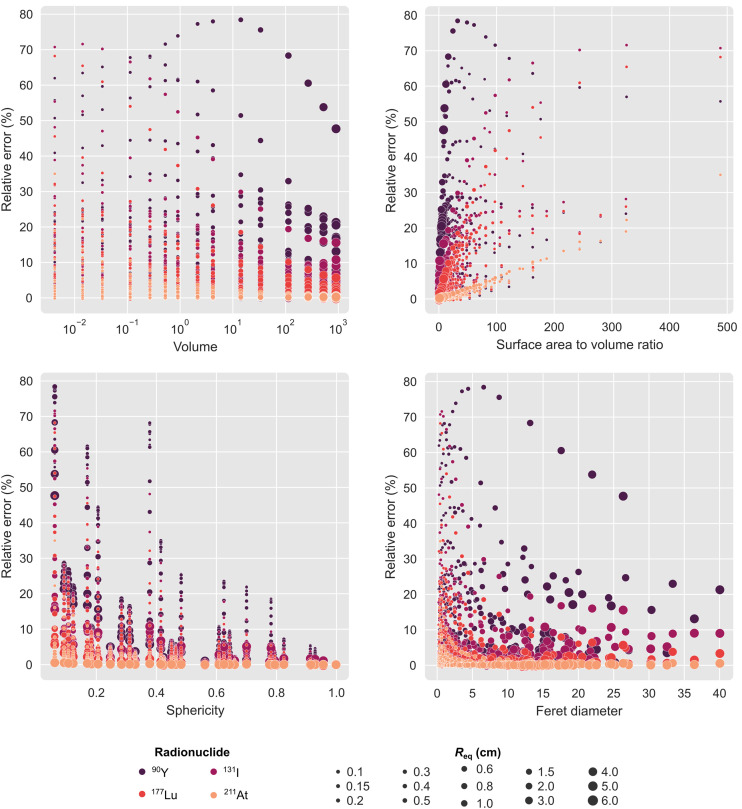
In radiopharmaceutical therapy, dosimetry-based treatment planning and response evaluation require accurate estimates of tumor-absorbed dose. Tumor dose estimates are routinely derived using simplistic spherical models, despite the well-established influence of tumor geometry on the dosimetry. Moreover, the degree of disease invasiveness correlates with departure from ideal geometry; malignant lesions often possess lobular, spiculated, or otherwise irregular margins in contrast to the commonly regular or smooth contours characteristic of benign lesions. To assess the effects of tumor shape, size, and margin contour on absorbed dose, an array of tumor geometries was modeled using computer-aided design software, and the models were used to calculate absorbed dose per unit of time-integrated activity (i.e., S values) for several clinically applied therapeutic radionuclides (90Y, 131I, 177Lu, 211At, 225Ac, 213Bi, and 223Ra). Methods: Three-dimensional tumor models of several different shape classifications were generated using Blender software. Ovoid shapes were generated using axial scaling. Lobulated, spiculated, and irregular contours were generated using noise-based mesh deformation. The meshes were rigidly scaled to different volumes, and S values were then computed using PARaDIM software. Radiomic features were extracted for each shape, and the impact on S values was examined. Finally, the systematic error present in dose calculations that model complex tumor shapes versus equivalent-mass spheres was estimated. Results: The dependence of tumor S values on shape was largest for extreme departures from spherical geometry and for long-range emissions (e.g., 90Y β-emissions). S values for spheres agreed reasonably well with lobulated, spiculated, or irregular contours if the surface perturbation was small. For marked deviations from spherical shape and small volumes, the systematic error of the equivalent-sphere approximation increased to 30%–75% depending on radionuclide. The errors were largest for shapes with many long spicules and for spherical shells with a thickness less than or comparable to the particle range in tissue. Conclusion: Variability in tumor S values as a function of tumor shape and margin contour was observed, suggesting use of contour-matched phantoms to improve the accuracy of tumor dosimetry in organ-level dosimetry paradigms. Implementing a library of tumor phantoms in organ-level dosimetry software may facilitate optimization strategies for personalized radionuclide therapies.
Figures


References
-
- Stabin MG, Sparks RB, Crowe E. OLINDA/EXM: the second-generation personal computer software for internal dose assessment in nuclear medicine. J Nucl Med. 2005;46:1023–1027. - PubMed
-
- Kesner A, Olguin E, Zanzonico P, Bolch W. MIRDCalc V 1.0: a community spreadsheet tool for organ-level radiopharmaceutical absorbed dose calculations [abstract]. J Nucl Med. 2018;59(suppl 1):473.
Publication types
MeSH terms
Grants and funding
LinkOut - more resources
Full Text Sources
Medical
Miscellaneous