. 2023 Jul 1;94(1):84-97.
doi: 10.1016/j.biopsych.2023.02.012. Epub 2023 Mar 14.

The Impact of Mmu17 Non-Hsa21 Orthologous Genes in the Ts65Dn Mouse Model of Down Syndrome: The Gold Standard Refuted

Affiliations

The Impact of Mmu17 Non-Hsa21 Orthologous Genes in the Ts65Dn Mouse Model of Down Syndrome: The Gold Standard Refuted

Faycal Guedj et al. Biol Psychiatry. .

Abstract

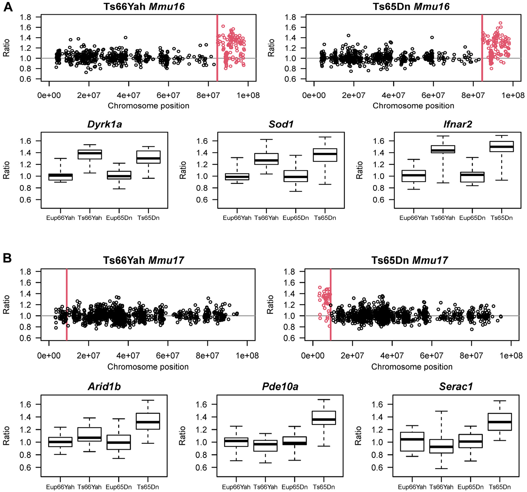
Background: Despite successful preclinical treatment studies to improve neurocognition in the Ts65Dn mouse model of Down syndrome, translation to humans has failed. This raises questions about the appropriateness of the Ts65Dn mouse as the gold standard. We used the novel Ts66Yah mouse that carries an extra chromosome and the identical segmental Mmu16 trisomy as Ts65Dn without the Mmu17 non-Hsa21 orthologous region.

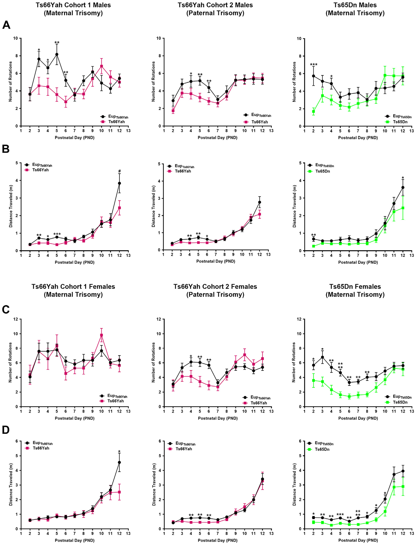
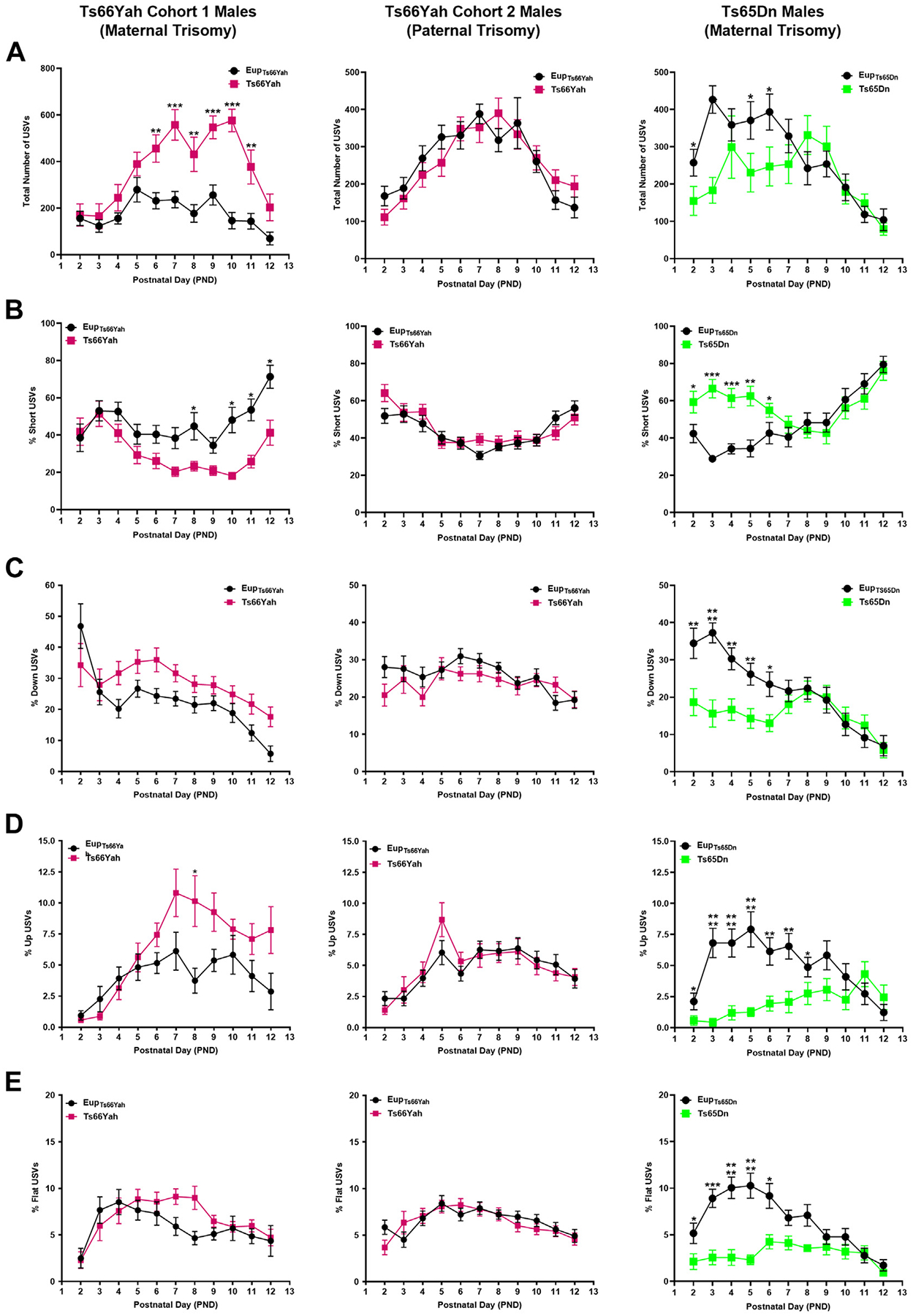
Methods: Forebrains from embryonic day 18.5 Ts66Yah and Ts65Dn mice, along with euploid littermate controls, were used for gene expression and pathway analyses. Behavioral experiments were performed in neonatal and adult mice. Because male Ts66Yah mice are fertile, parent-of-origin transmission of the extra chromosome was studied.

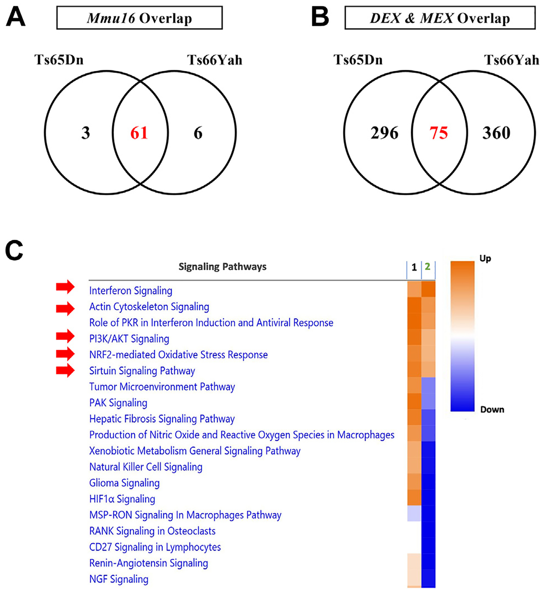
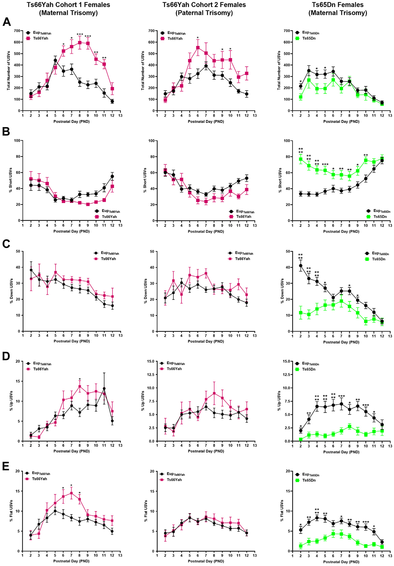
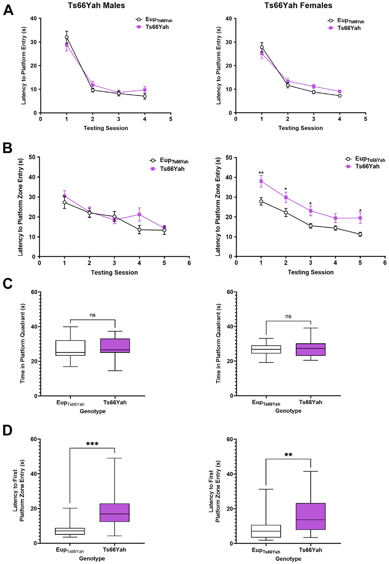
Results: Forty-five protein-coding genes mapped to the Ts65Dn Mmu17 non-Hsa21 orthologous region; 71%-82% are expressed during forebrain development. Several of these genes are uniquely overexpressed in Ts65Dn embryonic forebrain, producing major differences in dysregulated genes and pathways. Despite these differences, the primary Mmu16 trisomic effects were highly conserved in both models, resulting in commonly dysregulated disomic genes and pathways. Delays in motor development, communication, and olfactory spatial memory were present in Ts66Yah but more pronounced in Ts65Dn neonates. Adult Ts66Yah mice showed milder working memory deficits and sex-specific effects in exploratory behavior and spatial hippocampal memory, while long-term memory was preserved.

Conclusions: Our findings suggest that triplication of the non-Hsa21 orthologous Mmu17 genes significantly contributes to the phenotype of the Ts65Dn mouse and may explain why preclinical trials that used this model have unsuccessfully translated to human therapies.

Keywords: Down syndrome; Mmu17 non-Hsa21 orthologous genes; Mouse models; Phenotype; Trisomy 21; Ts65Dn; Ts66Yah.


Figures

Figure 1.
Figure 1.
Expression of Mmu16 orthologous and Mmu17 non-Hsa21 orthologous genes in the Ts66Yah and Ts65Dn E18.5 embryonic forebrain. (A) (Top) Chromosomal map showing the overexpression of Mmu16 trisomic genes (red open circles) in the Ts66Yah and Ts65Dn embryonic forebrain. Chromosomal position is represented on the x-axis as the distance (in base pair) from the start of Mmu16. For both Ts65Dn and Ts66Yah, the Mmu16 trisomic region starts at Mrpl39 located 84,514,464 bp (84.5 Mb) distally from the start of chromosome 16. (Bottom) Examples of Mmu16 orthologous genes that are overexpressed in both mouse models, including Dyrk1a, Sod1, and Ifnar2. (B) (Top) Chromosomal map showing the expression of the Mmu17 non-Hsa21 orthologous genes trisomic only in the Ts65Dn mouse model. As expected, these genes were only overexpressed in the Ts65Dn embryonic forebrain (open red circles) but not in the Ts66Yah embryonic forebrain. (Bottom) Expression of some key Mmu17 non-Hsa21 orthologous genes (Arid1b, Pde10a, and Serac1) in the Ts66Yah and Ts65Dn embryonic forebrain.
Figure 2.
Figure 2.
Overlap in dysregulated genes and pathways in the Ts65Dn and Ts66Yah E18.5 embryonic forebrains. (A) Overexpression of Mmu16 orthologous genes is conserved between the Ts65Dn and Ts66Yah mouse models. (B) Little overlap in the DEX and MEX genes is present between the Ts65Dn and Ts66Yah models, despite the conserved overexpression of Mmu16 trisomic genes. (C) Dysregulated pathways in the Ts65Dn and Ts66Yah embryonic forebrain. 1 = Ts66Yah and 2 = Ts65Dn. As a result of the distinct secondary genome-wide differences in dysregulated genes, Ts65Dn and Ts66Yah mice share very few dysregulated signaling pathways. DEX, differentially expressed; MEX, marginally expressed.
Figure 3.
Figure 3.
Neonatal motor development in the Ts66Yah and Ts65Dn mouse models. Comparison of the number of body rotations (A, C) and total distance traveled (B, D) in cohort 1 (from trisomic mothers) Ts66Yah, cohort 2 (from trisomic fathers) Ts66Yah, and Ts65Dn female and male pups between postnatal days 2 and 12. Significant differences are indicated as *p < .05, **p < .01, ***p < .001, and ****p < .0001.
Figure 4.
Figure 4.
Communication in Ts66Yah and Ts65Dn male neonates. Comparison of the total number of USVs (A) and the percent of short (B), down (C), up (D), and flat (E) USVs as a proxy for neonatal communication in cohort 1 (from trisomic mothers) Ts66Yah, cohort 2 (from trisomic fathers) Ts66Yah, and Ts65Dn male neonatal mice between postnatal days 2 and 12. Significant differences are indicated as *p < .05, **p < .01, ***p < .001, and ****p < .0001. USV, ultrasonic vocalization.
Figure 5.
Figure 5.
Communication in Ts66Yah and Ts65Dn female neonates. Comparison of the total number of USVs (A) and the percent of short (B), down (C), up (D), and flat (E) USVs as a proxy for neonatal communication in cohort 1 (from trisomic mothers) Ts66Yah, cohort 2 (from trisomic fathers) Ts66Yah, and Ts65Dn female neonatal mice between postnatal days 2 and 12. Significant differences are indicated as *p < .05, **p < .01, ***p < .001, and ****p < .0001. USV, ultrasonic vocalization.
Figure 6.
Figure 6.
Exploratory behavior and working memory in Ts66Yah and Ts65Dn adult mice. (A, B) Exploratory behavior measured as total distance traveled in the open field as well as the distance traveled in the center vs. periphery in adult cohort 1 and cohort 2 Ts66Yah female and male mice. (C, D) Percent of alternation and total number of arm entries in the Y-maze in adult cohort 1 Ts66Yah and cohort 2 Ts66Yah female and male mice. Significant differences are indicated as *p < .05 and **p < .01. ns, not significant.
Figure 7.
Figure 7.
Hippocampal-dependent spatial memory in Ts66Yah adult mice. Hippocampal-dependent spatial learning/memory was measured in the Morris water maze test in adult Ts66Yah female and male mice as latency to reach the platform during the visible platform phase (A) and the hidden platform phase (B). During the probe trial, the total time spent in the platform quadrant (C) and the latency to first enter the platform zone was also analyzed (D). Significant differences are indicated as *p < .05, **p < .01, and ***p < .001. ns, not significant.

Similar articles

Cited by

References

    1. Davisson MT, Schmidt C, Akeson EC (1990): Segmental trisomy of murine chromosome 16: A new model system for studying Down syndrome. Prog Clin Biol Res 360:263–280. - PubMed
    1. Davisson MT, Schmidt C, Reeves RH, Irving NG, Akeson EC, Harris BS, Bronson RT (1993): Segmental trisomy as a mouse model for Down syndrome. Prog Clin Biol Res 384:117–133. - PubMed
    1. Muñiz Moreno MDM, Brault V, Birling MC, Pavlovic G, Herault Y (2020): Modeling Down syndrome in animals from the early stage to the 4.0 models and next. Prog Brain Res 251:91–143. - PubMed
    1. Tosh J, Tybulewicz V, Fisher EMC (2022): Mouse models of aneuploidy to understand chromosome disorders. Mamm Genome 33:157–168. - PMC - PubMed
    1. Herault Y, Delabar JM, Fisher EMC, Tybulewicz VLJ, Yu E, Brault V (2017): Rodent models in Down syndrome research: Impact and future opportunities. Dis Model Mech 10:1165–1186. - PMC - PubMed

Publication types