. 2023 Aug;165(8):2299-2307.
doi: 10.1007/s00701-023-05581-y. Epub 2023 Apr 19.

Multi-layered repair of high-flow CSF fistulae following endoscopic skull base surgery without nasal packing or lumbar drains: technical refinements to optimise outcome

Affiliations

Multi-layered repair of high-flow CSF fistulae following endoscopic skull base surgery without nasal packing or lumbar drains: technical refinements to optimise outcome

Cathal John Hannan et al. Acta Neurochir (Wien). 2023 Aug.

Abstract

Aims: Post-operative CSF leak remains a significant problem following endoscopic skull base surgery, particularly when there is a high-flow intra-operative CSF leak. Most skull base repair techniques are accompanied by the insertion of a lumbar drain and/or the use of nasal packing which have significant shortcomings. Our aim was to review the results of a large series of endoscopic skull base cases where a high-flow intra-operative CSF leak rate was encountered and repaired to assess if modifications in technique could reduce the post-operative CSF leak rate.

Methods: A retrospective review of a prospectively maintained database of skull base cases performed by a single surgeon over a 10-year period was performed. Data regarding patient demographics, underlying pathology, skull base repair techniques and post-operative complications were analysed.

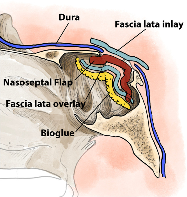
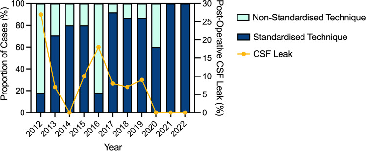
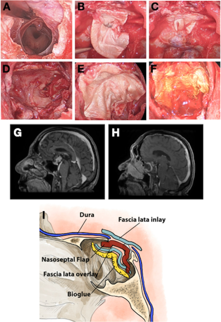
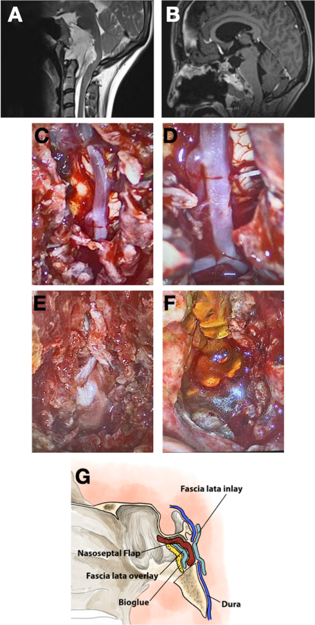
Results: One hundred forty-two cases with high-flow intra-operative CSF leak were included in the study. The most common pathologies were craniopharyngiomas (55/142, 39%), pituitary adenomas (34/142, 24%) and meningiomas (24/142, 17%). The CSF leak rate was 7/36 (19%) when a non-standardised skull base repair technique was used. However, with the adoption of a standardised, multi-layer repair technique, the post-operative CSF leak rate decreased significantly (4/106, 4% vs. 7/36, 19%, p = 0.006). This improvement in the rate of post-operative CSF leak was achieved without nasal packing or lumbar drains.

Conclusion: With iterative modifications to a multi-layered closure technique for high-flow intra-operative CSF leaks, it is possible to obtain a very low rate of post-operative CSF leak, without lumbar drains or nasal packing.

Keywords: CSF leak; Chordoma; Endoscopic skull base; Meningioma; Pituitary adenoma; Skull base.


Conflict of interest statement

The authors declare no competing interests.

Figures

Fig. 1
Fig. 1
Diagrammatic representation of the materials used in skull base repair and the order in which they are placed in the standardised technique. The most recent iteration consists of an inlay fascia lata graft, covered by a nasoseptal flap which is then covered with a further layer of fascia lata and secured using BioGlue® tissue sealant
Fig. 2
Fig. 2
Stacked column graph indicating the proportion of cases per year repaired using the standardised and non-standardised repair techniques (left y axis). The line graph overlain on the columns indicates the proportion of cases where a post-operative CSF leak was encountered (right y axis). There was an increase in the use of standardised techniques as the series progressed and a corresponding decrease in the rates of post-operative CSF leak
Fig. 3
Fig. 3
Stacked column graphs indicating the proportion of cases complicated by (A) post-operative CSF leak or (B) post-operative meningitis. ***p ≤ 0.001; ns, no statistically significant difference
Fig. 4
Fig. 4
Multi-panelled figure demonstrating the repair of a defect in the tuberculum sella/planum sphenoidale following the resection of a suprasellar craniopharyngioma. A Intra-operative endoscopic photograph demonstrating the skull base defect and view into the third ventricle following tumour resection. B Endoscopic photograph demonstrating the placement of an inlay fascia lata graft. Note that this graft is significantly larger than the dural defect. C Endoscopic photograph demonstrating the final position of the fascia lata graft, with the edges tucked in underneath the margins of the skull base defect. D Endoscopic photograph demonstrating placement of a pedicled nasoseptal flap to completely cover the skull base defect followed by the (E) placement of an onlay fascia lata graft which is in turn secured with (F) a layer of BioGlue. G Pre-operative T1-weighted MRI scan demonstrating the presence of a suprasellar craniopharyngioma with superior extension into the third ventricle. H Post-operative T1-weighted MRI scan demonstrating complete tumour resection and the enhancement of the pedicled nasoseptal flap overlying the skull base defect. I Diagrammatic representation of the Dublin technique as applied to approaches to sellar/suprasellar pathology
Fig. 5
Fig. 5
Multi-panelled figure demonstrating the repair of a clival defect following the resection of a chordoma. A Pre-operative T2-weighted MRI scan demonstrating the presence of an extensive clival chordoma, causing severe brainstem compression. B Post-operative T1-weighted MRI scan following contrast administration, demonstrating complete tumour resection with resolution of brainstem compression, along with enhancement of the nasoseptal flap. C Intra-operative endoscopic photograph following tumour resection, demonstrating a longitudinally extensive clival defect. The basilar artery and brainstem are clearly displayed. D Endoscopic view of the vertebro-basilar junction and emergence of the right anterior inferior cerebellar artery. E Intra-operative photograph demonstrating placement of the inlay layer of fascia lata within the dural/bony defect. F Final overview of the skull base repair, with a layer of BioGlue applied over an onlay fascia lata graft. G Diagrammatic representation of the Dublin technique as applied to approaches to clival pathology

References

    1. Ackerman PD, Spencer DA, Prabhu VC. The efficacy and safety of preoperative lumbar drain placement in anterior skull base surgery. J Neurol Surg Rep. 2013;74:1–9. doi: 10.1055/s-0032-1331022. - DOI - PMC - PubMed
    1. Cavallo LM, Solari D, Somma T, Cappabianca P. The 3F (fat, flap, and flash) technique for skull base reconstruction after endoscopic endonasal suprasellar approach. World Neurosurg. 2019;126:439–446. doi: 10.1016/j.wneu.2019.03.125. - DOI - PubMed
    1. Conger A, Zhao F, Wang X, Eisenberg A, Griffiths C, Esposito F, Carrau RL, Barkhoudarian G, Kelly DF. Evolution of the graded repair of CSF leaks and skull base defects in endonasal endoscopic tumor surgery: trends in repair failure and meningitis rates in 509 patients. J Neurosurg. 2018;130:861–875. doi: 10.3171/2017.11.Jns172141. - DOI - PubMed
    1. CSF rhinorrhoea after endonasal intervention to the skull base (CRANIAL): a multicentre prospective observational study (2022). Front Oncol 12:1049627. 10.3389/fonc.2022.1049627 - PMC - PubMed
    1. Esposito F, Dusick JR, Fatemi N, Kelly DF. Graded repair of cranial base defects and cerebrospinal fluid leaks in transsphenoidal surgery. Operative Neurosurgery. 2007;60:ONS295–ONS304. doi: 10.1227/01.Neu.0000255354.64077.66. - DOI - PubMed

Publication types

MeSH terms

LinkOut - more resources