. 2023 Jul 3;29(37):e202300663.
doi: 10.1002/chem.202300663. Epub 2023 May 10.

Photothermal Perylene Bisimide Hydrogels

Affiliations

Photothermal Perylene Bisimide Hydrogels

Lisa Thomson et al. Chemistry. .

Abstract

Gels formed using a perylene bisimide (PBI) as a low molecular weight gelator can show the photothermal effect. Formation of the PBI radical anion results in new absorption bands forming, meaning that subsequent irradiation with a wavelength of light overlapping with the new absorption band leads to heating of the gel. This approach can be used to heat the gel, as well as the surrounding milieu. We show how we can use electrochemical methods as well as multicomponent systems to form the radical anion without the need for UV light, and how we can use the photothermal effect to induce phase transitions in the solutions above the gels by exploiting photothermal behavior.

Keywords: PBI; gel; multicomponent; photothermal; radical anion.


Conflict of interest statement

The authors declare no conflict of interest.

Figures

Figure 1
Figure 1
(a) Chemical structure of PBI‐A; (b) Photograph of a gel formed from PBI‐A in 7 mL Sterilin vial. Scale bars represent 1 cm; (c) Normalized UV‐Vis spectrum of a gel formed from PBI‐A before (red) and after (black) irradiation with a 365 nm LED for 10 min.
Figure 2
Figure 2
Photographs of gels formed from PBI‐A using a thermal camera. In all cases, the upper temperature achieved is at the top left (red) and the minimum temperature on each image in the bottom left (dark blue). (a) shows two gels (highlighted by arrows), one which has been irradiated with a 365 nm LED for 30 min (bottom) and one which has not (top). This cannot be seen however due to the background and cuvette being the same temperature; (b) shows the gels 2 min after stopping irradiation with the 365 nm LED showing the samples have now cooled to room temperature; (c) shows the cooled gels 10 min after irradiation with a 810 nm LED which has increased in temperature; (d) shows three gels (indicated by arrows), two which have been irradiated with a 365 nm LED for 30 min (middle and bottom) and one which has not (top). This cannot be seen due to the background and cuvette being the same temperature; (e) 2 min after the subsequent 810 nm irradiation on the middle gel but stopping all irradiation on the bottom gel; (f) 10 min after the subsequent 810 nm irradiation on the middle gel but stopping all irradiation on the bottom gel. Scale bars represent 1 cm.
Figure 3
Figure 3
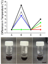
(Top) Temperature difference when irradiating PBI‐A gels with a 365 nm LED for 0 min (red); 30 seconds (orange); 1 minute (green); 5 min (blue); 30 min (grey) and 1 h (black). Temperatures were measured at four time points: (A) before any irradiation; (B) immediately after irradiation with the 365 nm LED; (C) after ten minutes of cooling post‐irradiation with the 365 nm LED; (D) after 15 min of irradiation with a 810 nm LED. (Bottom) Left to right shows a PBI‐A gel pre‐irradiated with a 365 nm LED and cooled with a PNIPAAm solution placed on top, the same gel after irradiation for 3 min with a near‐IR LED leading to heating of the PNIPAAm solution above the LCST, and a control sample without the pre‐irradiation with 365 nm LED but still with 3 min irradiation with the near‐IR LED showing no LCST transition. The scale bar represents 1 cm.
Figure 4
Figure 4
LED patterning of PBI‐A gels. (a) Photograph of gel formed in a 5 mm cuvette with light absorbing tape used as a mask; (b) normalised UV‐Vis data for the unmasked section of gel; (c) normalised UV‐Vis data for the masked section of the gel. For (b) and (c), data were collected before any irradiation (red); after 30 min of 365 nm irradiation (black); after 15 min relaxation (blue); after 30 min relaxation (green); after 1 h relaxation (grey); after 2 h relaxation (orange); (d) Photographs of the patterned gel using a thermal camera showing (i) the gel which has been irradiated with a 365 nm LED for 30 min; (ii) gel after cooling to room temperature. The mask was then removed after cooling; (iii) irradiation of the gel with an 810 nm LED for 1 min; (iv) irradiation of the gel with an 810 nm LED for 10 min. Scale bars for (d)–(g) represent 1 cm.
Figure 5
Figure 5
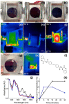
(a) Photograph of PBI‐A gel on an electrode; (b) gel after formation of the radical anion; (c) gel after electrochemical formation of the radical anion followed by irradiation with 810 nm LED. (d)–(f) show the same gels imaged using a thermal camera; the upper temperature achieved is at the top left and the minimum temperature on each image in the bottom left; (g) photograph of an electrochemically‐patterned gel and (h) the corresponding thermal image after irradiation with a 810 nm LED; (i) Chemical structure of Stilbene‐F; (j) normalised UV‐Vis spectrum of a mixed PBI‐A and Stilbene‐F gel before any irradiation (red), after 10 min of 365 nm radiation (black) and after 10 min of 450 nm radiation (blue); (k) temperature difference when irradiating mixed PBI‐A and Stilbene‐F gels with 810 nm. Black data shows gel which was first irradiated with 365 nm and cooled and blue data shows gel which was first irradiated with 450 nm and cooled before the 810 nm irradiation. Scale bars for (a) represent 1 cm.

References

    1. Bian W., Wang Y., Pan Z., Chen N., Li X., Wong W.-L., Liu X., He Y., Zhang K., Lu Y.-J., ACS Appl. Nano Mater. 2021, 4, 11353–11385.
    1. Yang Z., Sun Z., Ren Y., Chen X., Zhang W., Zhu X., Mao Z., Shen J., Nie S., Mol. Med. 2019, 20, 5–15. - PMC - PubMed
    1. Huang X., El-Sayed I. H., Qian W., El-Sayed M. A., J. Am. Chem. Soc. 2006, 128, 2115–2120. - PubMed
    1. Jung H. S., Verwilst P., Sharma A., Shin J., Sessler J. L., Kim J. S., Chem. Soc. Rev. 2018, 47, 2280–2297. - PMC - PubMed
    1. Zhao L., Liu Y., Xing R., Yan X., Angew. Chem. Int. Ed. 2020, 59, 3793–3801; - PubMed
    2. Angew. Chem. 2020, 132, 3821–3829.