. 2023 May 30;42(5):112409.
doi: 10.1016/j.celrep.2023.112409. Epub 2023 Apr 18.

Integrated glycoproteomic characterization of clear cell renal cell carcinoma

Affiliations

Integrated glycoproteomic characterization of clear cell renal cell carcinoma

T Mamie Lih et al. Cell Rep. .

Abstract

Clear cell renal cell carcinoma (ccRCC), a common form of RCC, is responsible for the high mortality rate of kidney cancer. Dysregulations of glycoproteins have been shown to associate with ccRCC. However, the molecular mechanism has not been well characterized. Here, a comprehensive glycoproteomic analysis is conducted using 103 tumors and 80 paired normal adjacent tissues. Altered glycosylation enzymes and corresponding protein glycosylation are observed, while two of the major ccRCC mutations, BAP1 and PBRM1, show distinct glycosylation profiles. Additionally, inter-tumor heterogeneity and cross-correlation between glycosylation and phosphorylation are observed. The relation of glycoproteomic features to genomic, transcriptomic, proteomic, and phosphoproteomic changes shows the role of glycosylation in ccRCC development with potential for therapeutic interventions. This study reports a large-scale tandem mass tag (TMT)-based quantitative glycoproteomic analysis of ccRCC that can serve as a valuable resource for the community.

Keywords: CP: Cancer; N-linked glycosylation; clear cell renal cell carcinoma; cross-correlation; glycoproteomics; mass spectrometry.


Conflict of interest statement

Declaration of interests The authors declare no competing interests.

Figures
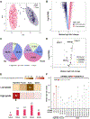
Figure 1
Figure 1
Comparative analysis to examine alter glycosylation in ccRCC. (A) Principal component analysis between ccRCC tumors and NATs. (B) Differential analysis between tumors and NATs. Significantly altered intact glycopeptides were defined as >2-fold changes with false discovery rate (FDR)<0.01. (C) Distribution of glycan types for upregulated and downregulated intact glycopeptides. (D) Differential analysis of glycosylation enzymes on global level. Significantly altered glycosylation enzymes were defined as >1.5-fold changes with FDR<0.05. (E) Glycan types observed in differentially expressed intact glycopeptides (FDR<0.05) between high-grade and low-grade tumors. (F) Glycosylation changes in NDRN249SSDETFLK of GPNMB in high-grade tumors relative to low-grade tumors (FDR<0.05 as significant changes). (B, D, E, and F) The p-values were computed using two-sided Wilcoxon rank sum test and adjusted (i.e., FDR) using Benjamini-Hochberg method. See also Figure S2 and Table S2.
Figure 2
Figure 2
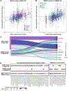
Alteration in glycosylation in BAP1-mutant and PBRM1-mutant tumors. (A) Median log2 fold change between intact glycopeptides and corresponding glycoproteins of BAP1-mutant and BAP1-WT tumors. (B) Median log2 fold change between intact glycopeptides and corresponding glycoproteins of PBRM1-mutant and PBRM1-WT tumors. (C) Distribution of glycan types for upregulated and downregulated intact glycopeptides in BAP1-mutant and PBRM1-mutant compared to wildtypes. (D) Biological processes of BAP1-mutant-associated and PBRM1-mutant-associated glycoproteins based on the differential analysis of intact glycopeptides of BAP1-mutant vs BAP1-WT, PBRM1-mutant vs PBRM1-WT, and BAP1-mutant vs PBRM1-mutant. Significantly altered intact glycopeptides were defined as >1.5-fold changes with FDR<0.05. The p-values were computed using two-sided Wilcoxon rank sum test and adjusted (FDR) using Benjamini-Hochberg method. See also Table S3.
Figure 3
Figure 3
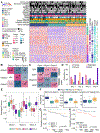
Inter-tumor heterogeneity of ccRCC. (A) Glycoproteomic subtyping of ccRCC. (B) Association between Glyco subtypes and global subtypes (* p-value<0.05, hypergeometric test). (C) Association between Glyco subtypes and immune subtypes (* p-value<0.05, hypergeometric test). (D) Glycan type distribution of each intact glycopeptide cluster (IPC). (E) Association among Glyco subtypes, glycan type, and IPC. (F) Examples of intact glycopeptide signatures of each glyco subtype. (G) Survival analysis of Glyco subtypes. Log-rank test p=0.015 (H) Survival analysis based on abundance of High-Man where samples with abundance above median were assigned to High group (n=51), otherwise assigned to Low group (n=52). Log-rank test p=0.013. See also Table S4.
Figure 4
Figure 4
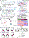
Cross-correlation between glycosylation and phosphorylation in ccRCC. (A) Correlation between glycan types and phosphoproteins. (B) Networks based on the linear models constructed using intact glycopeptides and phosphopeptides that were associated with complement and coagulation cascades. (C) Association between EGFR-N352 (N2H5) and EGFR-S1018 with p=0.037 (t-statistic). (D) Cross-Correlation (CC) clusters derived using NMF multi-omic clustering. (E) Distinct glycosylation (FLT1-N251, N4H5S1) and phosphorylation (ENO1-S373) events in CC 1 tumors relative to the other CC clusters. (F) Distinct glycosylation (CYBB-N240, N2H8) and phosphorylation (DDX3X-S102) events in CC 2 tumors relative to the other CC clusters. (G) Distinct glycosylation (TF-N630, N3H6S1) and phosphorylation (HBA2-T138) events in CC 3 tumors relative to the other CC clusters. (H) Summarized key observations for ccRCC in this study. See also Figure S3 and Table S5.

References

    1. Hsieh JJ, Purdue MP, Signoretti S, Swanton C, Albiges L, Schmidinger M, Heng DY, Larkin J, and Ficarra V (2017). Renal cell carcinoma. Nat Rev Dis Primers 3, 17009. 10.1038/nrdp.2017.9. - DOI - PMC - PubMed
    1. Siegel RL, Miller KD, Fuchs HE, and Jemal A (2022). Cancer statistics, 2022. CA Cancer J Clin 72, 7–33. 10.3322/caac.21708. - DOI - PubMed
    1. Blanco AI, Teh BS, and Amato RJ (2011). Role of radiation therapy in the management of renal cell cancer. Cancers (Basel) 3, 4010–4023. 10.3390/cancers3044010. - DOI - PMC - PubMed
    1. Diamond E, Molina AM, Carbonaro M, Akhtar NH, Giannakakou P, Tagawa ST, and Nanus DM (2015). Cytotoxic chemotherapy in the treatment of advanced renal cell carcinoma in the era of targeted therapy. Crit Rev Oncol Hematol 96, 518–526. 10.1016/j.critrevonc.2015.08.007. - DOI - PubMed
    1. Li QK, Pavlovich CP, Zhang H, Kinsinger CR, and Chan DW (2019). Challenges and opportunities in the proteomic characterization of clear cell renal cell carcinoma (ccRCC): A critical step towards the personalized care of renal cancers. Semin Cancer Biol 55, 8–15. 10.1016/j.semcancer.2018.06.004. - DOI - PMC - PubMed

Publication types