. 2023 May 19;18(5):1101-1114.
doi: 10.1021/acschembio.2c00805. Epub 2023 Apr 19.

New High-Throughput Screen Discovers Novel Ligands of Full-Length Nuclear Receptor LRH-1

Affiliations

New High-Throughput Screen Discovers Novel Ligands of Full-Length Nuclear Receptor LRH-1

M Merced Malabanan et al. ACS Chem Biol. .

Abstract

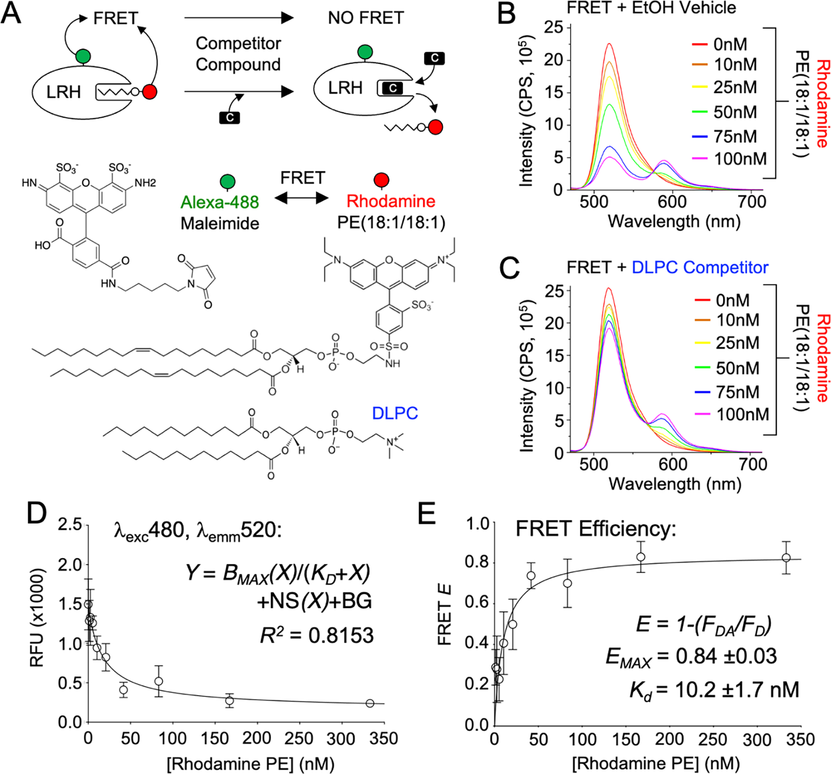
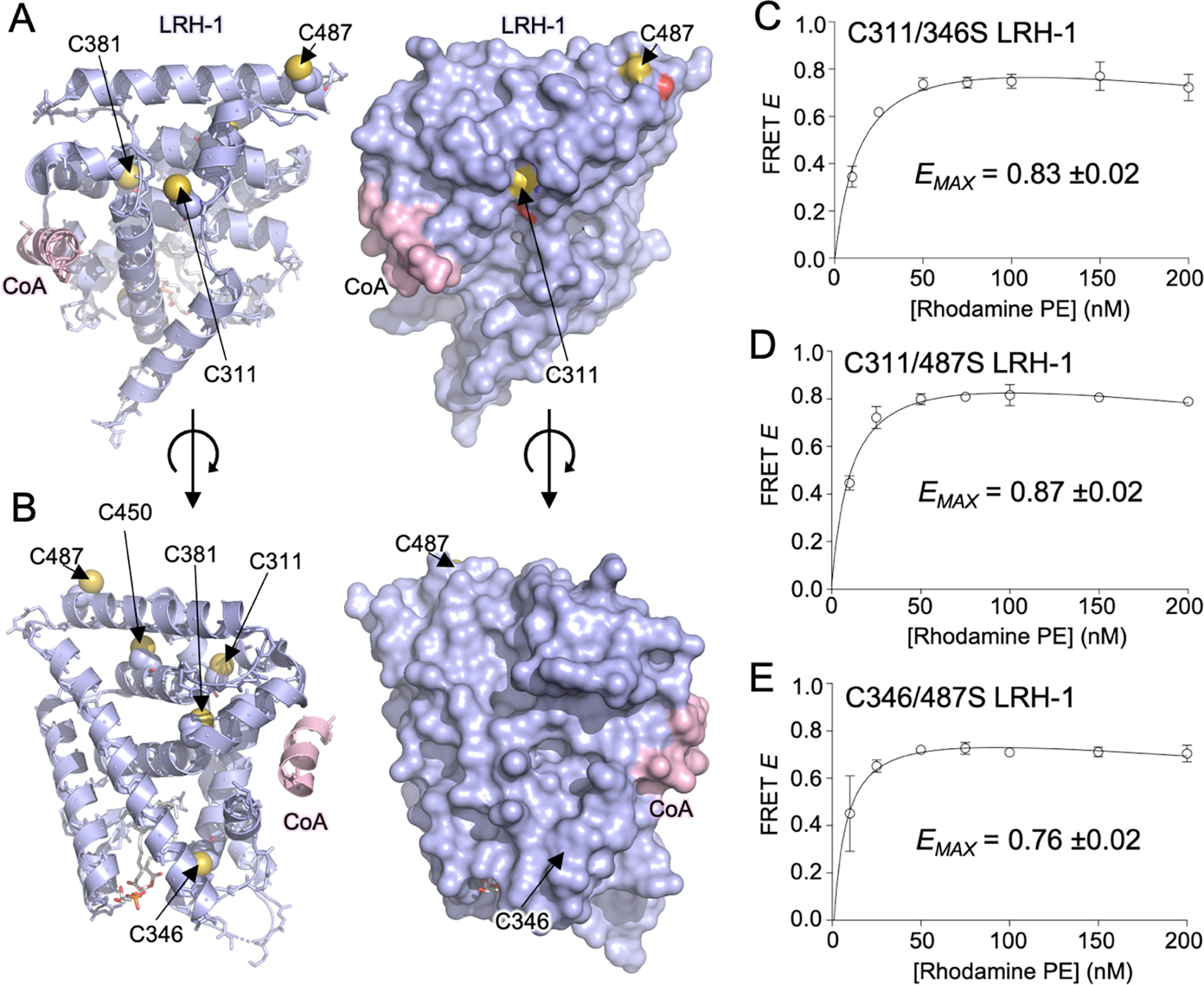
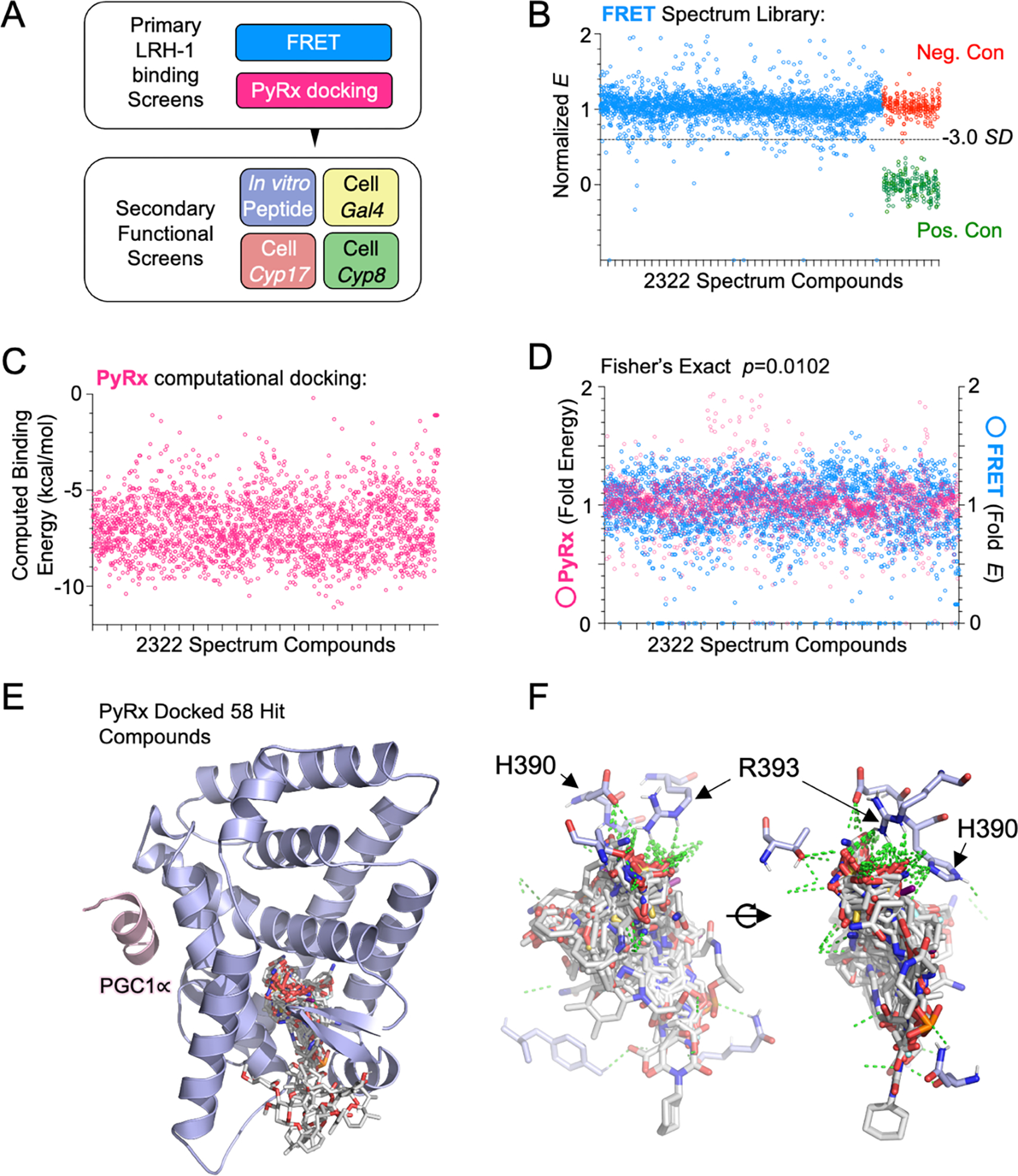
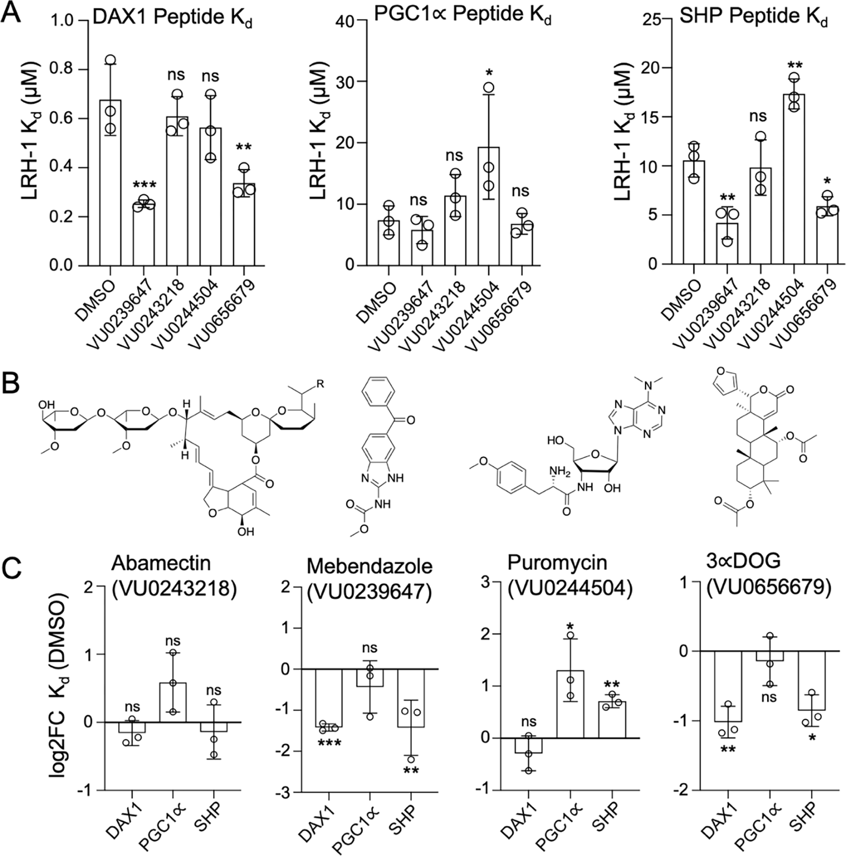
Nuclear receptor liver receptor homolog-1 (LRH-1, NR5A2) is a lipid-regulated transcription factor and an important drug target for several liver diseases. Advances toward LRH-1 therapeutics have been driven recently by structural biology, with fewer contributions from compound screening. Standard LRH-1 screens detect compound-induced interaction between LRH-1 and a transcriptional coregulator peptide, an approach that excludes compounds that regulate LRH-1 through alternative mechanisms. Here, we developed a FRET-based LRH-1 screen that simply detects compound binding to LRH-1, applying it to discover 58 new compounds that bind the canonical ligand-binding site in LRH-1 (2.5% hit rate), also supported by computational docking. Four independent functional screens identified 15 of these 58 compounds to also regulate LRH-1 function in vitro or in living cells. Although one of these 15 compounds, abamectin, directly binds LRH-1 and regulates full-length LRH-1 in cells, abamectin failed to regulate the isolated ligand-binding domain in standard coregulator peptide recruitment assays using PGC1α, DAX-1, or SHP. Abamectin treatment of human liver HepG2 cells selectively regulated endogenous LRH-1 ChIP-seq target genes and pathways associated with known LRH-1 functions in bile acid and cholesterol metabolism. Thus, the screen reported here can discover compounds not likely to have been identified in standard LRH-1 compound screens but which bind and regulate full-length LRH-1 in cells.


Figures

Figure 1.
Figure 1.. A scalable FRET-based LRH-1 compound screen detects a known compound that directly binds the single regulatory site in LRH-1 (DLPC).
A. Assay design for LRH-1 with structures of FRET donor (Alexa-fluor maleimide), FRET acceptor probe (Rhodamine-PE, Rh-PE) and known LRH-1 ligand DLPC (unlabeled). Compounds like DLPC that compete with the Rh-PE probe should decrease FRET. B. Fluorescence emission spectra (λexc 480nm) of 100nM LRH-1–488 with indicated concentrations of Rh-PE, in the presence of ethanol vehicle. Excitation of LRH-1–488 emits at 520nm, Rh-PE FRET decreases 520nm emission in a concentration dependent manner. C. Fluorescence emission spectra (λexc 480nm) of 100nM LRH-1-488 in the presence of 5μM DLPC competitor with indicated concentrations of Rh-PE. D. One-site model fit of emission intensity of 50nM LRH-1-488 (λexc480 nm, λemm520 nm) as a function of Rh-PE acceptor concentration, highlight saturability of the signal. E. Dependence of FRET efficiency (E) on Rh-PE probe concentration (probe Kd ≈ 10nM), further showing saturation of the signal at over 20X the Kd. These data suggest a FRET-based screen can be used to detect direct compound binding to LRH-1-488.
Figure 2:
Figure 2:. Efficiency of FRET is comparable between wild type and Cys-lite mutants of LRH-1.
A. Ribbon cartoon (left) and solvent-accessible surface models (right) of the human LRH-1 LBD (colored blue), identifying the positions of C381, C311 and C487 (yellow spheres) in the crystal structure (PDB:6OQX) with coactivator peptide (CoA, pink). B. Same models and representations as in A. but rotated 180 degrees about vertical axis to better show C450 and C346, along with another view of indicated residues and the coactivator peptide. C-E. FRET efficiency plotted as a function of Rh-PE probe concentration for indicated Cys-lite mutants of labeled LRH-1-488. Following labeling of indicated mutants of LRH-1 with the FRET acceptor, the Rh-PE probe was titrated and FRET measured. Indicated maximum FRET efficiencies were calculated identically as in Figure 1E (error represents +/− standard deviation from three independent determinations). These data suggest that FRET efficiency (E) with the Rh-PE probe was concentration-dependent and saturable independent of the LRH-1-488 Cys-lite mutant used. The wild-type human LRH-1-488 was used for screening.
Figure 3.
Figure 3.. 58 compounds from the Spectrum Library directly bind LRH-1.
A. Workflow to identify and validate LRH-1 regulatory compounds. B. Scatter plot of primary FRET screen of 2,322 compound Discovery Spectrum library (10μM each compound), hits defined as compounds that decreased FRET efficiency greater than 3 standard deviations (3σ) from the mean (E/E0 ≥ Mean–3σ) relative to DMSO control, hit compounds are blue points below indicated −3σ dotted line. C. PyRx computational docking of the same Spectrum library to a crystal structure of LRH-1 ligand binding domain (PDB:6OQX). D. Normalized (fold change over vehicle) comparison of docking and wet-lab primary FRET screens, suggesting a significantly different subset of hit compounds were identified, statistically shown by contingency analysis in Figure S3. E. Docked positions of all 58 hit compounds in the LRH-1 ligand binding domain crystal structure (PDB:6OQX). F. Close up of docked positions of hit compounds predicted to interact with LRH-1 residues H390 and R393, two residues previously shown to be important for LRH-1 function. Compounds predicted to bind at the mouth of the ligand binding pocket removed for clarity in this panel but shown in Panel E. These data identify 58 compounds that directly bind LRH-1 at the canonical ligand binding site, and computationally predict interactions with LRH-1 residues known to be important for LRH-1 function.
Figure 4.
Figure 4.. Functional screens identify 15 of 58 direct-binding LRH-1 compounds also regulate LRH-1 function.
A. Log2 fold change (Log2FC) of fluorescence anisotropy relative to DMSO vehicle of a FAM-labeled PGC1α coactivator peptide binding to isolated human LRH-1 LBD, induced by 10μM each of 50 hit compounds (8 of the 58 hit compounds with high inherent fluorescence were not analyzed), standard error from duplicate screens shown, 5 compounds changed peptide binding by Dunnett’s multiple comparisons following ordinary one-way ANOVA (padj<0.05). B. Log2FC relative to DMSO of luciferase reporter assay, 10μM each of 58 hit compounds in mammalian CHO cells expressing chimeric LRH-1 LBD fused to the Gal4 DNA-binding domain, standard error from four screens shown, 5 compounds changed LRH-1 activity (padj<0.05, as in A.). C. and D. Log2FC relative to DMSO of luciferase reporter in HEK293T cells transfected with full length human LRH-1, Renilla luciferase and either (C) Cyp17A1 or (D) Cyp8B1 driven luciferase reporters, 10μM each of 58 hit compounds, four Cyp17A1 and two Cyp8B1 independent experiments, showed 6 (Cyp17A1) or 6 (Cyp8B1) compounds changed LRH-1 activity (padj<0.05, as in panel A). E. Heatmap of responses relative to DMSO induced by the 15 hit compounds in indicated screens, centroid linkage clustering by Spearman rank suggests 3 groups of compounds with similar patterns of LRH-1 regulation in these four screens. F. Left, 15 hit compounds docked into human LRH-1 LBD (PDB:6OQX) using PyRx, compounds colored to heatmap clustering in panel E; Right, compounds without LRH-1 protein, showing 15 hit compounds populate the ligand binding pocket (Clusters 1 and 2) and the mouth of the ligand binding pocket (Cluster 3 only). These data identify 15/58 compounds that regulate LRH-1 function in at least one of four secondary screens that monitor LRH-1 function.
Figure 5.
Figure 5.. Detailed, lower-throughput validation that abamectin does not regulate LRH-1 interaction with well-known LRH-1 coregulator peptides.
A. Bar graphs of dissociation constants (Kd) for each of three FAM-labeled coregulator peptides (DAX1, PGC1α and SHP) for purified human LRH-1 LBD, induced by indicated compound, determined by fluorescence polarization/anisotropy. B. Chemical structures of hit compounds identified in the primary screen, from left to right, Abamectin (VU0243218, mixture of R=CH3, and R=CH2CH3), Mebendazole (VU0239647), Puromycin (VU0244504) and 3-α-acetoxy-dihydro-deoxy-gedunin (3∝DOG, VU0656679). C. Log2 fold change (Log2FC) relative to DMSO of indicated compound-induced Kd of DAX1, PGC1α and SHP peptides for LRH-1. In all panels *padj<0.05, **padj<0.01, ***padj<0.001 in an ordinary one-way ANOVA corrected for multiple comparisons (Dunnett’s); ns is not significant. These data suggest abamectin does not alter LRH-1 interaction with the three coregulator peptides tested, while other hit compounds could selectively regulate LRH-1 in the same assay, done in parallel.
Figure 6.
Figure 6.. Abamectin treatment of human HepG2 cells selectively activates genes that are direct targets of LRH-1.
A. Structure of abamectin, an 80/20 fermentation product mixture of avermectin b1a (R=CH2CH3) and avermectin b1b (R=CH3), which differ by one carbon. B. Volcano plot comparing RNA-seq from human liver HepG2 cells, an established model of LRH-1 regulation of endogenous human liver target genes, treated for 24hrs with either 50uM abamectin or DMSO vehicle control. Abamectin regulated 1,601 transcripts in these cells compared to DMSO control (padj<0.05 and Log2 fold change at least ±1.0). We noted several classic LRH-1 target genes were regulated by abamectin, these genes have been previously identified to recruit LRH-1 by ChIP-seq in HepG2 cells, labeled on the volcano plot. C. Enrichment of abamectin-regulated genes in the LRH-1 ChIP-seq target gene set, comparing all abamectin-regulated genes from RNA-seq (padj<0.05 and Log2 fold change at least ±1.0) vs. all quantifiable genes in the transcriptome. While only 1601 of all 28,636 genes (5.6%) are regulated by abamectin, 557 of all 5,646 (9.9%) LRH-1 ChIP-seq target genes are regulated by abamectin, a significant enrichment of abamectin genes amount all LRH-1 ChIP-seq target genes (****p<0.0001, Fisher Exact Test). D. Crystal structure of LRH-1 bound to RJW100 (PDB: 5L11), showing RJW100 occupancy in the ligand binding site. E. RTqPCR of CYP11A1 LRH-1 target gene from total HepG2 cell RNA after 24hr treatment with DMSO vehicle, 20uM RJW100 and/or 10uM abamectin, as indicated. These data suggest RJW100 attenuates the abamectin response. F. Same as in B., however RT-qPCR of the CYP8B1 LRH-1 target gene from total HepG2 cell RNA after 24hr treatment with DMSO vehicle, 20uM RJW100 and/or 10uM topotecan, as indicated. These data suggest RJW100 attenuates the response of these endogenous LRH-1 target genes to hit compounds, indicated padj values are one-way ANOVA Dunnett’s corrected for multiple comparisons. Together, these data suggest abamectin selectively regulates direct LRH-1 target genes in HepG2 cells, which express endogenous human LRH-1, consistent with direct activation of LRH-1.

Similar articles

Cited by

References

    1. Bell PA; Munck A Steroid Binding and Stabilization of Glucocorticoid-Receptor Proteins from Rat Thymus. Biochemical Journal 1972, 126 (Page 3). 10.1042/bj1260011pa. - DOI - PMC - PubMed
    1. Bocos C; Göttlicher M; Gearing K; Banner C; Enmark E; Teboul M; Crickmore A; Gustaffson JÅ Fatty Acid Activation of Peroxisome Proliferator-Activated Receptor (PPAR). Journal of Steroid Biochemistry and Molecular Biology 1995, 53 (1–6), 467–473. 10.1016/0960-0760(95)00093-F. - DOI - PubMed
    1. Schoonjans K; Staels B; Auwerx J The Peroxisome Proliferator Activated Receptors (PPARs) and Their Effects on Lipid Metabolism and Adipocyte Differentiation. Biochimica et Biophysica Acta - Lipids and Lipid Metabolism. Elsevier B.V. July 26, 1996, pp 93–109. 10.1016/0005-2760(96)00066-5. - DOI - PubMed
    1. Chakravarthy M. v.; Lodhi IJ; Yin L; Malapaka R. R. v; Xu HE; Semenkovich CF Identification of a Physiologically Relevant Endogenous Ligand for PPARa in Liver. Cell 2009, 138 (3), 476–488. 10.1016/j.cell.2009.05.036. - DOI - PMC - PubMed
    1. Kininis M; Kraus WL A Global View of Transcriptional Regulation by Nuclear Receptors: Gene Expression, Factor Localization, and DNA Sequence Analysis. Nucl Recept Signal 2008, 6 (1), nrs.06005. 10.1621/nrs.06005. - DOI - PMC - PubMed

Publication types

MeSH terms